

Background and aims:
Bone disease extension in symptomatic multiple myeloma (MM) has prognostic implications. However, methods better than the traditional ones are necessary to establish the diagnosis according to the updated International Myeloma Working Group criteria (IMWG).
Magnetic resonance imaging (MRI) is a novel technique to detect bone lesions at diagnosis. The IMWG has incorporated it to the diagnostic work-up. However, due to MRI false-positive results in non-viable lesions, PET-TC is preferred during follow-up to assess prognosis.
Diffusion-weighted magnetic resonance imaging (DW-MRI) uses the diffusion of water in the bone marrow, which is greatly influenced by cellularity and tumor burden. DW-MRI has been explored for the assessment of minimal residual disease (MRD) after autologous stem cell transplantation (ASCT) in a recently published study, showing better progression free survival (PFS) in patients with negative DW-MRI.
Our aim is to compare the PFS in patients with MM with negative and positive DW-MRI at 3 months of ASCT and to assess the differences with PET-CT.
Methods:
MRI and PET/CT were performed in patients diagnosed with symptomatic MM at 3 months after ASCT. All patients received a Bortezomib based three drugs combination as induction treatment. None received maintenance.
Any ≥5 mm focal lesion seen on PET-CT with standardized uptake value (SUVmax)>2.5 for bone tissue or >3.5 for soft tissues was considered positive.
Any ≥5 mm focal lesion seen on DW-MRI that restricted the diffusion, as well as the presence of extramedullary disease, was considered positive.
Statistical analysis was performed using the Kaplan Meier test and Fisher's exact test.
Results:
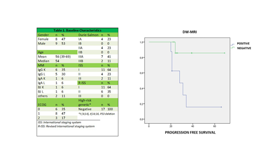
The baseline characteristics of the 17 patients included are shown in Table 1.
The median PFS in the positive DW-MRI group was 28 months, whereas the median PFS in the negative DW-MRI group was not reached (p=0.029) (image 1).
Both tests were coincidently positive in 3 patients (17%) and negative in 7 patients (41%). There was no coincidence in the remaining 7 (41%) patients, 5 (29%) being DW-MRI positive and 2 (11%) being PET-TAC positive. The difference was statistically non-significant.
Conclusions:
In our population, patients with negative DW-MRI after ASCT have a significantly higher PFS. These data are favorable to the use of DW-MRI in the follow-up of MM.
No relevant conflicts of interest to declare.

This feature is available to Subscribers Only
Sign In or Create an Account Close Modal