

Background: In secondary lymphatic organs (SLO), chronic lymphocytic leukemia (CLL) cells form characteristic pseudo-follicles, also called proliferation centers, in which proliferating CLL cells are in intimate contact with CD4+ T helper cells. Despite evidence for a co-evolution of CLL cells and counterpart T helper cells, the prevalence and dynamics of T cell subsets, such as T follicular helper cells (TFH), T regulatory helper cells (TRegs) and IL-17-producing T helper cells (Th17) has not been characterized in detail. The CLL nurselike cell (NLC) model recapitulates key cellular and molecular interactions between CLL cells and the microenvironment in SLO, based on functional and gene expression studies, and represents a valid in vitro model for the SLO microenvironment in CLL. To gain insight into the potential role of T cells in CLL SLO, we utilized the NLC model to characterize T helper cell subsets and their dynamics in NLC co-cultures.
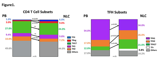
Methods and Results: First, we quantified CD4+ and CD8+ T cell subsets in fresh peripheral blood mononuclear cell (PBMC) samples from 14 patients and then placed 1x10⁷ PBMCs/ml in culture to establish NLC. We rechecked the relative proportion of CD4+ and CD8+ T cells, as well as absolute T cell counts, and their viability after 3, 6, 9, 12 and 14 days in NLC co-culture conditions. Interestingly, throughout the 14 days of culture, the number of T helper cells remained stable when compared to baseline samples. Next, we characterized T helper cell subsets in 25 different CLL samples, comparing CD4+ T cell subsets in freshly isolated CLL PBMC with matched samples harvested after 14 days of NLC culture. Samples were stained with subset-specific fluorescence-labeled antibody combinations, and analyzed by flow cytometry. NLC co-culture resulted in a significant expansion of TFH and TReg cells. TFH absolute cell counts assessed by flow cytometry using counting beads, increased from 9.4±2.2/μl at baseline to 29.0±5.9/μl in NLC co-cultures (n=14, p=0.001) and TReg from 17.0±3.3/μl to 51.0±15.0/μl (n=14, p=0.027). In contrast, Th17 absolute cell counts declined after 14 days of culture from 113.0±21.0/μl to 68.0±13.0/μl (n=14, p=0.001). Moreover, TH2 cells declined from 42.0±7.4/μl to 30.0±8.2/μl (n=14, p=0.005). Next, we analyzed for changes in TFH subsets (TFH1, TFH2 and TFH17). When compared to TFH cells from CLL PBMC, NLC culture resulted in a relative increase in TFH2 from 17.0±2.4% to 26.0±1.7% (n=25, p=0.013), and in TFH17 from 12.0±1.6% to 21.0±2.7% (n=25, p=0.006) In contrast, TFH1 frequency decreased after 14 days of culture (54.0±3.5% versus 34.0±2.9%, n=25, p=0.001). T follicular regulatory (TFr) cells also increase under co-culture conditions from undetectable to 4.8±1.9% (n=25, p=0.025)(Figure1). Looking at the maturity of CD4+ cells we noted a relative increase of central memory cells from 42.0±3.2% to 50.0±3.2% (n=25, p=0.023), whereas effector memory cells decreased from 34.0±3.4% to 26.0±2.7% (n=25, p=0.009), the fraction of naïve CD4+ cells remained unchanged.
Next, we assessed the activation status of co-inhibitory receptors on CD3+CD4+ cells. We found a significant relative increase in CD4+ T cells expressing PD1 and CTLA-4 after 14 days of culture. Additionally, the TFH and TReg subsets demonstrated a significantly higher CTLA-4 expression after culture while TReg cells also addressed an increase in PD1 frequency. Furthermore, we observed a significantly higher CD28 expression on TFH and TReg subsets after 14 days of co-culture. Following, to address the residence and migration capacity of T cells in NLC co-cultures, we analyzed the CD69 and CD62L expression. Our results revealed significantly higher expression of CD69 in NLC co-cultures, whereas CD62L levels remained unchanged.
Conclusion: The expansion of TFH and TReg cells in NLC co-cultures suggests the selection and clonal expansion of T cells that may support and engage in crosstalk with CLL cells. Additionally, the expansion of TFr and TFH17 cells could reflect a milieu of immune tolerance establishing in NLC co-cultures. Ongoing TCR sequencing of serial CD4+ T cell samples (baseline versus 14 days under NLC conditions) will characterize changes of clonal architecture in the T helper cell compartment, and will further improve our understanding of co-evolution of CLL and T helper cells.
Wierda:Gilead Sciences: Research Funding; Juno Therapeutics: Research Funding; KITE pharma: Research Funding; Sunesis: Research Funding; Miragen: Research Funding; Janssen: Research Funding; Xencor: Research Funding; Acerta Pharma Inc: Research Funding; Pharmacyclics LLC: Research Funding; Genentech: Research Funding; AbbVie: Research Funding; GSK/Novartis: Research Funding; Oncternal Therapeutics Inc.: Research Funding; Cyclcel: Research Funding; Loxo Oncology Inc.: Research Funding. Burger:Aptose Biosciences, Inc: Research Funding; BeiGene: Research Funding; Gilead Sciences: Research Funding; AstraZeneca: Honoraria; Janssen Pharmaceuticals: Consultancy, Honoraria; Pharmacyclics, an AbbVie company: Research Funding.

This feature is available to Subscribers Only
Sign In or Create an Account Close Modal