BACKGROUND: Angioimmunoblastic T cell lymphoma (AITL) has a unique histological phenotype with a relatively small number of malignant T-cells and an extensive infiltrate of normal cells including T-cells, B-cells, NK cells and FDCs. The malignant cell has a T follicular helper (Tfh) cell phenotype with increased expression of immune checkpoint molecules including PD-1 and ICOS. With the routine use of antibodies targeting immune checkpoint molecules, clinical results in limited numbers of AITL patients have thus far been mixed with some responding patients but also some who rapidly progress. Because both malignant T-cells and normal T-cells in AITL may be PD-1 positive, we conducted a study to compare the phenotype of normal T-cells and malignant T-cells within the tumor microenvironment. We also evaluated whether soluble immune checkpoint molecules in the peripheral blood differed between AITLs, controls, and other PTCL subgroups.
METHODS: We designed a novel CyTOF antibody (Ab) panel comprised of 37 markers to identify and characterize cells of T, B, NK, macrophage and FDC lineages within a cohort of biopsy specimens from 6 AITL patients compared to 6 normal controls (2 LN, 4 tonsil). Soluble immune receptors and ligands were then measured by multiplex ELISA on peripheral blood from 8 AITLs, 5 controls, and 32 additional samples from PTCL subtypes including PTCL-NOS, ALCL and CTCL.
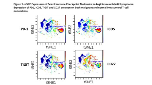
RESULTS: Malignant T-cells in AITL specimens could clearly be identified by viSNE mapping based on CyTOF expression of CD4, CD5, BCL6, CD10 (variable), CXCR5 (variable), PD-1 and ICOS. Aberrant loss of CD3 and CD7 on malignant T-cells was commonly seen. Expression of other immune checkpoint molecules was assessed. Malignant cells expressed high levels of TIGIT and moderate levels of CD27, but malignant T-cell populations were negative for expression of TIM3, LAG3, and CTLA-4. A substantial proportion of normal T-cells also expressed immune checkpoint molecules including PD-1 and CD27, and a subset of these cells also expressed TIGIT and ICOS. A small minority of normal T-cells expressed TIM3, LAG3, and CTLA-4. As others have described, the normal Tfh cell population had an immunophenotype that was otherwise relatively similar to the malignant population, but distinguishable via viSNE mapping with our CyTOF Ab panel. Expression of PD-L1 could be detected on NK-cells, macrophages, and a few select populations of normal T cells, but not on B-cells or FDCs. Malignant T-cells did not express PD-L1. Due to the potential importance of immune checkpoint molecules in this disease, we then measured soluble checkpoint receptors and ligands in the serum. Soluble PD-1 was significantly increased in the peripheral blood of AITL when compared to normal controls (average 2637 pg/mL (SE = 512.1) in AITL vs 57.87 (SE = 16.88) in controls). Similarly, soluble PD-L1 was significantly increased in AITL compared to normal controls (13.8 (SE = 10.5) in AITL vs 0.94 (SE = 0.25) in controls).
CONCLUSIONS: Malignant T-cells in AITL have a unique phenotype which was able to be reliably identified through use of viSNE mapping with our CyTOF Ab panel. Furthermore, malignant T-cells were found to consistently express additional immune regulatory molecules beyond PD-1 and ICOS, including TIGIT and CD27. However, a substantial subset of normal T-cells have a very similar phenotype suggesting that activation of T-cells in this disease will likely stimulate both normal and malignant T-cells. This may explain the rapid disease progression seen in a subset of AITL patients treated with PD-1 blockade. Furthermore, soluble PD-1 is significantly increased in the serum of AITL patients and may explain the overall lack of clinical efficacy of anti-PD-1 antibodies in treating AITL.
Cerhan:NanoString: Research Funding; Janssen: Membership on an entity's Board of Directors or advisory committees; Celgene: Research Funding. Ansell:LAM Therapeutics: Research Funding; Seattle Genetics: Research Funding; Trillium: Research Funding; Seattle Genetics: Research Funding; Affimed: Research Funding; Bristol-Myers Squibb: Research Funding; Bristol-Myers Squibb: Research Funding; Trillium: Research Funding; LAM Therapeutics: Research Funding; Trillium: Research Funding; Trillium: Research Funding; Trillium: Research Funding; Bristol-Myers Squibb: Research Funding; Seattle Genetics: Research Funding; Bristol-Myers Squibb: Research Funding; LAM Therapeutics: Research Funding; LAM Therapeutics: Research Funding; Regeneron: Research Funding; Affimed: Research Funding; Mayo Clinic Rochester: Employment; Regeneron: Research Funding; Affimed: Research Funding; Regeneron: Research Funding; Regeneron: Research Funding; Affimed: Research Funding; Affimed: Research Funding; Mayo Clinic Rochester: Employment; LAM Therapeutics: Research Funding; Affimed: Research Funding; Mayo Clinic Rochester: Employment; Seattle Genetics: Research Funding; Trillium: Research Funding; Mayo Clinic Rochester: Employment; Affimed: Research Funding; Bristol-Myers Squibb: Research Funding; Bristol-Myers Squibb: Research Funding; Regeneron: Research Funding; Seattle Genetics: Research Funding; Mayo Clinic Rochester: Employment; Seattle Genetics: Research Funding; Regeneron: Research Funding; LAM Therapeutics: Research Funding; LAM Therapeutics: Research Funding; Seattle Genetics: Research Funding; Affimed: Research Funding; Regeneron: Research Funding; Regeneron: Research Funding; LAM Therapeutics: Research Funding; Affimed: Research Funding; Trillium: Research Funding; Bristol-Myers Squibb: Research Funding; Bristol-Myers Squibb: Research Funding; Mayo Clinic Rochester: Employment; Trillium: Research Funding; Bristol-Myers Squibb: Research Funding; Mayo Clinic Rochester: Employment; Seattle Genetics: Research Funding; Seattle Genetics: Research Funding; Trillium: Research Funding; Mayo Clinic Rochester: Employment; Mayo Clinic Rochester: Employment; Regeneron: Research Funding; LAM Therapeutics: Research Funding.
Author notes
Asterisk with author names denotes non-ASH members.


This feature is available to Subscribers Only
Sign In or Create an Account Close Modal