Abstract
Peripheral T-cell lymphomas (PTCL) are a heterogeneous group of lymphoid malignancies with generally poor outcomes when treated with current regimens. The pro-survival BCL-2 family members BCL-2, BCL-xL, and MCL-1 contribute to tumor maintenance, progression, and chemoresistance across a range of cancers but their contributions in distinct subtypes of PTCL are poorly understood. Immunohistochemical analyses of PTCL specimens have revealed a distinct expression pattern of BCL-2 family members, most notably the high level expression of BCL-2 in up to 50% of certain PTCL entities (Rassidakis et al., J Pathol 2003). In fact, high BCL-2 expression has been associated with unfavorable prognosis (Ling et al., Biomed Environ Sci 2011).
We amassed a collection of 21 T-cell lymphoma cell lines (representing Alk+ ALCL, Alk- ALCL, PTCL-NOS, cutaneous T-cell lymphoma (CTCL) and rare subtypes) and 7 patient-derived xenograft (PDX) models of T-cell lymphoma. The latter include models of Alk+ ALCL, Alk- ALCL, ATLL, NK-T cell lymphoma and AITL (available at http://www.PRoXe.org) (Townsend et al. Cancer Cell 2016). To assess the expression level and protein abundance of BCL2 family members, we performed RNA-Seq and immunoblotting. To functionally characterize dependence on BCL-2 family members, we utilized BH3 profiling, a technique that allows for assessment for how "primed" or close to the cell death threshold cells are by evaluating the degree of mitochondrial outer membrane permeabilization (MOMP), induced by a panel of BH3 domain peptides (Ryan and Letai, Cell Death and Differentiation 2013). Binding specificity of BH3 domain peptides allows for determination of which pro-survival Bcl-2 family members cells are dependent on for survival and thus makes it a powerful tool to predict response to BH3 mimetics. Finally, we assessed in vitro the cytotoxicity induced by the BH3 mimetics venetocxlax (ABT-199, a BCL-2 specific agent) and navitoclax (ABT-263, which targets both BCL-2 and BCL-xL) in PTCL cell lines.
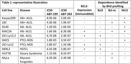
Gene expression and protein levels of the anti-apoptotic BCL-2 family members showed a distinct pattern in the cell lines that closely recapitulated immunohistochemical analysis of clinical samples (Rassidakis et al., J Pathol 2003). Specifically, both MCL-1 and BCL-xL were ubiquitously expressed, with higher levels of MCL-1 in ALCL cell lines and the PTCL-NOS cell line SMZ-1, while BCL-xL was highly expressed predominately in CTCL cell lines. While cell lines and PDX models from Alk+ ALCL and CTCL universally did not express BCL-2, approximately two-thirds of cell lines and PDX models representing Alk- ALCL, PTCL-NOS, AITL, NK/T-cell lymphoma, ATLL and rare subtypes of T-cell lymphomas did express BCL-2. Despite this expression, only 3 of 8 BCL2-expressing cell lines were sensitive to ABT-199 (IC50<1 µM), indicating that BCL2 expression is an inadequate biomarker for ABT-199 sensitivity. In contrast, BH3 profiling of these models identified either exclusive BCL-2 dependence, which correlated with sensitivity to ABT-199 in vitro, or exclusive MCL-1 dependence, which correlated with resistance to ABT-199. Alk+ and Alk- ALCL cell lines and PDX models were predominately MCL-1 dependent, but some also showed co-dependence on BCL-xL that correlated with sensitivity to ABT-263 in vitro. Among CTCL cell lines, we identified a dominant BCL-xL dependence that correlated with low nanomolar IC50 to ABT-263. In line with this, primary samples of CTCL (n=3) also showed BCL-xL dependence, offering a novel therapeutic strategy for this disease. Table 1 shows a representative illustration of these data in a selection of cell lines.
In summary, we have defined distinct classes of BCL-2 family member dependence that are revealed by BH3 profiling and predict sensitivity or resistance to available clinical agents.
Letai:AbbVie: Consultancy, Research Funding; Tetralogic: Consultancy, Research Funding; Astra-Zeneca: Consultancy, Research Funding. Weinstock:Novartis: Consultancy, Research Funding.
Author notes
Asterisk with author names denotes non-ASH members.


This feature is available to Subscribers Only
Sign In or Create an Account Close Modal