Abstract
Introduction: Sickle cell disease (SCD) is a genetic disorder characterized by sudden onset of painful vaso-occlusive episodes (VOC) which can be triggered by stress as reported by sickle cell patients. The exact mechanism of VOC origin is not well understood; however, it could result as a progression of microvascular blockade with rigid sickled red blood cells (RBC). Anything that decreases regional blood flow (RBF) will increase the transit time that it takes the RBC to escape the microvasculature before it gets rigid and entrapped, thus increasing the chances of vaso-occlusion that may progress to VOC. The RBF is controlled by the autonomic nervous system (ANS) which is modulated by stress, and as we have previously shown, SCD patients have augmented autonomic mediated vasoconstriction response (VCR) to sigh and pain. Our studies showed significant VCR when subjects were told they were about to experience pain, suggesting that VCR might be the physiologic link between common VOC triggers like stress and sickled RBC retention in the microvasculature.
Objectives: To study the effect of mental stress on autonomic parameters peripheral blood flow (PBF) and heart rate variability (HRV) in SCD.
Methods: 19 SCD and 16 control (healthy and sickle cell trait) subjects were studied. Two standard mental stress tasks with graded levels of difficulty (N-back test and Stroop test) were presented to subjects using E-prime (Psychological software). Subjects were also exposed to a novel pain anticipation task, previously shown to induce VCR.
PBF was measured using photo-plethysmography (PPG) on the left thumb. Reduction in PPG amplitude indicates vasoconstriction. Cardiac beat-to-beat variability (R-to-R interval;RRI) was extracted from electrocardiogram. ANS balance was derived from the following spectral indices of the RRI: high frequency power (HFP) ≈ parasympathetic activity, low frequency power (LFP), and the low/high ratio (LFP/HFP) representing sympathovagal balance.
Average changes in PBF and HRV from the baseline were taken as the responses to the mental tasks.
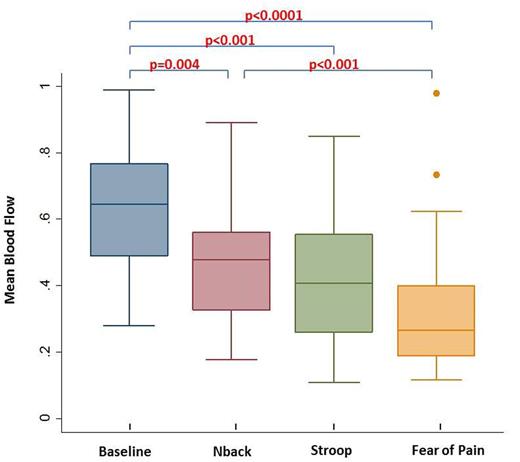
Results: There was a significant decrease in mean PBF, RRI and HFP during all the mental stressors (N-back test, Stroop test and pain fear) compared to the baseline (p<0.01). Based on task accuracy scores, there was clear difference in sublevels of task difficulty (p<0.0001). While PBF decreased during all the sublevels of N-back and Stroop compared to baseline (p<0.01), there was no PBF difference between the task sublevels. However, when subjects were told that they were about to experience severe pain from heat applied to their arm in a sham pain instruction that was given after N-back and Stroop, there was a significant decrease in PBF compared to baseline (p<0.001) and compared to the response during N-back (p<0.01). The parasympathetic withdrawal in response to mental tasks and fear of pain followed a similar pattern.
Conclusions: Graded mental stress and fear of pain causes significant decrease in regional blood flow and parasympathetic withdrawal in SCD subjects and normal controls. The vasoconstriction response to "fear of pain" was significantly greater than the VRC caused by N-back and to a lesser extent in Stroop. The pattern of responses were not significantly different between SCD and controls however the consequences of decreased blood flow could be quite different because of the resulted entrapment of sickle cells in the microvasculature. This could explain how stress may play a role in the initiation of VOC in SCD by enhancing ANS mediated vasoconstriction, prolonging microvascular transit time and increasing the likelihood of vaso-occlusion.
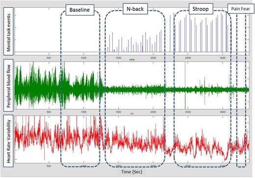
The 1st signal is mental tasks event marker, 2nd signal is photo-plethysmography (PPG) and the 3rd signal is heart rate variability derived from the electrocardiogram signal.
Peripheral blood flow and heart rate variability during baseline and various mental tasks
Peripheral blood flow and heart rate variability during baseline and various mental tasks
Box plot of mean peripheral blood flow (PBF) during baseline, mental tasks and fear of pain.
Box plot of mean peripheral blood flow (PBF) during baseline, mental tasks and fear of pain.
Wood:Celgene: Consultancy; AMAG: Consultancy; Ionis Pharmaceuticals: Consultancy; Vifor: Consultancy; Ionis Pharmaceuticals: Consultancy; Biomed Informatics: Consultancy; Vifor: Consultancy; World Care Clinical: Consultancy; World Care Clinical: Consultancy; Celgene: Consultancy; AMAG: Consultancy; Apopharma: Consultancy; Apopharma: Consultancy; Biomed Informatics: Consultancy.
Author notes
Asterisk with author names denotes non-ASH members.


This feature is available to Subscribers Only
Sign In or Create an Account Close Modal