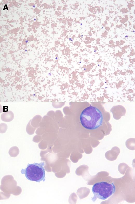
A 16-year-old boy presented with 2 weeks of fevers, night sweats, fatigue, and myalgia, in addition to an erythematous rash on the trunk and extremities after new findings of hepatosplenomegaly and inguinal lymphadenopathy. Monospot test was positive, confirming the diagnosis of infectious mononucleosis. Cytomegalovirus immunoglobulin (Ig)G and IgM antibodies were normal at <10 titers. Epstein-Barr virus (EBV) IgG antibody to early antigen was elevated at 20 titers (normal is <10 titers), and both the EBV IgG and IgM antibodies to capsid antigen were elevated at 640 titers (normal is <10 titers). Laboratory studies showed a hemoglobin level of 6.1 g/dL and a platelet count of 64 × 109/L. Direct antiglobulin test (DAT) was negative using anti-IgG and weakly positive using anti-C3d. In the peripheral blood smear, atypical reactive lymphocytes and striking agglutination of red blood cells were seen (panels A-B).
Red cell agglutinins are infrequently seen in infectious mononucleosis (∼1% of cases) and have been ascribed to polyclonal IgG/IgM cold agglutinins specific for the i antigen on the red cells. The weakly positive DAT using anti-C3d and the negative DAT using anti-IgG are characteristic for infectious mononucleosis, indicative of complement-mediated intravascular hemolysis that can occur 1 to 2 weeks after infection.
For additional images, visit the ASH IMAGE BANK, a reference and teaching tool that is continually updated with new atlas and case study images. For more information visit http://imagebank.hematology.org.


This feature is available to Subscribers Only
Sign In or Create an Account Close Modal