Abstract
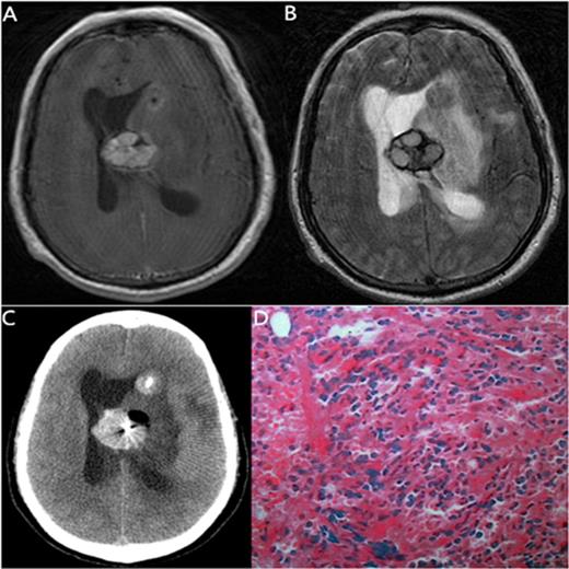
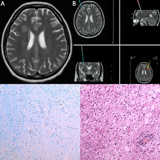
Lesions of the central nervous system (CNS) were seen during and after treatment of leukemia. We aimed to characterize the specific pathology and MRI findings observed in leukemia patients with CNS lesions and to determine their value in the management of such patients. The data from stereotactic biopsy for pathology (12 patients) and MRI examinations (14 patients) were retrospectively evaluated. Factors that predisposed to the lesions of CNS were reviewed from the medical records. Among the 14 patients, 4 had a CNS infection, 2 had a neurodegenerative disorder, 8 had CNS leukemia. The clinical diagnosis based on clinical presentation and MRI features was not consistent with the pathological diagnosis in 2 patients: in one patient, the clinical diagnosis was a CNS infection, though the patient's pathological diagnosis was CNS leukemia; in the other patient, the clinical diagnosis was CNS leukemia, but the pathological diagnosis was glial cell hyperplasia. CNS lesions in leukemia have a wide range of causes. Apart from the relapse of leukemia in CNS, there are treatment related neurotoxicities and infections that are caused by immunocompromised states. Stereotactic biopsy for pathological confirmation has the advantages of minimal invasion and convenience, which remains the gold standard for diagnosing the nature of CNS lesions. Because many CNS lesions of leukemia are treatable, early correctly diagnosis is essential.
Medical records of 14 leukemia patients with CNS lesions
| . | case . | Age, y /Sex . | Leukemia type . | A/I* . | Clinical presentation . | MRI findings . | clinical diagnosis . | Stereotactic biopsy for pathology diagnosis . | Outcome . |
|---|---|---|---|---|---|---|---|---|---|
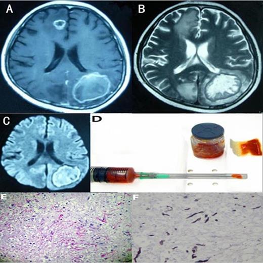
| 1 | 15/M | AML (M5) | 14/3 d after Cyclosporin | Seizure | MRI: The left occipital lobe, right frontal lobe low-density lesions with enhancement of capsule wall. | Infection | Fungal brain abscesses | Improved | |
| 2 | 38/M | AML | 34/9 mo after allo-HSCT | Fever | MRI: Scattered lesions at the right frontal lobe. Short T1, long T2 signal. | Infection | Not performed | Improved | |
| 3 | 20/M | ALL | 20/during 2nd course of chemo | Headache, limb tic | MRI: Bilateral cerebral hemisphere cortex multiple long T1 and T2 signal nodular lesions. | Infection | Fungal brain abscesses | Improved | |
| 4 | 54/M | APL | 53/2 mo after Retinoic acid | Headache, limb numb, seizure | MRI: Bilateral posterior parietal lobe showing patchy enhancement. | Leukemia | Leukemia | Improved | |
| 5 | 7/M | ALL (B cell) | 7/during the chemo | Headache | MRI: a cystic lesion in the right temporal lobe and cerebellum obvious edema. | Infection | Leukemia | Progressed | |
| 6 | 26/F | AML (M4) | 25/during 4th course of chemo | Fever, tic | MRI: Mixed signals at the left frontotemporal top border zone. | Infection | Brain abscesses | Improved | |
| 7 | 25/M | ALL (T cell) | 21/3 y after immuno-suppressive agent | Headache, limb weakness | MRI: Mixed signals at right hemisphere, perilesional mild enhancement. | Leukemia | T cell leukemia/lymphoma | Improved | |
| 8 | 16/M | ALL (T cell) | 16/ at the diagnosis of leukemia | Headache, blurred vision | MRI: Scattered, abnormal signal of sizes at the cerebellum. | Leukemia | Not performed | Progressed | |
| 9 | 49/M | ALL (B cell) | 49/1 mo after chemo | none | MRI: Glial cell proliferation around lesion at right parietal lobe. | Leukemia | Glial cell hyperplasia | Improved | |
| 10 | 60/M | CMML | 57/4 y after allo-HSCT | Nausea, right limb weakness | MRI: a 2.5×2 cm2 lesion in left basal ganglia. | Leukemia | Chronic myelomonocytic leukemia | Died | |
| 11 | 26/M | ALL (B cell) | 20/9 mo after last course of chemo | Headache | MRI: lesions at the left temporal lobe, perilesional with obvious edema. | Leukemia | B cell leukemia/lymphoma | Improved | |
| 12 | 29/F | AML (M5b) | 27/1 mo after last course of chemo | none | MRI: Bilateral cerebellum, left occipital lobe abnormal signal enhanced on T1 with Gd. | Leukemia | Leukemia | Improved | |
| 13 | 42/M | ALL | 41/3 mo after allo-HSCT | Dizziness, Walking instability | MRI: The left cerebellar hemisphere visible nodular enhancement lesions. | Leukemia | Leukemia | Improved | |
| 14 | 23/F | ALL (B cell) | 23/1 mo after allo-ASCT | headache | MRI: The right paracele white matter lesions visible long T1, long T2 signal, the boundary is not clear, edema is not obvious, no enhancemen. | Degenerative disease | Nerve cell degeneration | Improved |
*A, the age when the diagnosis was made for leukemia.I, the interval between the last treatment and the onset of neurological symptoms.
The MRI, biopsy material and pathology of a 15-year-old man with AML (M5)
No relevant conflicts of interest to declare.
Author notes
Asterisk with author names denotes non-ASH members.


This feature is available to Subscribers Only
Sign In or Create an Account Close Modal