Abstract
Background: Burkitt lymphoma (BL) is a B-cell lymphoma with high proliferating ability, derived from germinal center B-cells (GCB). The MUM-1/IRF4 gene has been identified as a myeloma-associated oncogene which is activated at the transcription level as a result of t(6;14)(p25;q32) chromosomal translocation. In normal lymphohematopoietic tissue, MUM1 protein has an important role in lymphocyte activation and in terminal B cell differentiation, and is expressed in plasma cells and in a small percentage of GCB. It is known that some of BL express MUM1, suggesting late GCB origin, but its characteristics still remain controversial.
Patients and methods: A total of 91 patients previously diagnosed as BL were retrospectively analyzed. Two patients were excluded from the study, because translocation or amplificationof BCL-2 was detected by FISH in addition to translocation of MYC. The other 89 cases were confirmed as BL based on morphology, immunophenotype and the result of FISH analysis for translocation of MYC and BCL-2. The expression of MUM-1 was examined by immunohistochemistry. Furthermore, to characterize MUM1-positive (MUM1+) BL, we have compared the clinicopathologic characteristics of MUM1+ BL and MUM1-negative (MUM1-) BL.
Results: Forty out of the 89 cases showed positivity for MUM1. The clinical characteristics of 40 MUM1+ BL were as follows. There were 20 men and 20 women, with a median age of 28 years, ranging from 3 to 83 years old. Thirty three cases (83%) showed extranodal involvement at presentation. The most frequent sites of extranodal involvement were bone marrow (n=14), lower gastrointestinal tract (n=14), central nervous system (n=5) and peripheral blood (n=5). Eight (20%) of MUM1+ BL patients had a bulky mass, 27 (69%) were categorized as stage III/IV, and 14 (36%) had B symptoms. With regard to laboratory data at presentation, 35 patients (90%) had elevated level of LDH. According to international prognostic index, 27 cases (69%) were identified into high/high-intermediate.
Compared with MUM1- BL, patients with MUM1+ BL showed significantly younger onset (p=0.0053) and a higher ratio of females (p=0.007). The MUM1+ BL was also featured by higher percentage of elevated level of LDH, and tended to have anemia (hemoglobin level <10g/dL) (p=0.049 and p=0.074, respectively). We have also highlighted the difference in the involved sites between the two groups. The MUM1+ group showed significantly lower ratio of stomach involvement (p=0.0060), and tended to more frequently involve peripheral blood, breast and kidney (p=0.088, p=0.090 and p=0.090, respectively).
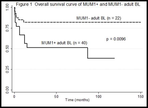
Regarding the therapy and prognosis, majority of the cases received intensive, short-duration chemotherapy, which is the standard therapy for BL. Patients with MUM1+ BL showed significantly worse overall survival curve compared with MUM1- BL (p=0.042). Of note, comparing MUM1+ and MUM1- BL cases of adults (age>20 years old), MUM1+ cases showed more significantly worse prognosis (p=0.0096). (Figure 1)
Conclusion: MUM1+ BL showed significantly worse prognosis, particularly in adult cases, compared with MUM1- BL. In addition, the difference of the onset age, sex ratio, and involved sites between the two groups was highlighted. Our results demonstrate that MUM1 expression might predict worse prognosis of BL, and MUM1+ BL should be distinguished from MUM1- BL.
No relevant conflicts of interest to declare.
Author notes
Asterisk with author names denotes non-ASH members.


This feature is available to Subscribers Only
Sign In or Create an Account Close Modal