Abstract
Background
Von Willebrand's disease (VWD) is the most common inherited bleeding disorder. Conventional assays cannot accurately detect all abnormalities in von Willebrand factor (VWF)-platelet-collagen interactions or predict a bleeding phenotype in patients with VWD.
Objective
We hypothesized that a microfluidic assay (MFA) could complement current assays and better correlate with bleeding.
Methods
Patients: Patients with VWD or mucocutaneous bleeding (MCB) are enrolled in an IRB-approved study. Bleeding scores (BS) are obtained via the Vincenza bleeding assessment tool. In this analysis, Type 1 VWD is defined by a VWF Antigen (VWF:Ag) level <40 IU/dL and a VWF activity (VWF:RCo)/VWF:Ag ratio >0.6, Type II VWD has a VWF:RCo/VWF:Ag ratio of <0.6, and Type III disease has VWF:Ag and VWF:RCo levels <10 IU/dL. MCB is defined as clinical mucocutaneous bleeding, a VWF:Ag level >40 IU/dL and no other laboratory evidence of a diagnosable bleeding diathesis.
Standard Assays: Samples are assayed for platelet count, platelet aggregometry (agonists: collagen/ristocetin/ADP), VWF:Ag, VWF:RCo, Type III collagen binding (VWF:CBIII) and PFA-100 closure time (ADP and EPI cartridges).
MFA: C ollagen (500 µg/mL) is patterned onto a glass substrate and whole blood is perfused at three distinct shear rates, low (150s-1), mid (750s-1), and high (1500s-1) for five minutes. Images of platelet adhesion are collected every 3 seconds at pre-defined points.
Data Processing: Total platelet surface coverage is determined and graphed over time. Two outputs are evaluated, (1) the lag time: the time to reach 5% surface coverage and (2) the slope: slope during the linear portion of platelet adhesion.
Results
Patients: 42 samples were obtained from 35 patients with VWD/MCB. 11/35 (31%) are from type 1 VWD patients, 2/35 (6%) are from type 2 VWD patients, 1/35 (3%) are from a Type 3 VWD patients, and 22/35 (60%) are from patients with MCB.
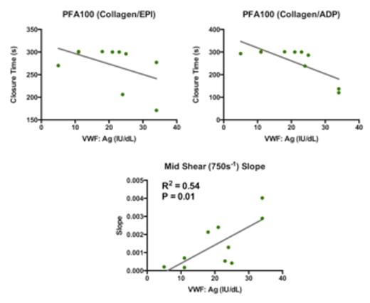
Assay Correlation: In all patients, the midshear slope correlated better with VWF:Ag (R2 = 0.52 P<0.0001) than the PFA100-ADP (R2 = 0.28 P=0.0008) and PFA100-EPI (R2 =0.32 P=0.0002) (Table 1). The midshear slope (R2 = 0.52 P=< 0.0001) also outperformed the PFA100-ADP (R2 = 0.33 P= 0.0003) and PFA100-EPI (R2 = 0.39 P= < 0.0001) in correlating with VWF:RCo levels (Table 1). In the Type 1 VWD cohort, the mid shear slope correlated with VWF:Ag (R2 = 0.54 P=0.01) (figure 1). In comparison, the PFA-100 ADP/EPI was relatively insensitive to VWF:Ag levels <40 IU/dL (figure 1). These results show that a single MFA assay correlates with VWF:Ag and VWF:RCo levels better than the PFA-100 closure time in a cohort of patients with VWD/MCB and is more sensitive to VWF:Ag levels <40 IU/dL in a cohort of individuals with Type 1 VWD.
BS Correlation: No clinical assays or MFA output significantly correlated with BS in any cohort analyzed.
Response to therapy: In 4 patients pre and 1hr post DDAVP, the MFA lag time remained the same or decreased (low=15.75s to 15.75s, mid=18s to 11.25s, high=30.75s to 9s) and the slope increased (low=0.0044 to 0.0057, mid=0.0044 to 0.0066, high=0.0037 to 0.0059). The increased low shear slope, 0.0044 to 0.0057, was statistically significant (p= 0.0244). This suggests that a MFA may be useful to determine response to DDAVP therapy.
Conclusions
A novel collagen-based MFA outperformed the PFA100 ADP/EPI in correlating with VWF:Ag and VWF:RCo in patients with VWD/MCB. In type 1 VWD patients, it also demonstrated a significant correlation with VWF:Ag levels and a more linear relationship than the PFA100 ADP/EPI. Finally, the MFA was able to determine a response to DDAVP therapy. Thus a MFA, with a small sample input (150 µL) as compared to other shear dependent assays such as the PFA100 (1000 µL), may be useful to determine multiple clinically relevant parameters in situations where blood samples are difficult to obtain such as in neonatal settings or poor venous access.
Comparison of R2 values Between Standard Assays and Shear-based Assays in All Patients with VWD/MCB.
| Standard Assay . | Shear-Based Assay . | R2 Value . | P Value . |
|---|---|---|---|
| VWF:Ag | PFA100 (Collagen/EPI) | 0.32 | 0.0002 |
| PFA100 (Collagen/ADP) | 0.28 | 0.0008 | |
| MFA Mid-shear Slope | 0.52 | <0.0001 | |
| VWF:RCo | PFA100 (Collagen/EPI) | 0.39 | < 0.0001 |
| PFA100 (Collagen/ADP) | 0.33 | 0.0003 | |
| MFA Mid-shear Slope | 0.52 | < 0.0001 |
Correlation of the PFA100 Collagen/Epinephrine, Collagen/ADP Cartridges, and MFA-Midshear Slope with VWF:Ag levels.
Correlation of the PFA100 Collagen/Epinephrine, Collagen/ADP Cartridges, and MFA-Midshear Slope with VWF:Ag levels.
Ng:CSL Behring: Consultancy; Baxter: Consultancy. Manco-Johnson:Baxter, bayer, biogen, CSL Behring, NovoNordish: Honoraria.
Author notes
Asterisk with author names denotes non-ASH members.


This feature is available to Subscribers Only
Sign In or Create an Account Close Modal