Abstract

Purpose: The aim of this multi-centre study was to retrospectively assess in thalassemia major (TM) if deferiprone (DFP) had a dose-dependent effect on liver iron concentration (LIC) assessed by quantitative magnetic resonance imaging (MRI).
Methods: Among the 958 TM patients enrolled in the MIOT (Myocardial Iron Overload in Thalassemia) network, we identified hose with an MRI follow up study at 18±3 months who had been received DFP monotherapy and had no changes in dose of DFP between the 2 MRI scans. Patients were divided into two groups according to the DFP dose: 79 patients with ≤ 75 mg/kg/d (group 1) and 39 with > 75 mg/kg/d (group 2).
Hepatic iron overload was measured by the T2* multiecho technique and T2* values were converted into LIC values using the calibration curve introduced by Wood et al.
Results: The two groups had comparable baseline MRI LIC values. The table shows the evolution of different iron overload risk classes between the baseline and the FU MRI. The percentage of patients that worsened their status was significantly higher in group 1 than in group 2 (26.6% vs 7.7%; P=0.016).
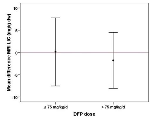
Subgroup analysis in patients with hepatic iron overload at baseline (MRI LIC > 3mg/g/dw) was conducted: 48 patients from group 1 (DFP dose: mean 70.6±11.2 mg/kg/d, median 75 mg/kg/d) and 30 from group 2 (DFP dose: mean 85.2±6.6 mg/kg/d, median 84 mg/kg/d). The two subgroups had comparable baseline MRI LIC values (10.2±8.1 mg/g dw vs 11.1±8.7 mg/g dw (P=0.314). While the mean change in subgroup 2 ( -1.8±6.3mg/g/dw, P=0.131) was more favourable than in subgroup 1 (+0.1±7.7 mg/g/dw, P=0.903), the change in MRI LIC values did not reach statistical significance between the two subgroups (P=0.579) (Figure 1), which may be due to small cohort evaluated.
Conclusions: In TM patients the worsening in MRI LIC can be prevented by increasing the dose of deferiprone above the widely used regimen of 75 mg/kg body weight. Our results are consistent with the iron balance studies performed by Grady RW et al.
Evolution of different iron overload risk classes between the baseline and the FU MRI. The underlined numbers represent the patients who remained in the same risk class.
| DFP dose ≤ 75 mg/kg/d (N=79) . | |||||
|---|---|---|---|---|---|
| FU LIC | |||||
| <3 mg/g dw | 3-7 mg/g dw | 7-15 mg/g dw | ≥15 mg/g dw | ||
| Baseline LIC | <3 mg/g dw (N=31) | 21 | 7 | 3 | 0 |
| 3-7 mg/g dw (N=22) | 10 | 4 | 6 | 2 | |
| 7-15 mg/g dw (N=14) | 0 | 6 | 5 | 3 | |
| ≥15 mg/g dw (N=12) | 1 | 0 | 5 | 6 | |
| Total at the FU | 32 | 17 | 19 | 11 | |
| DFP dose > 75 mg/kg/d (N=39) | |||||
| FU LIC | |||||
| <3 mg/g dw | 3-7 mg/g dw | 7-15 mg/g dw | ≥15 mg/g dw | ||
| Baseline LIC | <3 mg/g dw (N=9) | 6 | 2 | 1 | 0 |
| 3-7 mg/g dw (N=14) | 3 | 11 | 0 | 0 | |
| 7-15 mg/g dw (N=8) | 0 | 4 | 4 | 0 | |
| ≥15 mg/g dw (N=8) | 1 | 0 | 2 | 5 | |
| Total at the FU | 10 | 17 | 7 | 5 | |
Changes of MRI LIC values in patients with basal MRI LIC > 3 mg/g/dw.
Pepe:Chiesi: Speakers Bureau; ApoPharma Inc: Speakers Bureau; Novartis: Speakers Bureau.
Author notes
Asterisk with author names denotes non-ASH members.

This icon denotes a clinically relevant abstract


This feature is available to Subscribers Only
Sign In or Create an Account Close Modal