Abstract

BACKGROUND: Human herpesvirus type 6 (HHV-6) is increasingly recognized as an opportunistic and potentially life-threatening pathogen in recipients of allogeneic hematopoietic stem cell transplantation (AlloSCT). HHV-6 is a member of the beta herpesvirus subfamily (genus Roseolovirus). HHV-6 infection is recognized as the cause of a febrile disease and exanthem subitum in early childhood. Approximately 60% of solid organ transplant and 40% of patients after alloSCT experienced HHV-6 reactivation. Reported clinical manifestations of HHV-6 infection in transplanted patients are skin rash, interstitial pneumonia, bone marrow suppression and encephalitis. Moreover, some clinical reports suggest that HHV-6 can facilitate the occurrence of severe clinical complications of alloSCT, increasing transplant-related mortality.
METHODS: From January 2009 to February 2013, we retrospectively evaluated 54 consecutive adult patients (median age 50 years) who developed positivity to HHV-6 after alloSCT for high-risk hematological malignancies. Stem cell donors were family haploidentical (37), HLA identical sibling (8), unrelated volunteer (6), cord blood (3). The viral load was determined by quantitative PCR (Nanogen Advanced Diagnostic S.r.L) in cell-free body fluids such as plasma, bronchoalveolar lavage (BAL), cerebrospinal fluid (CSF), bone marrow (BM) aspirates or in gastrointestinal biopsies.
RESULTS: Median time from alloSCT to HHV-6 reactivation was 34 days (range: 0-705). Thirty-one patients presented HHV-6 positive in plasma, 9/54 in BM, 33/54 in gut biopsies or BAL, 7/54 in CSF. At the time of viral positivity all pts were receiving acyclovir as viral prophylaxis except five. Twenty-nine patients had acute graft versus host disease (GvHD). Twenty-two out of these twenty-nine patients experienced a grade III-IV acute GvHD, requiring high dose steroids in twenty-six cases. A concomitant CMV positivity was detected in 15/54 patients. The median absolute count of CD3+ lymphocytes was 207 cells/mcl. In 52/54 cases we reported HHV-6 clinical manifestations: fever (43), skin rash (22), hepatitis (19), diarrhoea (24), encephalitis (10), BM suppression (18), delayed engraftment (11). HHV-6 positivity led to antiviral pharmacological treatment in 37/54 cases, using as first choice therapy foscarnet. Amongst the total fifty-four patients with documented HHV-6 positivity thirty-one solved the clinical event. However the mortality rate was relatively high in this population (overall survival (OS) ±SE at 1 year after HHV-6 reactivation was 38% ± 7%), mainly related to severe infections or GvHD.
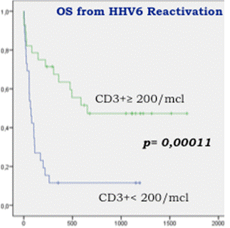
A better OS is significantly associated with CD3+ cells ≥200/mcl at the time of HHV-6 reactivation (fig 1) (OS at 1 year 63% compared to 11% for patients with CD3 <200/mcl; HR: 0.27, 95% CI 0.12-0.54, p=0.0002). The overall survival of these patients was also positively affected by the absence of acute GvHD grade III-IV at time of viral reactivation (HR: 0.03, 95% CI 1.08-4.03, p=0.03) and by the complete disease remission at time of HSCT (HR:0.26, 95% CI 0.07-0.89, p=0.03). In this analysis the overall survival was not significantly influenced by steroids administration (HR: 1.36, 95% CI 0.71-2.60, p=0.36), time after alloSCT (HR: 1.30, 95% CI 0.51-3.33, p=0.59), type of antiviral prophylaxis (HR: 1.02, 95% CI 0.45-2.33, p=0.96), plasma viral load (HR:1.18, 95% CI 0.51-2.76, p=0.69) and organ involvement (HR:1.14, 95% CI 0.59-2.20, p=0.70).
CONCLUSIONS: This retrospective study confirms a correlation of HHV-6 with high morbidity and mortality rates after alloSCT, thus suggesting a regular HHV-6 monitoring in alloSCT recipients. The regular monitoring of HHV-6 DNA, using a real-time PCR assay, may be useful for identifying active HHV-6 infection and for the introduction of a pre-emptive treatment, possibly reducing the incidence of the most severe clinical complications. Despite HHV-6 detection typically occurred early after alloSCT, a better immune reconstitution has the potential to improve clinical outcome.
Overall survival after alloSCT in HHV-6 positive patients: green line showed patients with more than 200/mcl CD3+ cells, blue line the ones with less than 200/mcl CD3+ cells at HHV-6 reactivation. P value is provided by Log Rank test.
Overall survival after alloSCT in HHV-6 positive patients: green line showed patients with more than 200/mcl CD3+ cells, blue line the ones with less than 200/mcl CD3+ cells at HHV-6 reactivation. P value is provided by Log Rank test.
Bonini:MolMed S.p.A.: Consultancy.
Author notes
Asterisk with author names denotes non-ASH members.

This icon denotes a clinically relevant abstract


This feature is available to Subscribers Only
Sign In or Create an Account Close Modal