Abstract
Context
Diagnosis of early-stage CTCL can be challenging because skin lesions contain a mixture of both diverse benign and clonal malignant T cells. Currently, clonality is assessed by TCR PCR but this method is qualitative, often fails to detect clones present at low numbers, and cannot be used to identify malignant T cells for subsequent study by flow cytometry or immunohistochemistry. High throughput sequencing (HTS) of the TCR gamma (TCRG) and TCR beta (TCRB) CDR3 regions provide comprehensive analysis of the total repertoire of T cell clones in a specimen, the breadth of repertoire diversity, and an exact quantitation of individual T cell clones.
Objective
In this study, we investigated whether the Adaptive Biotechnologies immunoSEQ assay of TCRG and TCRB could discriminate between benign and malignant T-cell skin dyscrasias.
Patients
This study involved a total of 132 patients. We analyzed skin biopsies from 39 patients with histopathologically and clinically suspicious or diagnostic CTCL (over 80% Stage I or Stage II), 9 patients with Lymphomatoid Papulosis, 4 treated CTCL patients in partial or complete remission, non-lesional biopsies from 3 CTCL patients, 6 biopsies from individuals without skin disease, 9 patients with atopic dermatitis, 12 patients with eczematous dermatitis, 24 patients with psoriasis, 15 patients with psoriasis after treatment with etanercept and clinical clearance, and non-lesional biopsies from 11 patients with psoriasis.
Methods
Genomic DNA was extracted from FFPE or OCT sections of skin biopsy specimens of patients. T cell receptor gamma (TCRG) and beta (TCRB) chain sequences were then independently amplified using multiplex PCR with optimized primer sets. Following HTS, a bioinformatics pipeline clusters the sequences into distinct clonotypes to determine overall frequencies and to identify diagnostic clones. V, (D), and J genes are also identified for each clonotype.
Results
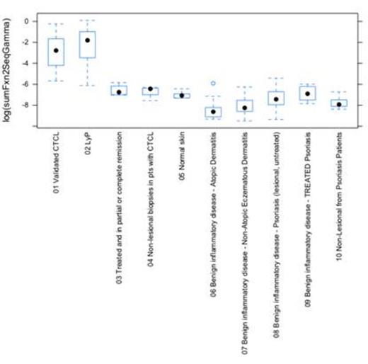
The assays defined the dominant clonal sequences in every case and distinguished likely gamma/delta from alpha/beta T cell lymphoma. Using the fraction of the top two TCRG sequences as a fraction of the total nucleated cell population defined a cut off of approximately 1/500 above which the biopsy approached 100% specificity for malignant disease and below which the assay approached 100% specificity for non-malignant or treated malignant disease. TCR PCR was negative in 10 out of 34 biopsies suspicious of CTCL—all of these were among those determined to be clonal by HTS. While there was a strong correlation between analyses using the top two TCRG sequences (because both TCRG alleles are VJ rearranged in most T cells) versus the top one TCRB sequence (because only one TCRB allele is V(D)J rearranged in most T cells), the TCRG analysis was more powerful in terms of discrimination and separation of disease categories (see figure; P<0.00001 between CTCL or LyP and any other category)
10 categories of histopathologically and clinically defined skin disease were studied by high-throughput sequencing of the TCRB and TCRG loci. “Box and whisker” plots of the TCRG analyses are shown. The boxed areas represent the second and third quartiles of the values of the top two TCRG sequences identified as a log fraction of the total nucleated cells in the study sample. Solid dots represent the median values. There is a clear demarcation between CTCL and LyP and the other categories of skin disease.
10 categories of histopathologically and clinically defined skin disease were studied by high-throughput sequencing of the TCRB and TCRG loci. “Box and whisker” plots of the TCRG analyses are shown. The boxed areas represent the second and third quartiles of the values of the top two TCRG sequences identified as a log fraction of the total nucleated cells in the study sample. Solid dots represent the median values. There is a clear demarcation between CTCL and LyP and the other categories of skin disease.
Conclusions
HTS analyses of DNA extracted from skin biopsies of patients with skin disorders can provide important quantitative data of T cell number, clonality, repertoire, and frequency and in so doing provide a useful discriminator of benign versus malignant disease. This same methodology may prove useful for other lymphoid malignancies in which the diagnosis is based on identification of clonal populations within a diverse admixture of cells. Moreover, identification of the TCR VB of the malignant clone allows it to be further characterized with anti-Vb antibodies, and assessed after treatment as MRD.
Kirsch:Adaptive Biotechnologies: Employment, Equity Ownership. Williamson:Adaptive Biotechnologies: Employment, Equity Ownership. Robins:Adaptive Biotechnologies: Consultancy, Equity Ownership, Patents & Royalties.
Author notes
Asterisk with author names denotes non-ASH members.


This feature is available to Subscribers Only
Sign In or Create an Account Close Modal