Abstract
Introduction. SIRT1 is a histone/protein deacetylase belonging to the family of Sirtuins and is the human homologue of the yeast Sir2 enzyme1. Histone deacetylases (HDACs) play a key role in several cancer-related mechanisms and the evidences of the efficacy of HDACs inhibitors in the treatment of lymphomas is rapidly growing2. SIRT1 binds and deacetylates the master transcriptional regulator Bcl-6 with implications in the pathogenesis of diffuse large B-cell lymphoma3. Ki67 is the proliferation index and is a diagnostic and useful marker currently used in the pathological evaluation of lymphoma samples4. Our aim is the characterization of SIRT1 in the B lymphocytes derived from follicular lymphomas (FL) or follicular hyperplasias and their comparison to Ki67, Bcl-6 and HIC-1.
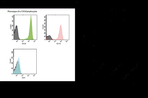
Methods. Immunohistochemical stainings (IHC) have been performed on a total of 61 formalin fixed paraffin embedded (FFPE) tissue sections including 36 FL and 25 follicular hyperplasias. All these FFPE samples were stained also for the proliferation index Ki67/MIB1. HIC-1 was also evaluated on 36 FL and 17 follicular hyperplasias. IHCs have been evaluated independently by two pathologists. B lymphocytes were immunomagnetically sorted starting from frozen biopsies collected in a Tissue Bank. Flow cytometry was used to determine the phenotype of the purified B populations. Genomic DNA and total RNA were extracted from purified B lymphocytes. Real-time, quantitative PCR (qPCR) was performed with Evagreen. qPCR was also performed on 4 samples of peripheral blood mononuclear cells (PBMCs) used as a reference. Data analysis and statistics were performed with GraphPad Prism 5.
Results. IHCs show that SIRT1 localizes preferentially in the centroblasts of the follicles. In the 36 FL analyzed, SIRT1 is expressed in 33/36 cases (91.7%) and is stronger in the centroblasts of 23/36 cases (63.9%). Interestingly, the Ki67 staining correlates positively with SIRT1 in the FL where SIRT1 localizes in the centroblasts. However, SIRT1 positivity does not correlate with FL grade in our cohort. HIC1 reactivity in the same cohorts of samples resulted only in a weak staining of the stroma but the lymphocytes were negative.
In order to investigate the expression levels of the two transcriptional variants of SIRT1 (SIRT1_001 and SIRT1_002) and Bcl-6 in the tumoral lymphocytes, we sorted and purified B lymphocytes from a total of 24 biopsies of FL and 10 follicular hyperplasias. qPCR revealed that FL have a statistically significant higher SIRT1_001 expression when compared to follicular hyperplasias and PBMCs. Furthermore, the FL having a Bcl-6 fold increase higher than 10 have also SIRT1_001 significantly higher than the FL with a Bcl-6 fold increase less than 10.
Conclusions. Collectively, our data show that Ki67 positivity correlates with SIRT1 localization in the nuclei of the centroblasts. Interestingly, the transcriptional isoform SIRT1_001 is higher in the B lymphocytes of FL than in follicular hyperplasias and seems also to be positively correlated to Bcl-6 overexpression in FL samples. Our investigations are now aimed at defining the significance of these data in terms of epigenetic regulation of key gene promoters in the tumoral B lymphocytes.
1 Heltweg, B. et al.Cancer research66, 4368-4377, doi:10.1158/0008-5472.CAN-05-3617 (2006).
2 Murawski, N. & Pfreundschuh, M. The lancet oncology11, 1074-1085, doi:10.1016/S1470-2045(10)70210-2 (2010).
3 Liu, T., Liu, P. Y. & Marshall, G. M. Cancer research69, 1702-1705, doi:10.1158/0008-5472.CAN-08-3365 (2009).
4 Yamamoto, E. et al.Cancer science104, 1670-1674, doi:10.1111/cas.12288 (2013).
No relevant conflicts of interest to declare.
Author notes
Asterisk with author names denotes non-ASH members.


This feature is available to Subscribers Only
Sign In or Create an Account Close Modal