Key Points
Only 62 (2.7%) of 2257 high-risk patients tested positive for LAC over 6 years; only 5 (0.02%) with early recurrent miscarriage tested positive.
The 2 assays recommended by ISTH guidelines were less effective than our 4-assay panel at capturing and describing LAC-positive patients.
Abstract
Routine investigation for recurrent pregnancy loss includes measurement of antiphospholipid antibodies under the perception that the lupus anticoagulant (LAC) is prevalent in this population. Our tertiary clinic sees ∼250 new patients with recurrent pregnancy loss annually, in addition to those with systemic lupus erythematosus and/or antiphospholipid syndrome. We measure LAC using a 4-assay panel that expands on the 2 assays recommended by the International Society on Thrombosis and Haemostatis (ISTH) guidelines. Of 2257 patients tested for LAC during a 6-year period, 62 (2.7%) repeatedly tested positive. Only 5 patients (0.2%) had both a history of early recurrent miscarriage and LAC positivity. Patients with LAC had a significantly more frequent history of thrombosis (35.5% vs 2.4%). LAC was absent in an overwhelming majority of women with exclusively early recurrent pregnancy loss but was associated with sporadic stillbirth. Among our panel of assays, none was predominant, and an increasing number of positive assays was associated with an increased history of morbidity. Therefore, our results do not support the ISTH contention that 2 assays are sufficient to identify and describe patients with LAC. We found that a confirmed, repeated LAC was very infrequent even in a high-risk setting.
Continuing Medical Education online
This activity has been planned and implemented in accordance with the Essential Areas and policies of the Accreditation Council for Continuing Medical Education through the joint sponsorship of Medscape, LLC and the American Society of Hematology.
Medscape, LLC is accredited by the ACCME to provide continuing medical education for physicians.
Medscape, LLC designates this Journal-based CME activity for a maximum of 1.0 AMA PRA Category 1 Credit(s)™. Physicians should claim only the credit commensurate with the extent of their participation in the activity.
All other clinicians completing this activity will be issued a certificate of participation. To participate in this journal CME activity: (1) review the learning objectives and author disclosures; (2) study the education content; (3) take the post-test with a 70% minimum passing score and complete the evaluation at http://www.medscape.org/journal/blood; and (4) view/print certificate. For CME questions, see page 466.
Disclosures
Nancy Berliner, Editor, has received grants for clinical research from GlaxoSmithKline. The authors and CME questions author Laurie Barclay, freelance writer and reviewer, Medscape, LLC, declare no competing financial interests.
Learning objectives
Upon completion of this activity, participants will be able to:
Describe the prevalence of lupus anticoagulant (LAC) and its associations with obstetric outcomes in a high-risk population, based on laboratory database study.
Compare the diagnostic value of a 2-assay vs a 4-assay panel to detect the presence of LAC in a high-risk population, based on a laboratory database study.
Release date: July 18, 2013; Expiration date: July 18, 2014
Introduction
The lupus anticoagulant (LAC) has long been associated with increased risks for thrombosis and adverse obstetric outcomes including early, recurrent pregnancy loss,1-4 although the mechanisms involved in its pathogenicity have yet to be elucidated.5 It is generally recommended that investigation of recurrent pregnancy loss include testing for antiphospholipid antibodies (aPL) including anticardiolipin antibodies (aCL), anti-β2 glycoprotein I antibodies (anti-β2GPI), and the LAC.6-9 However, it has also been observed that few patients with exclusively early recurrent miscarriage have sufficiently high titers of aCL or anti-β2GPI to fulfill classification criteria for the antiphospholipid syndrome (APS)10 and that the LAC is seldom found.11-13
The International Society on Thrombosis and Haemostasis (ISTH) updated its recommendations for detection of the LAC and proposed the use of 2 assays to measure LAC: the dilute Russell viper venom time (dRVVT) and a sensitive partial thromboplastin time (PTT-LA), as well as mixing studies with normal plasma and confirmatory testing results to ensure PL-dependence of prolonged clotting times.14 Earlier recommendations have included the kaolin clotting time (KCT) and a dilute prothrombin time (dPT) as sensitive measures of the LAC.15,16 The updated guidelines no longer support their use, citing difficulties in performing the KCT in some laboratories and increased chance of false-positive results with the use of more than 2 assays.14
Our tertiary clinic, the T.E.R.M. Program (Treatment and Evaluation of Recurrent Miscarriage), established ∼25 years ago in Toronto, Canada, sees ∼250 new patients annually with recurrent pregnancy loss, in addition to hundreds of patients with systemic lupus erythematosus (SLE) and/or APS. Our laboratory has been using a panel of 4 assays for the LAC for 20 years (dRVVT, KCT, dPT, and a PTT-LA).17-19 Therefore, we were able to retrospectively evaluate the prevalence of LAC in a large sample of women at variable risks for thrombosis and adverse obstetric events as well as identify any relationships between the different assays used to measure LAC and evaluate the usefulness of the ISTH recommendations.
Methods
Study design
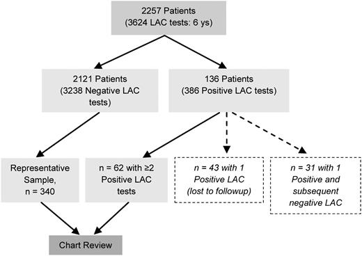
All patients who tested positive for LAC at least twice between January 2005 and February 2011 were identified from a laboratory database (Figure 1). The LAC-negative population was too large for chart review from a logistic point of view. Using the following formula n = N/[1 + N(e)2] here, n is the sample size, N is the population size, and e is the desired level of precision (set at 0.05), a sample size of 340 was calculated to be representative of the larger LAC-negative sample of 2121, with 95% confidence interval (CI).20 We randomly selected LAC-negative patient selection for review by converting the consecutive list of LAC-negative panels run (by date) to an alphabetical list of patients and reviewed the first 340 charts available in the clinic. We excluded from the LAC-negative sample any healthy obstetric patients who were participating as controls in other clinical studies. From clinic charts, we collected demographic data and medical histories.
Laboratory analysis
LAC was measured in plasma that was centrifuged twice and stored at −80°C until tested. The following screening, mixing, and confirmation assays were used to detect LAC on a Start 4 Analyzer (Diagnostica Stago, Abbott Labs, Mississauga, ON, Canada): (1) dPT using a human recombinant thromboplastin reagent (Hemosil recombiplastin 2G; Beckman Coulter, Fullerton, CA); (2) dRVVT (American Diagnostica, Greenwich, CT); (3) a LAC-sensitive PTT-LA (Diagnostica Stago, Abbott Labs, Mississauga, ON); and (4) KCT. Upper limits of normal for the 4 assays (mean + 2 SD) were established using plasma from 129 healthy volunteers, and positive and negative plasma control samples were included with each assay. Prolonged clotting times in any of these 4 assays were rerun at ratios of 1:1 and 4:1 (patient plasma: normal plasma). The phospholipid dependence of plasma samples with prolonged clotting times at a 1:1 dilution (in at least 1 of the 4 assays) was confirmed using Sta-Clot LA (Diagnostica Stago) if the corrected clotting time (T1-T2) was ≥8.1 seconds (mean + 1.96 SD of 41 normal plasma samples). Only when prolonged clotting times were present in screening, mixing, and confirmation assays was LAC determined to be present. For the purposes of this study, LAC had to be present on more than 2 occasions, not less than 12 weeks apart, and within a period of 6 years. aCL IgG and IgM (aCL) were measured using INOVA Quantalite kits (Intermedico, Mississauga, ON). In our laboratory, levels less than 15 IgG antiphospholipid units (aCL IgG units) and 25 IgM antiphospholipid (MPL) units (aCL IgM units) are considered negative.
Clinical manifestations
Medical charts were reviewed for age, diagnosis, and a history of thrombotic events (TEs) including any concurrent risk factors.21 The following obstetric data were also reviewed: the number of pregnancies, live births, early spontaneous abortions, stillbirths, therapeutic abortions, neonatal deaths, ectopic pregnancies, intrauterine growth restriction (IUGR), and therapeutic intervention; hemolysis, elevated liver enzymes and low platelets (HELLP); eclampsia; and preeclampsia. Recurrent pregnancy loss was defined as ≥3 consecutive spontaneous losses before 32 weeks’ gestation. Early spontaneous abortion was defined as pregnancy loss before 12 weeks’ gestation, late spontaneous abortion was defined as pregnancy loss between 12 and 20 weeks’ gestation, and stillbirth was defined as pregnancy loss after 20 weeks’ gestation.
Statistical analysis
Proportional data were compared using the z test (SigmaPlot 11.0). For normally distributed data, the Student t test was used to determine statistically significant differences. For nonnormally distributed data, the Mann-Whitney rank-sum test was used. Odds ratios (ORs) were calculated using VassarStats Statistical Computation.22 In all cases, P values < .05 with an α of 0.5 and a power of >80% were considered significant.
Results
LAC was measured 3624 times in 2257 female patients (many underwent multiple tests). Of those, 136 patients had 386 positive test results (Figure 1): 62 (2.7%) had repeatedly tested positive for LAC; 31 (1.4%) tested positive for LAC on only 1 occasion and subsequently tested negative; 43 (1.9%) tested positive with no subsequent tests, as they were lost to follow-up; and 2121 (94.0%) tested negative for LAC at least once.
Comparison of LAC-positive and LAC-negative patients
Patients in the LAC-positive group were significantly older than those in the LAC-negative group (median, 40 vs 37 years, respectively; P < .001, Mann-Whitney rank-sum test). No difference was observed in the prevalence of patients with SLE in either group (21.0% vs 19.0%, respectively) (Table 1). However, there were significantly more patients with either primary or secondary APS in the LAC-positive group (60% vs 1%) and significantly more patients with a history of exclusively early recurrent pregnancy loss in the LAC-negative group (70% vs 8%).
Distribution of diagnoses among LAC-positive and LAC-negative patients
| Diagnosis . | LAC negative, n = 340 (%) . | LAC positive, n = 62 (%) . | P (95% CI) . |
|---|---|---|---|
| SLE | 66 (19.4) | 13 (21.0) | NS |
| 1° APS | 0 (0) | 24 (38.7) | <.001 (0.322-0.452) |
| SLE/APS | 3 (0.9) | 13 (21.0) | <.001 (0.147-0.255) |
| Early RPL | 238 (70.0) | 5 (8.0) | <.001 (0.489-0.755) |
| Other | 33 (9.7) | 7 (11.3) | NS |
RPL, early recurrent pregnancy loss; NS, not significant; SLE/APS, lupus with secondary APS.
Distribution of clinical events
There were significantly more patients with a history of TEs in the LAC-positive group compared with patients in the LAC-negative group (33.9% vs 2.9%; P = .001; 95% CI for the difference, .238-.382; OR, 21.2; 95% CI, 8.8-51.1). There were significantly more LAC-positive patients with a history of stillbirth (38.0% vs 7.1%; P < .001; 95% CI, .283-.477; OR, 8.6; 95% CI, 4.2-17.5) and significantly fewer with a history of ≥2 early spontaneous losses (28.0% vs 71.8%; P < .001; 95% CI, .287-.569) compared with LAC-negative patients, respectively (Table 2). Significantly more LAC-positive women had pregnancies complicated by IUGR and HELLP compared with LAC-negative women, but no apparent difference in the frequency of preeclampsia was observed between the 2 groups. There was no difference in the incidence of live birth ever (70.6% vs 72.0%) and no adverse obstetric events ever (9.5% vs 10.0%) (Table 2). Of those LAC-positive women whose treatment protocols during pregnancy were available from chart review (n = 33), 4 received no anticoagulant therapy; 3 received unfractionated heparin and aspirin (ASA, 81 mg); 4 received ASA only; and 23 received low-molecular-weight heparin and ASA. Evaluation of the efficacy of these therapies was beyond the scope of our analysis. In addition, almost 70% of LAC-positive women with a history of stillbirth had at least 1 subsequent live birth.
Distribution of clinical events among LAC-positive and LAC-negative patients
| Clinical event . | LAC negative, n = 340 (%) . | LAC positive, n = 62 (%) . | P (95% CI) . |
|---|---|---|---|
| TE (idiopathic) | 4 (1.2) | 10 (16.1) | <.001 (0.085-0.182) |
| TE (with risk factor*) | 6 (1.8) | 11 (17.7) | <.001 (0.113-0.217) |
| ≥1 stillbirth | 23/326 (7.1) | 19/50 (38.0) | <.001 (0.283-0.477) |
| ≥2 early SA | 234/326 (71.8) | 14/50 (28.0) | <.001 (0.287-0.569) |
| IUGR | 3/326 (0.9) | 9/50 (18.0) | <.001 (0.119-0.223) |
| HELLP | 1/326 (0.3) | 6/50 (12.0) | <.001 (0.077-0.157) |
| Preeclampsia | 6/326 (1.8) | 3/50 (6.0) | .186 (−0.087-0.003) |
| ≥1 live birth | 230/326 (70.6) | 36/50 (72.0) | .972 (−0.149 to 0.121) |
| No adverse obstetric events | 31/326 (9.5) | 5/50 (10.0) | .884 (−0.092 to 0.082) |
SA, spontaneous abortion.
Predisposing events for thrombosis: oral contraception, surgery, and postpartum period.
Regarding patients with a history of TEs, 14 of 29 of events among 21 LAC-positive patients had a predisposing risk factor compared with 2 of 9 events among 8 LAC-negative patients. Eighteen of the 29 events in the LAC-positive group were venous; 8 of 9 events in the LAC-negative group were venous. There were recurrent TEs in 7 of 21 patients in the LAC-positive group and in 1 of 8 patients in the LAC-negative group. Among patients with a history of TEs, there was no apparent significant difference in the time since last TE in either group (median, 9.5 vs 6.8 years), number of patients with recurrent events, or prevalence of idiopathic events between LAC-positive and LAC-negative patients. Small sample sizes reduced the power of the statistical analyses to below that required to detect a difference if one exists. Therefore, these similarities must be interpreted with caution.
Distribution of aCL IgG and aCL IgM
Significant differences were found between LAC-positive and LAC-negative patients regarding the prevalence and level of aCL positivity (Table 3). More than 90% of LAC-negative patients also tested negative for both aCL IgG and aCL IgM compared with 30% and 60% of LAC-positive patients, respectively. Only 0.9% and 1.5% of LAC-negative patients had levels of aCL IgG and aCL IgM, respectively, that fulfilled APS criteria (>40 GPL or MPL), in contrast to 45% and 27% of LAC-positive patients, respectively. No significant differences were observed between the frequencies of aCL IgG and aCL IgM in the LAC subgroup (n = 340) compared with the entire LAC-negative sample (n = 2121), indicating that our subgroup was representative of the whole.
Distribution of aCL IgG (GPL units) and aCL IgM (MPL units) among LAC-positive and LAC-negative patients
| aCL variable . | LAC negative (n = 2121) . | P (95% CI) LAC-negative vs LAC-negative subgroup . | LAC-negative subgroup (n = 340) . | LAC positive (n = 62) . | P (95% CI) LAC-positive vs LAC-negative subgroup . |
|---|---|---|---|---|---|
| Mean (median) GPL | 10.8 (9.3) | .485** | 11.0 (9.3) | 33.1 | <.001** |
| n < 15 GPL (%) | 1972 (93.0) | .962 (−0.028 to 0.030) | 316 (92.9) | 19 (30.6) | <.001 (0.515-0.721) |
| n > 40 GPL (%)* | 16 (0.8) | .893 (−0.011 to 0.009) | 3 (0.9) | 28 (45.2) | <.001 (0.370-0.516) |
| Mean (median) MPL | 12.8 (9.8) | .649** | 13.2 (9.7) | 37.5 (22.5) | <.001** |
| n < 25 MPL (%) | 2047 (96.5) | .686 (−0.027 to 0.015) | 330 (97.1) | 37 (59.7) | <.001 (0.225-0.411) |
| n > 40 MPL (%)* | 29 (1.4) | .918 (−0.015 to 0.013) | 5 (1.5) | 17 (27.4) | <.001 (0.197-0.321) |
Less than 15 GPL or 25 MPL is considered negative in our laboratory. GPL or MPL units greater than 40 fulfill APS criteria.10 P values are given comparing the LAC-positive group vs the LAC-negative subgroup. The LAC subgroup was representative of the entire group of LAC-negative patients, as there were no statistical differences between median aCL levels or prevalence of aCL-positive patients. In contrast, all aCL variables were significantly different between the LAC-negative subgroup and the LAC-positive group.
Fulfills classification criteria for APS; **Mann-Whitney rank-sum test.
Number of positive LAC assays per panel
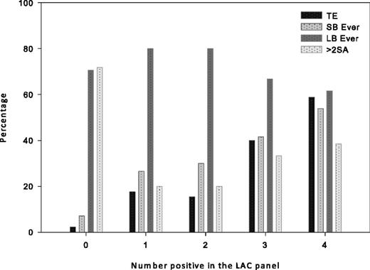
There was a significant increase in the history of TEs with increasing number of positive LAC assays per panel (P = .032; 95% CI, .089-.741) (Figure 2). A significant difference was observed between 1 or 2 positive LAC assay results compared with 3 or 4 positive results per panel and a history of TEs (P = .12; 95% CI, .097-.569). There were trends toward increasing frequency of stillbirth (1+: 26.6% vs 4+: 53.8%), recurrent TEs (1+: 1.6% vs 4+:8.1%), and decreasing frequency of live births (1+:80.0 vs 4+: 64.0) with the increasing number of positive LAC assay results (Figure 2). However, sample sizes were small, and these results did not reach statistical significance. There was an apparent difference in a history of adverse clinical outcomes between 1-2+ LAC assays compared with 3-4+ assays (Figure 2). No apparent correlation was observed between aCL level or prevalence and increasing LAC positivity (data not shown). If we had used only the DRVVT assay to determine LAC positivity, 20 (32.3%) of 62 patients would have been missed; if we had used PTT-LA, only 25 (40.3%) of 62 patients would have been missed. If we had used both DRVVT and PTT-LA but no other assays, we would have determined that 14 (22.6%) of 62 patients tested negative for the LAC.
Change in prevalence of clinical events with increasing LAC positivity. The frequency of TEs, stillbirth (SB), and live birth (LB) varied with increasing number of positive results on LAC assays: patients with 0 of 4 assays positive (ie, LAC negative) had the lowest prevalence of TEs and SB but the highest prevalence of recurrent pregnancy loss. In contrast, patients with 4 of 4 positive results on LAC assays had the highest prevalence of TEs and SB. Patients with either 1 of 4 or 2 of 4 positive LAC assay results appeared to have the same event frequencies.
Change in prevalence of clinical events with increasing LAC positivity. The frequency of TEs, stillbirth (SB), and live birth (LB) varied with increasing number of positive results on LAC assays: patients with 0 of 4 assays positive (ie, LAC negative) had the lowest prevalence of TEs and SB but the highest prevalence of recurrent pregnancy loss. In contrast, patients with 4 of 4 positive results on LAC assays had the highest prevalence of TEs and SB. Patients with either 1 of 4 or 2 of 4 positive LAC assay results appeared to have the same event frequencies.
Association of a specific LAC assay with TEs
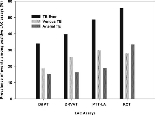
We did not find a predominant assay among the 4 assays in our LAC panel regarding an association with any specific clinical event. There were trends toward an increasing prevalence of TEs and decreasing live births ever with prolonged KCT compared with the other 3 assays (Figure 3), but these findings did not reach significance. Venous TE was the most frequent in patients with prolonged dPT, dRVVT, and PTT-LA assays; in the group with prolonged KCT, arterial TE was more frequent, but there were too few TEs in each group to draw any statistically significant conclusions.
Distribution of TEs among 4 LAC assays. KCT was the least frequently positive assay in our LAC sample but had the highest association with TEs, particularly arterial TE, compared with the other LAC assays. However, the sample size and event frequency were both low, so results must be interpreted with caution.
Distribution of TEs among 4 LAC assays. KCT was the least frequently positive assay in our LAC sample but had the highest association with TEs, particularly arterial TE, compared with the other LAC assays. However, the sample size and event frequency were both low, so results must be interpreted with caution.
Within the group of LAC-positive patients, we compared not only the number of positive assay results but also the degree of positivity within each assay by comparing clotting times to determine any association with a history of TEs. The median 1:1 dilution clotting times of patients with prolonged dPT and/or dRVVT were significantly higher in patients with a history of TEs compared to those without such a history: dPT: 101.3 vs 85.4 seconds; P = .045; dRVVT: 83.2 vs 70.1 seconds, P = .031, respectively (Mann-Whitney rank-sum test). We found no significant differences between the median clotting times of prolonged PTT-LA or KCT assays that differentiated a history of TEs (data not shown).
Discussion
We confirmed the associations between LAC positivity with venous TEs and adverse obstetric outcomes that have been reported previously.1-4 We did not find an increased history of preeclampsia with LAC positivity compared with reported population prevalence (2%-8%).23 Interestingly, regardless of an increased history of stillbirth and IUGR, our LAC-positive group had the same overall incidence of live birth ever (70%) and of at least 1 adverse obstetric event (90%) as the LAC-negative group, apparently indicating an inconsistent correlation between the presence of LAC and obstetric morbidity or mortality in the high-risk population attending our clinic. This lack of association was also observed by Cervera et al who, in a 5-year prospective study, found no immunologic predictor of pregnancy morbidity or mortality among 1000 patients with APS, and no differences in their clinical presentation according the presence or absence of LAC (or aCL).24 The same group also found that 74% of women with APS had 1 or more live births at the outset of the study and 80% of women who were pregnant at least once during the 5-year follow-up had at least 1 or more live births.24
Within the LAC-negative subgroup whose charts were reviewed, 70% of patients were diagnosed with early recurrent pregnancy loss. Approximately 250 new patients with recurrent miscarriage are referred to our clinic annually, resulting in ∼1500 patients seen during a 6-year period, which represents ∼70% of the 2200 patients included in this study. Therefore, the distribution of diagnoses in our study, particularly with respect to recurrent pregnancy loss, was representative of our population as a whole during the last 6 years. Only 5 patients with exclusively early recurrent miscarriage also tested positive for LAC, which represents a very low prevalence (0.3%). Other authors have also noted this trend in their populations.10-12,25,26 Given this rarity, it is perhaps somewhat surprising that determination of LAC (and other aPL) continues to be included in the recommended investigation of early recurrent pregnancy loss,6-9 although our results do confirm an association with later gestational morbidity and mortality.
Several studies have reported that “triple positivity” (the combination of positive aCL, LAC, and anti-β2GPI) is strongly associated with adverse clinical outcomes.27,28 On the basis of previous work that found a very low prevalence in our patient population,29 our laboratory does not routinely screen for anti-β2GPI. However, only one third of the LAC-positive patients with a history of TEs in our present study also had moderate to high aCL IgG, and fewer than a quarter had moderate to high aCL IgM. Therefore, it was evident that even without the inclusion of anti-β2GPI testing, most patients with a history of TEs would not have tested “triple positive” for aPL and that positivity for either aCL or anti-β2GPI was not necessary for venous or arterial TEs in our patients. Lockshin et al observed that the simultaneous positivity for aCL, anti-β2GPI, and LAC was not superior for the prediction of adverse obstetric outcomes than positive LAC results alone.19 Cervera et al found that no aPL or combination of aPL was a predictor of TEs or pregnancy morbidity or mortality in a 5-year study of 1000 patients with APS.24
The ISTH guidelines suggest that the risk for false-positive results is increased if more than 2 LAC assays are performed, and also that no single assay is sensitive for all LAC.14 Our low LAC-positive prevalence despite use of a panel of 4 assays indicates that a wider array of assays is not necessarily disadvantageous for the detection of LAC, as it does not necessarily result in a spuriously high prevalence. We were also unable to identify a single assay that was associated with either stillbirth or a history of TEs. This lack of association between any one particular assay and a clinical manifestation might indicate a need for more than 1 or 2 assays, contrary to the ISTH recommendations. In addition, our finding that an increased number of positive assay results per panel was associated with an increased occurrence of clinical events (and therefore a poorer prognosis) suggests that limiting the assessment of LAC to only 2 assays may not be the optimal choice.
Two of our tests, the dPT and the KCT, are specifically not recommended in the ISTH guidelines,14 but references cited in the update do not reflect that recommendation. For example, Galli et al15 concluded that the KCT result was “most abnormal” in those plasmas with LAC activity, whereas dRVVT was “most abnormal” in plasmas with high aCL activity. In the 20 years we have been performing this LAC panel in our laboratory, we have not experienced the methodologic issues raised with respect to performing the KCT.30 Furthermore, Pengo et al reported that a combination of both dRVVT and KCT was more useful than either alone or with thrombin time in detection of a higher level of LAC “potency.”31 In addition, the UK National External Quality Assessment Scheme (NEQAS) proficiency program evaluated dPT, KCT, dRVVT, and PTT-LA and noted that the “majority of centres reported strongly positive KCT screens for spiked and patient samples.”32 A positive plasma result was correctly identified by all 17 centers that used dPT as part of their LAC screen, from which it might be inferred that the dPT was a useful adjunct to LAC determination.33 Use of recombinant human tissue thromboplastin has decreased the variability seen earlier in dPT assays when thromboplastin was extracted from animal tissue.33 Recent evaluation of the updated ISTH guidelines by 2 independent groups both reported that in agreement with the guidelines, use of at least 2 assays increases association between a history of LAC and thrombosis. In contrast to conclusions in the guidelines, these reports also observed no superior combinations among dRVVT, dPT, and PTT-LA, confirming that dPT remains a valuable addition to the LAC test panel repertoire.16,34
Although many centers rely solely on the dRVVT,32,35,36 the UK NEQAS noted that even with that assay, dependent on methodology, discrepancies were reported. Therefore, it is essential that each laboratory develop its own controls and calibrators, and that when results are presented, not only the type of assay but also the precise methodology is included. Simply stating that the assay was run “according to ISTH guidelines” is inadequate because the guidelines do not specify methodology for each assay, or even necessarily which assay(s) to run. Such clarity might help dispel some of the confusion around results from different centers and allow useful correlations to be drawn with clinical events. This is particularly important when so few patients test positive for LAC.
After completion of a 6-year review of more than 3000 LAC assays, perhaps this conclusion was our most striking: patients with a repeated LAC assay result are rarely found, even among those referred to a high-risk clinic. A brief review of the literature reveals that this rarity is actually consistent with the prevalence in other large or longterm aPL-related studies, although it has not been previously highlighted. Consider the following: (1) Petri et al recently reported dRVVT frequency in SLE pregnancies in the Hopkins Lupus Cohort from 1987 to 2011. In 24 years, only 177 women were reported to have ever tested positive, and conclusions regarding the best predictor of pregnancy loss in SLE were based on 6 of 15 patients with a “high” dRVVT result (“>45 seconds”) during the first-trimester visit.26 (2) Cervera et al used the national registries of 20 university centers in 15 European countries to accrue 1000 patients with APS, of whom ∼half tested positive for LAC. This finding would suggest that on average, ∼25 patients per center had a confirmed LAC.24,37 (3) De Laat et al reported on a novel assay for the measurement of anti-β2 GPI–dependent LAC in 325 LAC-positive samples collected from 6 countries.38 (4) In a study of aPL-associated intrauterine fetal death, Helgadóttir et al retrospectively identified a total of 379 women attending 2 university hospitals in Norway with a documented intrauterine fetal death during a 13-year period: 5 tested positive for LAC.39 (5) It took 8 years for the PROMISSE trial to accrue 64 pregnant LAC-positive patients from more than 6 specialized clinics in 5 centers around North America.19 Given these data, our present sample of 62 patients during a 6-year period at a single center appears quite representative of the frequency of LAC positivity among high-risk patients.
Our study had several weaknesses that qualified our findings. We did not stratify patients on the basis of anticoagulant therapy during pregnancy, as patients may have been treated before attending our clinic at their physicians’ discretion; neither did we evaluate those regimens for efficacy. In addition, the small sample size of LAC-positive women restricted the power of some of our findings, and any interpretation of therapeutic efficacy should be circumspect. However, the very large sample of high-risk pregnancy patients tested during the 6-year study period and our use of a 4-assay panel did allow a realistic and informative perspective on the frequency of LAC, the distribution and association of each LAC assay with clinical events, and the difficulties of studying such a rare phenomenon.
In conclusion, we found that a confirmed, repeated LAC result was a very infrequent finding even in a high-risk setting. It was absent in an overwhelming majority of women with exclusively early recurrent pregnancy loss but was associated with sporadic stillbirth. Among our panel of assays, none was predominant, and an increasing number of positive assays were associated with an increased history of morbidity. Our results do not support the ISTH contention that 2 assays are sufficient for the detection of the LAC, and we will continue to use a 4-assay panel in our ongoing assessment of patients at risk for thrombosis and adverse late-gestational events.
The publication costs of this article were defrayed in part by page charge payment. Therefore, and solely to indicate this fact, this article is hereby marked “advertisement” in accordance with 18 USC section 1734.
Authorship
Contributions: C.A.C. designed the study, reviewed charts, collated and analyzed the data, and wrote the paper; J.D. performed chart reviews and reviewed the paper; K.A.S. contributed to the study design, reviewed charts, reviewed the paper, and suggested additional statistical analysis; and C.A.L. revised the study design, reviewed the statistical analysis, and revised the final version of the paper.
Conflict-of-interest disclosure: The authors declare no competing financial interests.
Correspondence: C. A. Clark, Suite 1800, 655 Bay St, Toronto, ON, Canada, M5G 2K4; e-mail: cclarksolo@aol.com.


