Abstract
A substantial portion of patients with inherited blood disorders such as beta thalassemia, or bone marrow failure syndromes such as aplastic anemia(AA), myelodysplastic syndromes(MDS) require frequent transfusions of red blood cells. Frequent blood transfusions may lead to the excess of plasma non-transferrin -bound iron(NTBI) and iron overload occurs, which will significantly injure bone marrow (BM) function as well as induce organ dysfunctions such as liver cirrhosis, diabetes and cardiac diseases. However, the exact mechanism behind this effect remains elusive and ideal treatment needs to be explored. In our preliminary studies, we have demonstrated free iron catalyzes oxidative damage to hematopoietic cells/ mesenchymal stem cells in vitro and suppresses hematopoiesis in iron overload patients (Zhao et al.,blood, 2010 abstract; Lu et al.,blood,2012 abstract; Lu et al., Eur J Haematol, 2013). Here we observed the hematopoiesis inhibitory effects of iron overload on the basis of estabalished iron overload mice model and preliminarily disscussed the mechanism.
In this study, we first established an iron overload mice model by administering different doses(12.5mg/ml,25mg/ml,50mg/ml) iron dextran by intraperitoneal injection every three days for four weeks. To confirm the efficacy of the mice model, the BM, hepatic and splenic iron deposits were observed by morphological study and the labile iron pool level (LIP) of bone marrow mononuclear cells(BMMNCs) was detected using the calcein-AM fluorescent dye. It was found that iron deposits in BM cells of iron overload mice, liver and spleen were markedly increased and the BMMNCs LIP level was much higher than that of normal control mice. The above results showed that the iron-overloaded mice model has been established successfully.
Next we observed whether iron overload (25mg/ml) could affect the hematopoiesis of BM. The colony-forming cell assay was performed by culturing BMMNCs in MethoCult M3434 methylcellulose medium to evaluate hematopoietic progenitor cells(HPCs) proliferation function. The competitive repopulation assay and single-cell colony cultures of sorted hematopoietic stem cells (HSCs,CD34-Lin- sca1+c-kit+cells,LSK+)were used to validate HSCs function.
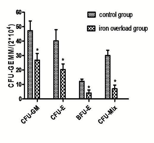
The counts of BMMNCs have no significant difference. However, It was found that hematopoietic colony-forming unit (CFU-E, BFU-E, CFU-GM and CFU-mix) was much lower than that of normal control(P<0.05)(Fig.1). Notely, the number of LSK+ cells (*103/femur) was decreased significantly in iron overload mouse (26.43±3.28) compared with normal control(40.12±5.21) and the single-cell colony formation(/60wells) was reduced significantly in iron overload mouse(28.54±3.33) compared with normal control(47.93±4.82) (P<0.05). The long-term and multilineage engraftment capability of the iron-overloaded HSCs was weaken after transplantation.
We then explored the possible mechanism of this inhibitory effects. Our previous studies have shown that iron overload could elevated reactive oxygen species (ROS) levels of mesenchymal stem cells and HSCs in vitro. Similarly, the intracellular ROS levels were analyzed by a flow cytometer. It was found that ROS level in iron overload BM was increased by 3.32 folds in erythroid cells, 1.51 folds in granulocytes and 4.80 folds in LSK+ cells,respectively. And also, the expression of p53, p38MAPK and p16Ink4a mRNA remained significantly elevated, which indicated that ROS related signal pathway was involved in the deficient hematopoiesis of iron overload BM.
In addition, we also observed the effects of iron overload on the mice with deficient hematopoiesis exposed to 4Gy total body irradiation(TBI), which was more similar to clinical pathological conditions such as AA or MDS. It was found that BM damage caused by iron overload was aggravated in pathological conditions (primary findings were not shown).
Results of hematopoietic colony forming unit of different groups(*P<0.05)
No relevant conflicts of interest to declare.
Author notes
Asterisk with author names denotes non-ASH members.


This feature is available to Subscribers Only
Sign In or Create an Account Close Modal