Abstract
The amyloidoses are heterogeneous diseases associated with the deposition of insoluble proteins or peptides with a characteristic beta-diffraction pattern extracellularly. At the current time, over 27 different extracellular fibril proteins are known to cause disease in humans. Iatrogenic amyloidosis is a rare, often not sought diagnosis. On occasion, patients with drug induced amyloidosis can present with other systemic features reminiscent of systemic immunoglobulin-derived (AL) amyloidosis, and may present a diagnostic challenge. Treatment of the latter often involves chemotherapy and/or stem cell transplantation, while the former tends to remain localized and needs local therapy; thus accurate diagnosis is critical. To this end, proteomic analysis of amyloid tissue has proven to be an invaluable tool in amyloid typing. We have analyzed the biochemical composition of iatrogenic amyloid using laser capture/tandem mass spectrometry (LC-MS/MS)-based proteomic analysis in 52 cases of insulin and enfuvirtide (Fuzeon¨) associated amyloidosis.
In brief, 10-μm-thick sections of formalin-fixed paraffin-embedded tissues were stained with Congo red. Congo red positively staining tissue as viewed with a fluorescent light source appeared bright red. Positive areas were dissected using laser microdissection to a volume of at least 60,000 μm2; three microdissections were analyzed for each case. The microdissected material was collected into 0.5-ml microcentrifuge tube caps containing 35 μL Tris/EDTA/0.002% Zwittergent buffer. Microdissected fragments were subjected to a heat-mediated antigen retrieval method (98C for 90minutes) before being denatured via sonication and subsequently digested into tryptic peptides overnight using 0.5ug of trypsin. The resulting digests were then analyzed with nanoflow LC-MS/MS. The MS/MS spectra of each case were matched against a composite protein sequence database using three different search algorithms (Sequest, X!Tandem, and Mascot). The composite database contained the human SwissProt entries but was also augmented with known immunoglobulin variant domains, known amyloidogenic mutations from literature, the enfuvirtide amino acid sequence, and common contaminants. Reversed protein sequences were appended to the database for estimating the false discovery rates of the identifications. The peptide identification results were filtered using Scaffold software (Proteome Software, Portland, OR) and then filtered peptides were assembled into protein identifications. Candidate proteins with at least one high-confident (probability of identification >90%) unique peptide identification and at least four MS/MS spectral matches were considered for clinical interpretation.
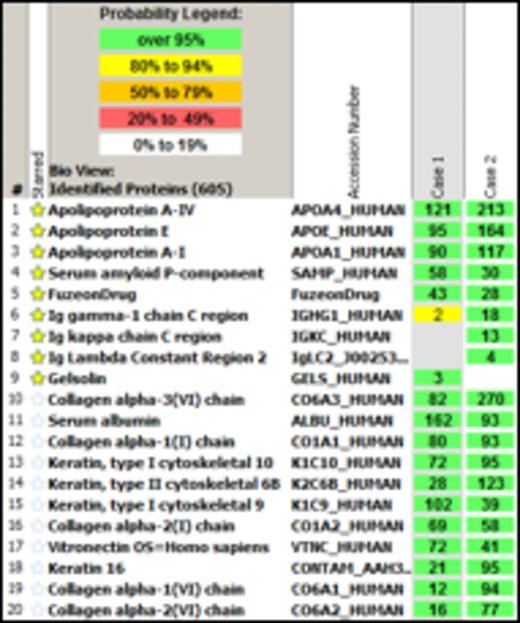
For each case, we created a personalized proteomic profile that lists all the confident protein identifications in each of the microdissection along with their respective MS/MS spectral counts. The number of MS/MS spectra matching to a protein is considered as a semi-quantitative measure of its abundance. The most abundant amyloidogenic protein detected across all microdissections and as interpreted in the context of the clinical history is considered to be the amyloid subtype.
Figure 1. Insulin-associated amyloidosis
In conclusion, we show the biochemical composition of all known drug-induced iatrogenic amyloidosis and provide the utility of proteomic analysis in elucidating amyloid subtyping for accurate diagnosis and management.
Legend: A spectral count number of greater than 4 is significant. The green boxes denote protein identification at a probability of over 95%, and yellow 80-94%. In figure 1, insulin (# 2) and in figure 2, enfuvirtide (#5) are shown in abundance, additionally other amyloid precursors such as apolipoproteins A-IV, E, A-I and SAP are also seen.
No relevant conflicts of interest to declare.
Author notes
Asterisk with author names denotes non-ASH members.


This feature is available to Subscribers Only
Sign In or Create an Account Close Modal