Abstract
Patients (pts) with TrIL have inferior outcomes compared with de novo diffuse large B-cell lymphoma (DLBCL), and their optimum follow up is not well defined. We sought to determine the utility of surveillance PET-CT in pts with TrIL achieving CMR after primary therapy and identify patterns of relapse.
We performed a retrospective analysis of pts with TrIL treated at Peter MacCallum Cancer Centre between 2002 and 2012 who achieved CMR after primary therapy who had ³1 subsequent surveillance PET-CT. In the period analysed, departmental protocol recommended 6-monthly scans for pts in CMR for the first 2 years, then annually until 5 years after completion of therapy, if there was intention to intervene on detection of subclinical relapse. A positive scan suggested relapsed lymphoma, with true positive results requiring either biopsy confirmation or unequivocal scan progression. A false positive scan was refuted by biopsy and/or follow up showing resolution of areas of increased FDG uptake. A negative scan was interpreted as negative for relapsed lymphoma: true negatives had no clinical relapse and false negatives manifest relapse within three months from the date of the scan. Indeterminate scans were recorded if determination could not be made.
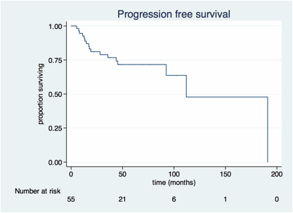
The cohort included 55 pts with TrIL: 38 underwent stem cell transplant (autologous, n= 37; allogeneic, n=1) as consolidation; 17 did not. (Table 1). After a median follow-up of 34 (range 3 – 101) months, the actuarial 3-year progression free (PFS) and overall survival (OS) were 77% (95% CI 62 – 86%) and 88% (75 – 94%) respectively. Multiple potential prognostic factors including IPI, stage, serum LDH, timing of transformation (simultaneous diagnosis of transformation versus delayed) and type of indolent histology were not predictive of PFS.
Patient characteristics
| patient characteristics . | . |
|---|---|
| number of pts | 55 |
| median age (range) years | 59 (35 – 83) |
| female (%) | 22 (40%) |
| histologically transformed at initial diagnosis | 31 (56%) |
| indolent histology FL MALT/MZL CLL/SLL ALCL | 46 (65%) 5 (9%) 3 (5%) 1 (2%) |
| IPI at transformation 0 - 1 2 3 - 5 | 14 (27%) 18 (34%) 20 (39%) |
| median serum LDH:ULN (range) | 1.03 (0 – 4.7) |
| Extranodal sites 0-1 ³32 | 33 (62%) 20 (38%) |
| ECOG performance status 0-1 ³32 | 51 (95%) 3 (5%) |
| stage I-II III-IV | 11 (20%) 44 (80%) |
| B symptoms | 12 (23%) |
Results of surveillance PET-CT scans, by time elapsed since completion of therapy.
| . | 0-6 mo . | 6-12 mo . | 12-18 mo . | 18-24 mo . | 24-36 mo . | 36-48 mo . | 48+ mo . | total . |
|---|---|---|---|---|---|---|---|---|
| true positives (subclinical) | 1 | 4 | 1 | 1 | 0 | 0 | 0 | 7 |
| true positives (suspected) | 1 | 2 | 2 | 0 | 2 | 1 | 0 | 8 |
| false positives | 1 | 1 | 1 | 0 | 0 | 1 | 0 | 4 |
| false negatives | 0 | 0 | 0 | 0 | 0 | 1 | 0 | 1 |
| indeterminate | 0 | 0 | 6 | 1 | 0 | 0 | 0 | 7 |
| true negatives | 33 | 37 | 26 | 21 | 21 | 10 | 5 | 153 |
| total | 36 | 44 | 36 | 23 | 23 | 13 | 5 | 180 |
| % of PET scans detecting subclinical relapse | 3% | 9% | 3% | 4% | 0% | 0% | 0% |
Of 180 surveillance PET-CT scans, there were 153 true negatives, 4 false positives, 1 false negative, 7 indeterminate and 15 true positives (Table 2). Considering indeterminate scans as false positives, the specificity of PET-CT for detecting relapse was 93%, sensitivity 93%, positive predictive value 54% and negative predictive value 99%. Of the 15 pts who experienced disease relapse, 7 (47%) were subclinical (i.e. detected by surveillance PET-CT scans) whilst 8 (53%) were suspected on the basis of clinical symptoms. Although 5% of scans in the first 2 years detected a subclinical relapse, all of these were either biopsy or clinically shown to be low-grade lymphoma. All 8 symptomatic relapses (at 2 – 42 months), in contrast were DLBCL.
In pts with TrIL achieving CMR, PET-CT detects subclinical relapses of low-grade histology with high sensitivity but with a moderate false-positive rate. This is of limited clinical benefit as the initiation of further therapy in these circumstances is rarely based on imaging findings alone. In contrast, all DLBCL relapses in our cohort were accompanied by clinical symptoms. Thus, surveillance imaging of pts with TrIL achieving CMR is not indicated. PET-CT should be reserved for evaluation of suspected relapse only.
No relevant conflicts of interest to declare.
Author notes
Asterisk with author names denotes non-ASH members.


This feature is available to Subscribers Only
Sign In or Create an Account Close Modal