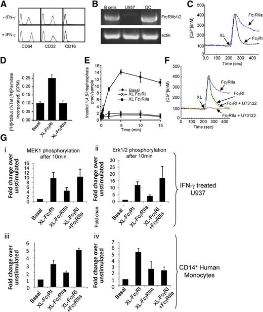
On page 320 in the 9 July 2009 issue, the original Figure 1B had an irrelevant lane spliced out without disclosure of this digital manipulation in the figure or figure legend. A repeat experiment has been performed to document the veracity of the results, and a replacement Figure 1 is now provided. The corrected Figure 1 and the original figure legend are shown.
FcR expression and signaling in U937 and primary monocytes. (A) U937 cells express FcγRI and FcγRII, but not FcγRIII, as determined by flow cytometry using anti-CD32PE and anti-CD64PE and anti-CD16PE (BD Pharmingen). (B) RT-PCR was used to assess the expression of FcγRIIb1/2 on U937 compared with B lymphocytes and dendritic cells. (C) FcγRI and FcγRIIa trigger differential cytosolic Ca2+ signals. Cytosolic calcium was measured in U937 by cuvette fluorimetry over 8 minutes after cross-linking of the individual FcRs using antibody clones 10.1 and 3D3 (BD Pharmingen), respectively (XL). (D) FcγRI triggers PLD activity, whereas FcγRIIa does not. PLD activity measured in resting U937 cells (Basal) or in cells after FcγR aggregation (XL FcγRI or XL FcγRIIa) for 30 minutes. (E) FcγRIIa triggers PLC activation in U937 cells. InsP3 generation was measured in resting cells (basal) or in cells after FcγR aggregation (XL FcγRI or XL FcγRIIa) for 15 minutes. (F) The PLC inhibitor U73122 blocks the FcγRIIa-mediated cytosolic calcium signal in U937 cells. Cells pretreated with the PLC inhibitor U73122 for 45 minutes were assayed for cytosolic calcium over 8 minutes after FcR cross-linking (XL FcγRI or XL FcγRIIa). (G) In IFN-γ–treated U937 (i-ii) and primary human monocytes (iii-iv), MEK1 and ERK1/2 are activated by FcγRI ligation by antibody clone 10.1 to a greater extent than by FcγRIIa ligation by antibody clone 3D3. Phosphoprotein array (Biorad) was used to measure ERK1/2 and MEK1 phosphorylation in cell lysates. MEK1, ERK, PLC, and PLD activity is expressed as a mean ± SD from 3 independent experiments. All intracellular calcium measurements were carried out in the presence of 1.5 M extracellular calcium and results shown are typical of 3 independent experiments.
FcR expression and signaling in U937 and primary monocytes. (A) U937 cells express FcγRI and FcγRII, but not FcγRIII, as determined by flow cytometry using anti-CD32PE and anti-CD64PE and anti-CD16PE (BD Pharmingen). (B) RT-PCR was used to assess the expression of FcγRIIb1/2 on U937 compared with B lymphocytes and dendritic cells. (C) FcγRI and FcγRIIa trigger differential cytosolic Ca2+ signals. Cytosolic calcium was measured in U937 by cuvette fluorimetry over 8 minutes after cross-linking of the individual FcRs using antibody clones 10.1 and 3D3 (BD Pharmingen), respectively (XL). (D) FcγRI triggers PLD activity, whereas FcγRIIa does not. PLD activity measured in resting U937 cells (Basal) or in cells after FcγR aggregation (XL FcγRI or XL FcγRIIa) for 30 minutes. (E) FcγRIIa triggers PLC activation in U937 cells. InsP3 generation was measured in resting cells (basal) or in cells after FcγR aggregation (XL FcγRI or XL FcγRIIa) for 15 minutes. (F) The PLC inhibitor U73122 blocks the FcγRIIa-mediated cytosolic calcium signal in U937 cells. Cells pretreated with the PLC inhibitor U73122 for 45 minutes were assayed for cytosolic calcium over 8 minutes after FcR cross-linking (XL FcγRI or XL FcγRIIa). (G) In IFN-γ–treated U937 (i-ii) and primary human monocytes (iii-iv), MEK1 and ERK1/2 are activated by FcγRI ligation by antibody clone 10.1 to a greater extent than by FcγRIIa ligation by antibody clone 3D3. Phosphoprotein array (Biorad) was used to measure ERK1/2 and MEK1 phosphorylation in cell lysates. MEK1, ERK, PLC, and PLD activity is expressed as a mean ± SD from 3 independent experiments. All intracellular calcium measurements were carried out in the presence of 1.5 M extracellular calcium and results shown are typical of 3 independent experiments.


This feature is available to Subscribers Only
Sign In or Create an Account Close Modal