Key Points
Priming of CMV-specific CD4+ and CD8+ T cells occurs as early as day 42 in patients undergoing UCBT.
Lack of CMV control in UCBT patients could be related to low absolute frequency of T cells and lack of in vivo expansion of T cells.
Abstract
A disadvantage of umbilical cord blood transplantation (UCBT) is the delay in immune reconstitution, placing patients at increased risk for infections after transplant. Cytomegalovirus (CMV) in particular has been shown to cause significant morbidity in patients undergoing UCBT. Here, we comprehensively evaluate the development of CD4+ and CD8+ T-cell responses to CMV in a cohort of patients that underwent double UCBT. Our findings demonstrate conclusively that a diverse polyclonal CMV-specific T-cell response derived from the UCB graft is primed to viral antigens as early as day 42 after UCBT, but these T cells fail to achieve sufficient numbers in vivo to control CMV reactivations. This is not due to an inherent inability of UCB-derived T cells to proliferate, as these T cells underwent rapid proliferation in vitro. The TCR diversity and antigen specificity of CMV-specific T cells remained remarkably stable in the first year after transplant, suggesting that later control of virus replication results from improved function of T cells primed early after transplant and not from de novo responses derived from later thymic emigrants. Ex vivo expansion and adoptive transfer of CMV-specific T cells isolated from UCBT recipients early after transplant could augment immunity to CMV.
Introduction
Umbilical cord blood (UCB) is increasingly used as a source of hematopoietic stem cells (HSCs) for transplantation and has advantages compared with bone marrow or peripheral blood stem cells (PBSCs) including availability, low risk of transmitting infections, and less stringent HLA matching. Leukemia relapse after umbilical cord blood transplant (UCBT) is comparable to other HSC products, and may be reduced when 2 UCB units are used.1-5 The rate of acute graft-versus-host disease (GVHD) is also comparable, with suggestion of a lower incidence of chronic GVHD.4,6 A disadvantage of UCB is that low numbers of CD34+ HSCs and CD3+ T cells are infused, which delays engraftment and reconstitution of T-cell immunity, respectively.7-10 The rate of engraftment is improved by infusion of 2 UCB units,11 however, the delay in T-cell immune reconstitution leads to higher rates of infections and contributes to nonrelapse mortality.3,12
The number of T cells transferred with an UCB graft is approximately a log10 less than a PBSC graft, and T cells in UCB are naive. Thus, there is no transfer of protective memory T cells, which are important for controlling latent viruses like cytomegalovirus (CMV).13-16 At our institution, nearly 100% of CMV-seropositive UCBT patients reactivate CMV early posttransplant and require antiviral drug therapy.17 Previous studies suggest that CMV-specific CD8+ T cells cannot reliably be detected in UCBT recipients until >100 days after transplant, when thymopoiesis recovers.14,18 In the few cases where CMV-specific T cells were detected before 100 days, the origin (cord blood or recipient) of these T cells and the breadth of viral antigens recognized were not determined.
Here, we use sensitive assays to evaluate the kinetics, origin, and specificity of CMV-specific T cells in patients that received double UCBT (dUCBT). The data show that in a majority of patients, UBC CD8+ and CD4+ T cells are primed to CMV antigens early after transplant, but low numbers of functional T cells are present in vivo. These CMV-specific T cells readily proliferate ex vivo, and can be shown to recognize multiple CMV antigens and use diverse T-cell receptors (TCRs) even at early times after UCBT. These results demonstrate that priming of CMV-specific T cells after UCBT is not defective, and suggests the inability to control viral reactivation results from the failure of the T cells to achieve sufficient numbers in vivo.
Methods
Patients and samples
Patients receiving dUCBT or peripheral blood stem cell transplant (PBSCT) from a CMV-seronegative donor at the Fred Hutchinson Cancer Research Center were eligible for this study. A skin biopsy was obtained from each patient to generate fibroblasts, and blood was collected prior to and at intervals after transplant. The Fred Hutchinson Cancer Research Center Institutional Review Board approved study activities, and participants provided written informed consent according to the Declaration of Helsinki.
CMV prophylaxis, monitoring, and antiviral therapy
CMV prophylaxis was administered to all UCBT patients and consisted of acyclovir (800 mg twice daily) beginning pretransplant and continuing until CMV reactivation occurred or day 365 posttransplant (7 patients), or of ganciclovir until 2 days prior to transplant followed by acyclovir (500 mg/m2 intravenously every 8 hours) until CMV reactivation occurred or day 365 (12 patients). Patients were monitored twice weekly until day 100 using polymerase chain reaction (PCR) to detect CMV DNA in serum, and then weekly for 1 year after UCBT.19 CMV reactivation was defined as any detection of CMV DNA in serum by PCR. Preemptive antiviral therapy was initiated with ganciclovir or foscarnet for any positive CMV PCR through day 100, and for any positive PCR of >1000 DNA copies per mL after day 100. Patients were monitored for viral load twice weekly during antiviral therapy until negative. Virus reactivation data were collected on all patients until day 400 or death.
Virus and cell lines
Cytokine flow cytometry
Blood was obtained up to 4 times (days 42-56, 80, 180, and 365) after UCBT or PBSCT, and peripheral blood mononuclear cells (PBMCs) were separated over Ficoll-Hypaque and cryopreserved. For analysis of CD8+ T-cell responses, PBMCs were thawed, washed in medium, suspended at 2 × 10⁶ cells per mL, and cocultured in a 3.3:1 ratio with patient fibroblasts either mock-infected or infected with RV798 CMV at a multiplicity of infection of 5 for 48 hours. Interferon-γ (IFN-γ)–producing CD8+ cells were enumerated by flow cytometry as described.21 Cytokine flow cytometry (CFC) to detect pp65-specific responses was performed by incubating T cells with peptide pools (0.5 µg/mL, Peptivator; Miltenyi) consisting of 15-mer peptides with an 11 amino acid overlap and spanning the pp65 protein, in the presence of antibodies to CD28 and CD49d (5 µL/mL; BD Biosciences). Cells were incubated for 6 hours, followed by CFC for IFN-γ.
CFC to detect CD4+ T-cell responses was performed by stimulating CD4+ T-cell lines with γ-irradiated (3000 cGy) autologous PBMCs previously pulsed with CMV antigen (12.5 µg/mL; Microbix Biosystems) for 2 hours at a 1:1 ratio. CFC staining of IFN-γ was performed after 4 hours.
Generation of CMV-specific T cells from UCBT recipients
CMV-specific CD8+ T-cell lines were generated by stimulating posttransplant PBMCs with autologous fibroblasts infected for 48 hours with RV798 CMV at a multiplicity of infection of 5. On days 2, 5, and 8, interleukin-2 (IL-2; 5 IU) was added to the media. On day 10, cells were harvested, an aliquot was restimulated with RV798-infected autologous fibroblasts for 4 hours, and the CMV-specific subset was enriched using IFN-γ capture as described,22 except that CD8+/IFNγ+ cells were sorted with a FACSAria (Becton Dickinson). The enriched CMV-specific CD8+ T cells were expanded as described.21 Chimerism analysis was performed on CMV-specific CD8+ T cells using the PowerPlex16 system (Promega) consisting of 16 markers (15 short tandem repeats and Amelogenin) to determine origin (host or UCB). Adaptive Biotechnologies performed deep sequencing of the TCR Vb locus of CMV-specific T-cell lines.
To enrich CD4+ CMV-specific T cells, PBMCs (2 × 10⁶ cells per mL) were stimulated with CMV antigen. The cell suspension was distributed into 96-well plates (100 µL per well), and cultured at 37°C for 4 days, at which time 100 µL of fresh media with IL-2 (50 IU/mL) was added to each well. After an additional 3 days of incubation, cells were harvested, resuspended at 2 × 10⁶/mL, and used in functional assays. In some experiments, an aliquot was restimulated for an additional 7 days with irradiated autologous PBMCs pulsed with CMV antigen and IL-2.
Lymphoproliferative and chromium release assays
PBMCs from different time points after UCBT were washed and resuspended in T-cell medium at a concentration of 2 × 10⁶/mL. Triplicate wells of 96-well plates were seeded with 4 × 10⁵ cells and CMV antigen (12.5 µg/mL) and cultured at 37°C for 96 hours. 3H thymidine was added for the final 18 hours, and the cells were harvested and analyzed for 3H-thymidine incorporation. T cells plated with media alone and phytohemagglutinin-L (5 μg/mL; Sigma) were negative and positive controls, respectively. CMV-specific T-cell lines were assayed for cytotoxicity in a chromium-release assay using 51Cr-labeled autologous or HLA-mismatched fibroblasts that were infected with RV798 CMV or mock infected.21
Viral genome scan and epitope mapping
COS-7 cells (5 × 10³ per well) were incubated in 96-well plates overnight, and then transfected using Fugene (0.35 µL per well) with plasmids encoding 1 or more of the HLA molecules (50 ng per well) expressed by the recipient together with pools or individual plasmids encoding 134 CMV open reading frames (ORFs) (100-200 ng per well). CMV-specific T cells were added to the transfected COS-7 cells, supernatant was harvested after 48 hours and analyzed for IFN-γ by enzyme-linked immunosorbent assay.23 Poly-HRP Blocking solution and poly-hrp20 streptavidin antibody were purchased from Research Diagnostics, and primary and secondary IFN-γ antibodies from Pierce Endogen.
The coding sequences of CMV ORFs identified as targets of CMV-specific CD8+ T cells by the genome scan were analyzed using algorithms (Bimas, SYFPEITHI, and Immune Epitope Database) for peptides predicted to bind to the HLA-restricting allele. Peptides with the highest binding scores were synthesized (GenScript), pulsed onto LCL expressing the HLA allele for 2 hours at 37°C, and assayed for recognition by CMV-specific CD8+ T-cell lines using CFC to detect intracellular IFN-γ.
Results
Patient characteristics
The characteristics, conditioning regimen, and GVHD prophylaxis of the 19 recipients of dUCBT are shown in Table 1.
Patient characteristics
| Patients . | Age, y . | Disease . | Conditioning regimen . | GVHD prophylaxis . | HLA match of cord to patient . | Donor CD3 chimerism, <d 30 . |
|---|---|---|---|---|---|---|
| CMV positive | ||||||
| 1 | 11 | AML | Flu, Cy, TBI 14.2 Gy | CsA, MMF | 4/6, 4/6 | 100% CB2 |
| 2 | 56 | AML | Flu, Cy, TBI 200 cGy | CsA, MMF | 5/6, 4/6 | 100% CB2 |
| 3 | 30 | AML | Flu, Cy, TBI 13.2 Gy | CsA, MMF | 5/6, 4/6 | 100% CB2 |
| 4 | 14 | ALL | Flu, Cy, TBI 13.2 Gy | CsA, MMF | 5/6, 4/6 | 100% CB1 |
| 5 | 17 | T-ALL | Flu, Cy, TBI 13.2 Gy | CsA, MMF | 4/6, 4/6 | 100% CB2 |
| 6 | 14 | CML | Flu, Cy, TBI 13.2 Gy | CsA, MMF | 4/6, 4/6 | 100% CB2 |
| 7 | 46 | AML | Flu, Treo, TBI 200 cGy | CsA, MMF | 4/6, 4/6 | 100% CB1* |
| 8 | 13 | T-ALL | Flu, Cy, TBI 13.2 Gy | CsA, MMF | 5/6, 4/6 | 98% CB1, 2% host† |
| 9 | 33 | AML | Flu, Cy, TBI 13.2 Gy | CsA, MMF | 4/6, 4/6 | 100% CB2 |
| 10 | 29 | AML | Flu, Cy, TBI 200 cGy | CsA, MMF | 4/6, 4/6 | Mixed CB1/2, 0% host‡ |
| 11 | 26 | AML | Flu, Cy, TBI 13.2 Gy | CsA, MMF | 5/6, 4/6 | 100% CB2 |
| 12 | 4 | AML | Flu, Cy, TBI 13.2 Gy | CsA, MMF | 4/6, 4/6 | 100% CB2 |
| 13 | 38 | MDS | Flu, Treo, TBI 200 cGy | CsA, MMF | 4/6, 4/6 | 100% CB1 |
| 14 | 73 | Pro-T-ALL | Flu, Cy, TBI 13.2 Gy | CsA, MMF | 5/6, 4/6 | Mixed CB1/2, host§ |
| 15 | 33 | AML | Flu, Cy, TBI 13.2 Gy | CsA, MMF | 4/6, 4/6 | 100% CB1|| |
| CMV negative | ||||||
| 16 | 56 | AML | Flu, Treo, TBI 200 cGy | CsA, MMF | 5/6, 4/6 | 91% CB1¶ |
| 17 | 39 | AML | Flu, Cy, TBI 13.2 Gy | CsA, MMF | 4/6, 4/6 | 100% CB1 |
| 18 | 26 | Biphenotypic AL | Flu, Cy, TBI 13.2 Gy | CsA, MMF | 4/6, 4/6 | 100% CB1 |
| 19 | 21 | ALL | Flu, Cy, TBI 13.2 Gy | CsA, MMF | 4/6, 4/6 | 100% CB1 |
AL, acute leukemia; ALL, acute lymphoblastic leukemia; AML, acute myeloid leukemia; CB, cord blood donor; CML, chronic myeloid leukemia; CsA, cyclosporine A; Cy, cyclophosphamide; Flu, fludarabine; MDS, myelodysplastic syndrome; MMF, mycophenolate mofetil; TBI, total body irradiation; Treo, treosulfan.
*2% host at d 56 and d 83, 100% CB1 at d 176; †100% CB1 at d 365; ‡100% mixed CB1/2 >d 165; §0% host d 84; ‖2% host d 80, ¶mixed CB1/2 >d 30, 100% CB1 at d 328.
CMV reactivation is common after UCBT
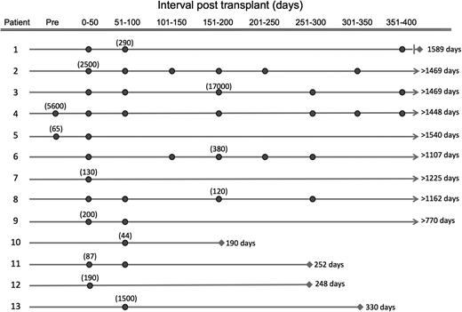
CMV reactivation occurred in 13 of the 15 CMV-seropositive patients at a median of 17 days (range, 2-84) posttransplant. Nine of these 13 patients exhibited repeated CMV reactivations (median = 4) after clearance of the initial episode with drug therapy (Figure 1). Patients 1 to 4 received low-dose acyclovir prophylaxis and had more frequent CMV reactivations with higher CMV DNA copy numbers than patients that received high-dose acyclovir prophylaxis. Three of the 4 CMV-negative recipients had no CMV reactivations; 1 patient had a single low-level positive PCR that may be a false positive, and remained negative thereafter.
CMV reactivations during the first 400 days posttransplant. The occurrence of each reactivation in each 50-day interval through day 400 after transplant is depicted by a dark circle on the timeline for the 13 of 15 CMV-seropositive patients that reactivated CMV. The number in parentheses indicates the maximum CMV DNA copy number per microliter observed for each patient. The survival (days) after transplant is shown in the column to the right of each patient timeline.
CMV reactivations during the first 400 days posttransplant. The occurrence of each reactivation in each 50-day interval through day 400 after transplant is depicted by a dark circle on the timeline for the 13 of 15 CMV-seropositive patients that reactivated CMV. The number in parentheses indicates the maximum CMV DNA copy number per microliter observed for each patient. The survival (days) after transplant is shown in the column to the right of each patient timeline.
Recovery of CD8+ CMV-specific T cells
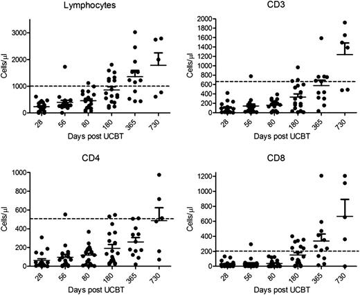
Consistent with previous reports,7,8,24 lymphocyte and absolute CD3+, CD8+, and CD4+ T-cell numbers did not reach the lower level of normal in a majority of UCBT patients until 1 year posttransplant (Figure 2). Seventeen of 19 patients had >90% CD3+ T-cell chimerism from 1 of the cord blood units at or before day 30, and 2 patients had mixed chimerism from both cord blood units (Table 1). By day 365, 100% of the CD3+ cells in 17 patients were derived from 1 donor cord, and 2 patients remained with mixed CD3+ T-cell chimerism from both cords. None of the patients had detectable host CD3+ T cells at day 365.
Recovery of lymphocytes and T-cell subsets after UCBT. The absolute number of lymphocytes, and CD3+, CD4+, and CD8+ T cells in the peripheral blood at intervals after UCBT is shown. The dashed line in each graph indicates the lower level of normal for each cell subset.
Recovery of lymphocytes and T-cell subsets after UCBT. The absolute number of lymphocytes, and CD3+, CD4+, and CD8+ T cells in the peripheral blood at intervals after UCBT is shown. The dashed line in each graph indicates the lower level of normal for each cell subset.
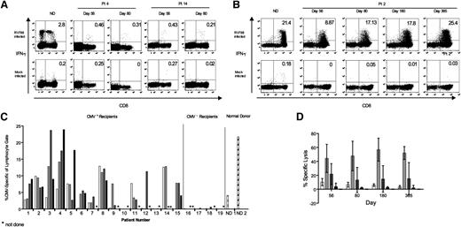
We examined whether and with what kinetics T cells were primed to CMV antigens in these lymphopenic UCBT recipients. In prior studies, enzyme-linked immunospot assay after stimulation with a small panel of viral peptides detected only rare CMV-specific CD8+ T cells early after UCBT in a subset of patients, without delineating the origin of responding T cells. Approaches to detect CMV-specific T cells using only a few immunogenic epitopes vastly underestimate CD8+ T-cell responses to CMV in normal donors,21,25,26 and may not be sufficiently sensitive to detect responses in UCBT recipients. Thus, we evaluated CD8+ T cells to CMV in patients undergoing UCBT using the RV798 CMV strain that has a deletion of the viral genes that block class I major histocompatibility complex expression in infected antigen-presenting cells (APCs).20 We and others have shown that stimulation of PBMCs from normal individuals infected with wild-type CMV enables class I presentation of all potential epitopes in the viral proteome, and is more sensitive for analyzing CD8+ T-cell immunity than using wild-type virus-infected cells, peptide panels of a few proteins, or tetramers to individual epitopes.20,21,26 PBMCs from normal CMV+ donors, from 3 CMV+ recipients of PBSCT from a CMV− donor, and 19 UCBT recipients were stimulated with fibroblasts infected with RV798 CMV, and stained for intracellular IFN-γ. IFN-γ–producing CD8+ T cells were readily detected in PBMCs of normal donors in response to RV798-infected but not uninfected fibroblasts (Figure 3A), and in PBMCs from each of the 3 recipients of PBSCs from a CMV− donor, demonstrating that CMV-specific CD8+ T cells are primed early after transplant in CMV+ recipients of PBSCs from a CMV− donor and expand in vivo (supplemental Figure 1, available on the Blood website). Surprisingly, we observed a low frequency of CD8+ T cells that produced IFN-γ in response to RV798-infected fibroblasts in blood samples obtained from 6 of 15 patients 42 to 56 days after UCBT (Figure 3A), suggesting that priming may also occur in UCBT recipients.
Detection of IFN-γ+ CMV-specific CD8+ T cells after UCBT by CFC of PBMCs and after in vitro expansion of T cells. (A) PBMCs from a normal donor (ND) and from 2 representative CMV-seropositive UCBT recipients were analyzed for CMV-specific CD8+ T cells by CFC. The number in the top right quadrant indicates the percentage of IFN-γ+ T cells in the CD8+ subset. (B) CD8+ T-cell lines were generated from PBMCs obtained at the indicated times by a single in vitro stimulation with RV798-infected fibroblasts and then assayed for IFN-γ by CFC. Data are shown for a normal donor and a representative UCBT patient at multiple time points. Mock-infected autologous fibroblasts served as a negative control in the assay. (C) IFN-γ CFC assay for CMV-specific T cells in T-cell lines from each of 19 UCBT recipients at intervals after transplant (15 CMV positive and 4 CMV negative) and from 2 normal donors. The data shows the percentage of IFN-γ+ T cells in the gated lymphocyte population after restimulation with RV798-infected fibroblasts. White bars, day 56; light gray bars, day 80; dark gray bars, day 180; black bars, day 365; asterisk (*) indicates not done. (D) Lysis of autologous fibroblasts infected with RV798 (light gray bars), mock-infected (white bars), or HLA-mismatched fibroblasts either RV798-infected (dark gray bars) or mock-infected (black bars) by CMV-specific T-cell lines from UCBT recipients. The graph shows the mean specific lysis of cell lines from different time points after UCBT in 5 patients. The effector-to-target (E:T) ratio was 10:1.
Detection of IFN-γ+ CMV-specific CD8+ T cells after UCBT by CFC of PBMCs and after in vitro expansion of T cells. (A) PBMCs from a normal donor (ND) and from 2 representative CMV-seropositive UCBT recipients were analyzed for CMV-specific CD8+ T cells by CFC. The number in the top right quadrant indicates the percentage of IFN-γ+ T cells in the CD8+ subset. (B) CD8+ T-cell lines were generated from PBMCs obtained at the indicated times by a single in vitro stimulation with RV798-infected fibroblasts and then assayed for IFN-γ by CFC. Data are shown for a normal donor and a representative UCBT patient at multiple time points. Mock-infected autologous fibroblasts served as a negative control in the assay. (C) IFN-γ CFC assay for CMV-specific T cells in T-cell lines from each of 19 UCBT recipients at intervals after transplant (15 CMV positive and 4 CMV negative) and from 2 normal donors. The data shows the percentage of IFN-γ+ T cells in the gated lymphocyte population after restimulation with RV798-infected fibroblasts. White bars, day 56; light gray bars, day 80; dark gray bars, day 180; black bars, day 365; asterisk (*) indicates not done. (D) Lysis of autologous fibroblasts infected with RV798 (light gray bars), mock-infected (white bars), or HLA-mismatched fibroblasts either RV798-infected (dark gray bars) or mock-infected (black bars) by CMV-specific T-cell lines from UCBT recipients. The graph shows the mean specific lysis of cell lines from different time points after UCBT in 5 patients. The effector-to-target (E:T) ratio was 10:1.
A rigorous determination of whether T-cell priming to CMV occurred early after UCBT required amplifying the T cells, and definitively demonstrating they both originated from the UCB donor and exhibited major histocompatibility complex–restricted recognition of CMV-infected cells. Thus, we stimulated a second aliquot of PBMCs from each of the UCBT recipients with RV798-infected fibroblasts for 10 days, and reassayed the T cells from these cultures by CFC using autologous mock and RV798-infected or HLA-mismatched RV798-infected fibroblasts as stimulator cells. With this approach, 11 of 15 CMV-seropositive patients including the 6 patients that responded in the direct assay had easily measurable CMV-specific CD8+ T-cell responses between day 42 and 56, and 13 of the 15 patients had responses by day 80 (Figure 3B-C). Several patients had expansion of CMV-specific T cells in vitro to a similar frequency as cultures of PBMCs obtained from healthy CMV+ donors and, once present, the responses were sustained in all evaluable patients through day 365 (Figure 3C). Analysis of donor/host chimerism of the CMV-specific CD8+ T cells in 5 patients demonstrated that 100% of the T cells in the culture were from the engrafted cord (data not shown). Only 2 CMV-positive patients (nos. 10, 13) did not demonstrate a T-cell response in this assay, and both patients had a marked deficiency in CD8 T-cell numbers. Patient 10 had an absolute CD8+ T-cell count in PBMCs of 1 cell per µL on day 56, and 0 cells/μL thereafter, and expired 190 days posttransplant. Importantly, CMV-specific CD8+ T-cell responses were not elicited in any of the 4 CMV-seronegative patients at any time after transplant, excluding the possibility that the single stimulation with RV798-infected fibroblasts primed CMV-specific T-cell precursors in vitro (Figure 3C).
We evaluated in a subset of 5 patients whether the CMV-specific CD8+ T cells were capable of lysing CMV-infected cells. The T-cell lines from all 5 patients specifically lysed RV798-infected and not mock-infected autologous fibroblasts. Consistent with the CFC data, there was minimal recognition of HLA-mismatched RV798-infected and mock-infected fibroblasts (Figure 3D). Collectively, these results demonstrate that HLA-restricted CMV-specific CD8+ T cells are primed to CMV early after UCBT in a majority of patients, and are capable of differentiating into cytolytic effector cells and rapid proliferation in vitro. However, the measured frequency and absolute numbers of functional CMV-specific cells in vivo remained low for months after UCBT.
Recovery of CMV-specific CD4+ T cells
Priming of antigen-specific CD8+ T cells in the absence of CD4+ T-cell help leads to “helpless” CD8+ T cells that are unable to proliferate in vivo upon subsequent antigen encounter.27-30 Thus, failure to prime a CMV-specific CD4+ T-cell response might explain the inability of CD8+ CMV-specific T cells to expand in vivo. To evaluate this possibility, we performed a lymphoproliferative assay (LPA), which although not specific for CD4+ T cells, is generally used to assess CD4+ T-cell proliferation to CMV antigens. Lymphoproliferative responses were detected in 4 of 13 CMV-seropositive patients at days 42 to 56, 6 of 12 patients at day 80, 10 of 12 at day 180, and 8 of 8 patients at day 365 (Table 2). The recovery of CMV-specific T cells as measured by the LPA appeared to be delayed, although this assay may be less sensitive than the 10-day stimulation used to detect CD8+ T-cell responses. To confirm that CD4+ T cells were responding in the LPA and investigate whether CMV-specific CD4+ T cells were present at low frequency even in patients with a negative LPA, we stimulated PBMCs from a subset of patients with CMV antigen at 7-day intervals in the presence of IL-2. Cells were tested after the second stimulation for CMV-specific CD4+ cells using CFC for IFN-γ. In all patients, including 3 who had a negative LPA response on days 42 to 56, and 4 with a negative response on day 80, we detected a significant frequency of CMV-specific CD4+ T cells using this more sensitive assay (supplemental Figure 2). These results demonstrate that priming of CD4+ T cells also occurs early after UCBT.
CMV-specific lymphoproliferative responses at intervals after UCBT
| . | Values . | |||
|---|---|---|---|---|
| Days post-UCBT | 56 | 80 | 180 | 365 |
| Mean CMV SI | 1.47 | 12.7 | 15.9 | 11.5 |
| Range | 0.08-3.05 | 0.6-68.3 | 1.2-39 | 2.1-42 |
| No. of patients with SI >2 (%) | 4/13 (31) | 6/12 (50) | 10/12 (83) | 8/8 (100) |
| . | Values . | |||
|---|---|---|---|---|
| Days post-UCBT | 56 | 80 | 180 | 365 |
| Mean CMV SI | 1.47 | 12.7 | 15.9 | 11.5 |
| Range | 0.08-3.05 | 0.6-68.3 | 1.2-39 | 2.1-42 |
| No. of patients with SI >2 (%) | 4/13 (31) | 6/12 (50) | 10/12 (83) | 8/8 (100) |
SI, stimulation index.
Our data demonstrated that both CD8+ and CD4+ T cells are primed to CMV antigens in a majority of UCBT recipients in the first 56 days after transplant, but remained at low absolute numbers in the blood, unlike CMV+ recipients of CMV− PBSCT. Several possibilities could explain the low numbers of CMV-specific T cells after UCBT including reduced proliferation in vivo related to immunosuppression with cyclosporine A (CsA) and mycophenolate mofetil (MMF), or enhanced death of activated T cells. To examine the former possibility, we repeated stimulations of cryopreserved T cells from 4 UCBT recipients with RV798-infected fibroblasts in the presence or absence of CsA/MMF, and determined the numbers of CMV-specific T cells recovered from these cultures. We did not derive sufficient T cells for analysis if IL-2 was not added to the culture; however, even if a low dose of IL-2 was added, we observed a reduced absolute number of CMV-specific T cells in cultures with MMF/CsA (supplemental Figure 3). These results suggest the possibility that immunosuppressive drugs may contribute in part to the failure of UCBT recipients to mount an effective CMV-specific T-cell response.
CMV-specific CD8+ T cells after UCBT recognize diverse CMV antigens
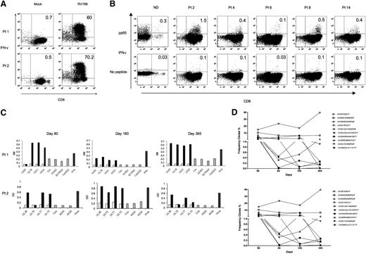
The low number of T cells infused with UCB compared with PBSCs limits TCR diversity early after transplant. Moreover, thymic production of naive T cells is often delayed and depends on the patient’s age, conditioning regimen, and GVHD.31-33 As a consequence, the CMV-specific T cells that develop early after UCBT might be focused on only a few antigens, and might not be sufficient to control CMV replication. To provide insight into the diversity of the T-cell response to CMV after UCBT, we examined the specificity and HLA restriction of CMV-specific T-cell lines in samples (of 8 patients) from days 80, 180, and 365 that contained 30% to 60% CMV-specific cells by CFC (Figure 4A). The T cells were first tested for recognition of overlapping peptides corresponding to pp65 because pp65 is commonly a target of the T-cell response to CMV in normal donors. In 4 of the 8 patients, we identified a clear response to pp65 (Figure 4B), and 2 additional patients had a response that was above control values. To obtain broader coverage of the CMV proteome, we recursively screened 4 T-cell lines for recognition of COS-7 cells that had been cotransfected with a plasmid library encoding >130 CMV ORFs and 1 or more of the individual patient HLA molecules. All 4 patients showed responses to epitopes encoded by at least 3 different CMV ORFs and 3 of the 4 patients used multiple class I HLA alleles as restricting elements (Figure 4C; Table 3). The T-cell responses to antigens encoded by specific CMV ORFs at day 80 were also present in day 180 and 365 samples in 3 of the 4 patients. One patient developed responses to antigens encoded by additional CMV ORFs (Table 3). We used prediction algorithms to identify candidate epitopes encoded by CMV ORFs identified in the genome scan. Peptides with the highest scores were pulsed onto autologous LCL, and assessed for recognition by CMV-specific cell lines by measuring intracellular IFN-γ. We identified 5 epitopes, including UL83 and UL44 epitopes previously described in healthy CMV-positive donors (supplemental Figure 4).
Expansion of CMV-specific CD4+ and CD8+ T cells from UCBT recipients and characterization of CD8 T-cell specificity. (A) Frequency of CMV-specific CD8+ T cells by CFC for IFN-γ performed on 2 representative T-cell lines expanded after IFN-γ capture. (B) CMV-specific T cells derived from UCBT recipients recognize pp65 peptides. Polyclonal T-cell lines were incubated with a peptide pool of 15-mer peptides with 11 amino acid overlaps spanning the entire pp65 protein and evaluated by CFC to detect IFN-γ+ cells. Results of 5 patients and 1 ND are shown. (C) Viral genome scan to determine the antigen specificity of CD8+ T cells derived from UCBT recipients at different times after transplant. Polyclonal T-cell lines were incubated with COS cells that were transfected without or with a plasmid encoding a patient specific HLA DNA alone (gray bars), with a plasmid encoding a single CMV-ORF alone (white bars) or cotransfected with both plasmids encoding the CMV ORF and the HLA allele (black bars). Supernatants were collected at 24 hours and assayed for IFN-γ by enzyme-linked immunosorbent assay. Results for the genome scan for T-cell lines derived from blood obtained on day 80, 180, and 365 post-UCBT from 2 representative patients are shown. (D) Deep sequencing of TCR Vb receptor genes in CMV-specific polyclonal T-cell lines from UCBT recipients (patient no. 2 top graph and patient 4 bottom graph) was performed. Results show the percentage of total clones of the top 10 clones tracked over time in both patients.
Expansion of CMV-specific CD4+ and CD8+ T cells from UCBT recipients and characterization of CD8 T-cell specificity. (A) Frequency of CMV-specific CD8+ T cells by CFC for IFN-γ performed on 2 representative T-cell lines expanded after IFN-γ capture. (B) CMV-specific T cells derived from UCBT recipients recognize pp65 peptides. Polyclonal T-cell lines were incubated with a peptide pool of 15-mer peptides with 11 amino acid overlaps spanning the entire pp65 protein and evaluated by CFC to detect IFN-γ+ cells. Results of 5 patients and 1 ND are shown. (C) Viral genome scan to determine the antigen specificity of CD8+ T cells derived from UCBT recipients at different times after transplant. Polyclonal T-cell lines were incubated with COS cells that were transfected without or with a plasmid encoding a patient specific HLA DNA alone (gray bars), with a plasmid encoding a single CMV-ORF alone (white bars) or cotransfected with both plasmids encoding the CMV ORF and the HLA allele (black bars). Supernatants were collected at 24 hours and assayed for IFN-γ by enzyme-linked immunosorbent assay. Results for the genome scan for T-cell lines derived from blood obtained on day 80, 180, and 365 post-UCBT from 2 representative patients are shown. (D) Deep sequencing of TCR Vb receptor genes in CMV-specific polyclonal T-cell lines from UCBT recipients (patient no. 2 top graph and patient 4 bottom graph) was performed. Results show the percentage of total clones of the top 10 clones tracked over time in both patients.
CMV ORF and HLA-restricting allele for CD8+ T-cell responses after UCBT
| Patient . | CMV ORF/HLA-restricting allele . | ||
|---|---|---|---|
| D 80 . | D 180 . | D 365 . | |
| 1 | UL83/A*2402 | UL83/A*2402 | |
| US21/B*2705 | US21/B*2705 | US21/B*2705 | |
| US24/B*2705 | US24/B*2705 | US24/B*2705 | |
| UL78/Cw*0202 | UL78/Cw*0202 | UL78/Cw*0202 | |
| 2 | UL70/A*0201 | UL70/A*0201 | UL70/A*0201 |
| UL100/A*0201 | UL100/A*0201 | UL100/A*0201 | |
| UL36/A*0301 | UL36/A*0301 | UL36/A*0301 | |
| UL77/A*0301 | UL77/A*0301 | UL77/A*0301 | |
| 4 | UL36/B*4403 | UL36/B*4403 | UL36/B*4403 |
| UL105/B*4403 | UL105/B*4403 | UL105/B*4403 | |
| US24/B*4403 | US24/B*4403 | US24/B*4403 | |
| 6 | UL84/A*0201 | UL105/Cw*0602 | UL105/Cw*0602 |
| US12/A*2402 | UL37/Cw*0602 | US23/Cw*0602 | |
| US23/Cw*0602 | |||
Diversity of the CD8+ T-cell response to CMV after UCBT was confirmed by deep sequencing of the Vb TCR locus in T-cell lines derived from the day 56 PBMC sample from 2 patients. This analysis showed expansion of 16 and 7 distinct clonotypes above a 1% frequency, and these TCRs were present in cell lines derived from all later time points in these patients, consistent with the formation of a CD8+ memory T-cell response (Figure 4D). Interestingly, few new clones emerged at a frequency of >0.5% on day 365 (data not shown). The data demonstrate that a diverse polyclonal CD8+ T-cell response is primed to CMV early after UCBT, and that control of virus replication coincides with increased absolute T-cell numbers and reduction of immunosuppressive drugs.
Discussion
UCBT is an alternative for patients who lack a suitably matched marrow or PBSC donor. A drawback of UCBT is the delay in reconstitution of T-cell immunity, which increases opportunistic infections.34-36 Treatment of Adenovirus, BK virus, and CMV is challenging, and many UCBT recipients have complications from all 3 viruses. Prior work suggested that recovery of protective T-cell immunity to CMV after UCBT depended on de novo thymopoiesis, which is typically measurable after day 100.18,24 The implication is that the T-cell repertoire infused with a UCB graft is insufficient to respond to CMV, or that APCs or T cells that initially repopulate the host are not sufficiently mature for priming a response to CMV. Here, we provide definitive evidence that CD8+ and CD4+ T-cell responses to CMV antigens are primed early after UCBT, and suggest that 1 possible mechanism for the failure to control virus relates to the inability of functional virus-specific T cells to accumulate to sufficient numbers in vivo.
To evaluate CMV-specific CD8+ T cells, we used a strain of CMV that is deleted of the genes that downregulate HLA class I molecules, and used both direct assays of PBMCs and in vitro–expanded T cells for detailed characterization of their origin, diversity, and specificity. Our analysis used patient fibroblasts as APCs and may underestimate the overall CD8 response to CMV because responses restricted by nonshared HLA alleles of the UCBT donor would be missed. Whether such responses develop after UCBT will be the subject of future studies. An additional caveat of our approach is that we measured responses by IFN-γ secretion, which may be altered in cord blood cells.37 Nevertheless, we detected CMV-specific CD8+ T cells in the majority of patients as early as days 42 to 56 after UCBT, demonstrating that priming of T cells to CMV occurs before thymopoiesis contributes significantly to the T-cell repertoire.24 Chimerism analysis of a subset of patients demonstrated definitively that the CMV-specific T cells were of UCB origin, removing a caveat of prior studies of CMV immune reconstitution.
Our analysis of CMV antigen specificity demonstrated that CD8+ T-cell responses recognized multiple antigens in individual patients. The genome scan interrogated only a portion of the CMV genome and half of the HLA molecules in each patient, and likely underestimates the repertoire of CMV-specific CD8+ T cells after UCBT. TCR Vb sequencing of CMV-specific T-cell lines in a subset of patients identified a large number of expanded clones that are present at all time points after UCBT, supporting this conclusion. Both TCR Vb gene usage and the specificity for distinct CMV antigens observed early after UCBT remained remarkably stable over the first year posttransplant, consistent with the formation of T-cell memory and providing evidence that further diversification of the T-cell response to CMV may be limited, although we cannot exclude the presence of CMV-specific T cells that are too rare to be detected by specificity analysis or as dominant clones in TCR Vb sequencing. The data suggest that the control of CMV that is established later after UCBT is related to the increased number and/or improved function of CMV-specific T cells primed early after UCBT, rather than priming of new T cells derived by thymopoiesis.
An important question arises from our data: why does the frequency of CMV-specific CD8+ T cells remains so low in the first several months after UCBT? CD4+ T cells are important for the development of a strong CD8+ T-cell response29 ; however, our data show CD4+ T cells specific for CMV are also primed early after UCBT but functional CD4 T cells are present in very low numbers in vivo. Thus, it is possible that CD4 deficiency contributes to the CD8+ CMV-specific T cells remaining low. Alternatively, CMV-specific T cells may not expand due to immunosuppressive drugs or other environmental factors present after UCBT. The UCB-derived T cells proliferated briskly and remained viable in vitro when stimulated with antigen, making an inherent defect in their ability to proliferate or an increased susceptibility to cell death less likely. Studies addressing the exact mechanism by which the T cells fail to expand will be important for our understanding of viral control in these patients.
To our knowledge, this is the first assessment of reconstitution of T-cell immunity to CMV after UCBT that includes comprehensive data for both CD4+ and CD8+ T cells. The methods applied allowed us to detect low-frequency functional responses and to characterize stability of responses over time, which has not previously been done. Our findings suggest that control of CMV is related to factors other than the T-cell repertoire that is capable of responding. The results also have therapeutic implications since the CMV-specific T cells that develop early after UCBT can be expanded ex vivo, and it is foreseeable that expanded populations could be enriched for CMV specificity and re-infused to augment quantitatively deficient responses and obviate the need for prolonged antiviral drug therapy.15,38
The online version of this article contains a data supplement.
The publication costs of this article were defrayed in part by page charge payment. Therefore, and solely to indicate this fact, this article is hereby marked “advertisement” in accordance with 18 USC section 1734.
Acknowledgments
This work was supported by grants from the National Institutes of Health CA136551, CA114536, and AI053193 (S.R.R.). S.M.M. was supported by a fellowship grant from St. Baldrick’s Foundation. M.E.B. is the Damon Runyon-Richard Lumsden Foundation Clinical Investigator, supported in part by the Damon Runyon Cancer Research Foundation (CI-57-11), and in part by K23CA154532-01 from the National Cancer Institute.
Authorship
Contribution: S.M.M. designed and performed research, analyzed data, and wrote the manuscript; A.G., M.E.B., T.N.Y., and S.E.P. designed and performed research and analyzed data; C.J.T. and C.S.D. analyzed data and provided expert advice; and S.R.R. designed and analyzed experiments and wrote the manuscript.
Conflict-of-interest disclosure: The authors declare no competing financial interests.
Correspondence: Stanley R. Riddell, D3-100, 1100 Fairview Avenue NE, Seattle, WA 98109; e-mail: sriddell@fhcrc.org.


