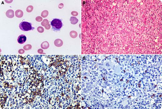
A 26-year-old Indian male presented with a history of dyspnea, fatigue, and jaundice of a month’s duration. He had icterus, pallor, hepatosplenomegaly, severe anemia, thrombocytopenia, and leucocytosis (37 × 109/L) with lymphocytosis. A majority (51%) of the lymphoid cells were large and had folded nuclei, indistinct nucleoli, finely granular basophilic cytoplasm, and circumferentially placed hairy projections (panel A). These cells were also seen in the marrow aspirate. They were positive for CD3, CD7, CD2, and T-cell receptor γδ, and negative for the B-cell markers. The cells were intrasinusoidal in location in the trephine biopsies (panel B). Immunohistochemistry was positive for CD3 (panel C) and negative for CD20 (panel D). The characteristic immunophenotype and intrasinusoidal pattern was consistent with the diagnosis of hepatosplenic T-cell non-Hodgkin lymphoma. Cytogenetics and cytotoxic markers could not be obtained.
T-cell large granular lymphocyte leukemias are usually CD8 positive and CD7 negative, lack the characteristic intrasinusoidal pattern of infiltration, and have an indolent course. Leucocytosis, although uncommon, has been reported previously in this lymphoma. Morphological variations, including blastlike lymphoid cells in the terminal phase, have been described; however, hairy projections, as seen here, are a rarity in this neoplasm.
For additional images, visit the ASH IMAGE BANK, a reference and teaching tool that is continually updated with new atlas and case study images. For more information visit http://imagebank.hematology.org.


This feature is available to Subscribers Only
Sign In or Create an Account Close Modal