Abstract
Abstract 4051
Imaging is playing an increasing role in the diagnosis, staging and treatment response monitoring of patients with multiple myeloma (MM) because of limitation of hematological parameters and uneven distribution of MM lesions in the bone marrow. Although MRI and 18F-FDG-PET/CT have been utilized for visualization of medullary and extra-medullary lesions in patients with MM, both are too time-consuming and expensive for routine clinical use. Whole body Low-dose multidetector CT (WBLD MD-CT) can provide information on the degree of myeloma cell infiltration, especially in the appendicular skeleton. This study was performed to determine the incidence and prognostic implications of abnormal medullary lesions in the appendicular skeleton detected by WBLD MD-CT in patients with monoclonal gammopathy of undetermined significance (MGUS), and asymptomatic and symptomatic MM. We also compared the various hematological parameters with the development of these abnormalities.
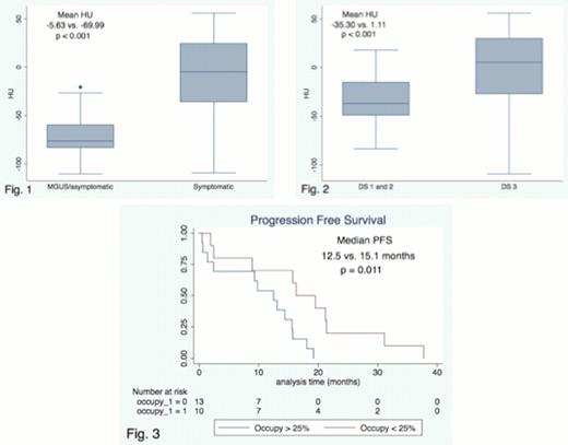
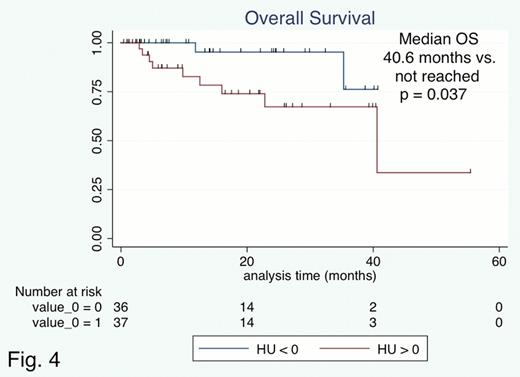
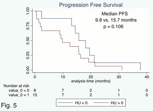
Between January 2008 and July 2012, WBLD MD-CT was performed in 89 patients with MM and MGUS as an initial evaluation of lytic bone lesion and medullary and extra-medullary MM lesions. CT image acquisition and analysis were as follows. WBLD MD-CT was performed in an non-enhanced manner on a 16—Salmon staging system (DS), and were 11, 26 and 37 in International Staging System (ISS), respectively. Medullary abnormalities were found in 61 of 73 patients (83.6%). Patients with symptomatic MM had significantly higher mean HU than patients with MGUS/asymptomatic MM (−5.64 vs. −69.99, p < 0.001, Fig. 1). In the symptomatic MM, laboratory variables such as albumin, hemoglobin, creatinine, beta-2-microglobulin and presence of poor cytogenetic abnormalities (del17p, IGH-FGFR3, or IGH/MAF on FISH) were not correlated with the mean HU values. Positive correlations between the mean HU value and the amount of intact immunoglobulin (Ig) with IgG and IgA subtypes were observed (correlation coefficient: 0.330 and 0.694, p = 0.049 and 0.005, respectively), but it was not observed in patients with light chain only MM (correlation coefficient: 0.133, p = 0.598). Patients with DS stage 3 showed significantly high HU values compared to those with DS stage 1 and 2 (−35.30 vs. 1.11, p < 0.001, Fig. 2) whereas the mean HU value was not significantly different between ISS 3 and 1 or 2 (p = 0.186). Patients with abnormal medullary lesions occupying >25% of boney canals had significantly poorer progression-free survival compared to those with < 25% (12.5 vs. 15.1 months, p = 0.011, Fig. 3). Moreover, patients with mean HU value >0 had significantly shorter median overall survival (40.6 months vs. not reached, p = 0.040, Fig. 4) and tended to have shorter progression-free survival (9.8 vs. 15.7 months, p = 0.106, Fig. 5) compared to those with mean HU value <0. Complete disappearance of abnormal medullary lesion did not usually achieved even after obtaining complete response, marked decrease of HU value was observed in most of the patients.
This study indicated that medullary abnormalities in the appendicular skeleton detected by WBLD MD-CT might be associated with high tumor burden of MM independent of ISS stage and laboratory variables. In addition, it also has possible prognostic relevance in patients with newly diagnosed MM.
No relevant conflicts of interest to declare.
Author notes
Asterisk with author names denotes non-ASH members.


This feature is available to Subscribers Only
Sign In or Create an Account Close Modal