Abstract
Abstract 3633
Classical Hodgkin lymphoma (cHL) is a malignant neoplasm of the immune system, characterized by the presence of an abundant reactive infiltrate and a minority of Hodgkin Reed-Sternberg cells (HRS cells). HRS cells retain their professional antigen presenting phenotype in most cases. In EBV- cHL, membranous HLA class I expression is retained in HRS cells in approximately 30% of the cases, whereas in EBV+ cHL HLA class I expression is retained in HRS cells in 79% of the cases. Moreover, a proportion of the EBV+ cHL cases shows an enhanced HLA class I expression as compared to the surrounding infiltrating cells.
The mechanism of the enhanced or lost HLA class I expression is unknown. Special AT-rich region binding protein 1 (SATB1) and promyelocytic leukemia protein (PML) are two proteins that have been shown to regulate HLA class I expression. Downregulation of SATB1 in Jurkat cells results in an enhanced expression of HLA-A, HLA-G, HLA-H and HCG4P6, whereas downregulation of PML results in a reduced HLA-A and HLA-G expression. PML is the main component of nuclear bodies (NBs) that organize the chromatin structure into loops by anchoring matrix attachment regions to the nuclear matrix. SATB1 has been shown to be associated with the PML-NBs in the HLA region.
To investigate the possible role of SATB1 and PML-NBs in the regulation of HLA class I expression in EBV+ cHL.
We analyzed 64 EBV+ cHL cases and as a control included 29 EBV- cHL cases. HLA class I membranous staining (HC10 antibody) by HRS cells was scored as positive or strongly positive, cases that lacked membrane staining were scored negative. ß2-microglobulin served as an additional marker for membranous HLA class I expression. For SATB1 (14/SATB1), we scored the percentage of HRS cells with nuclear SATB1 staining. For PML (PG-M3), we scored HRS cells based on the number of PML nuclear bodies in two categories; 10 or less NBs per cell or >10 NBs per cell (per 4 um tissue section).
In the EBV+ group 24 cases stained strongly positive, 22 positive and 18 negative for HLA class I. In the EBV- group, none of the cases showed a strong positive staining, 7 cases stained positive and 22 cases were negative for HLA class I. HLA class I staining results were consistent with the ß2-microglobulin staining results in all cases. The percentage of SATB1 positive HRS cells varied from 0–100% in both EBV+ and EBV- cHL. The number of PML-NBs was between 0–10 in 46 and >10 in 18 EBV+ cHL cases and between 0–10 in 23 and >10 in 6 EBV- cHL cases.
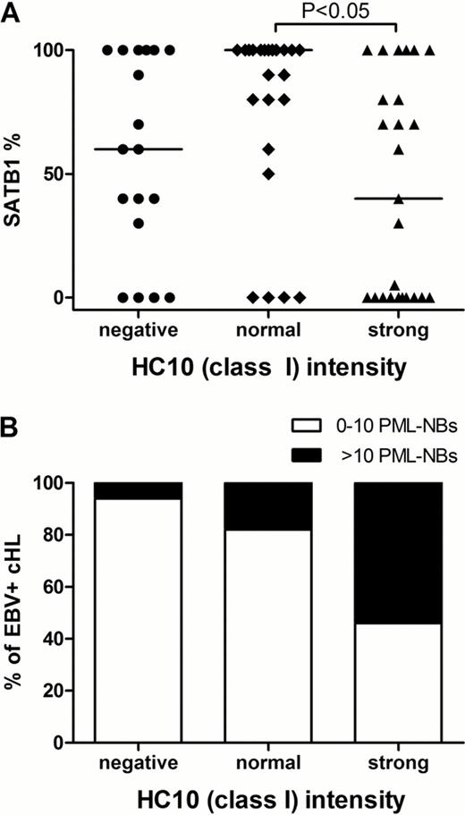
We observed no correlation between HLA class I staining in the EBV- cHL cases and the percentage of SATB1 positive HRS cells or the number of PML-NBs in the HRS cells. In EBV+ cHL cases we observed significant differences in the percentages of SATB1 positive cells between the HLA class I negative, normal and strongly positive groups (p=0.0412). The cases with normal HLA class I staining had significantly higher percentages of SATB1 positive cells as compared to the cases with strong HLA class I staining pattern (p<0.05) (Figure 1). There was no significant difference between negative and normal or negative and strongly positive HLA class I groups with respect to the percentage of SATB1. The percentage of EBV+ cHL cases with >10 PML-NBs significantly increased from HLA class I negative (1 out of 18, i.e. 5%) to normal (4 out of 22, i.e. 18%) and strong (13 out of 24, i.e. 54%) positive cHL cases (p=0.0011).
We found an inverse correlation between the percentages of SATB1 positive HRS cells in the HLA class I strong and normal EBV+ cHL groups. The number of cases with >10 PML-NBs were significantly increased in HLA class I strong as compared to the HLA class I normal and negative EBV+ cHL groups. Thus SATB1 and PML may play an important role in the regulation of HLA class I expression levels in EBV+ cHL.
SATB1 and PML staining results in EBV+ cHL. A) SATB1 percentages in EBV+ cHL cases with negative, normal or strong HLA class I staining pattern differ significantly (p=0.0412, Kruskal-Wallis test). Dunn's multiple comparison test indicated a significant difference specifically between the HLA class I normal and strong staining groups (P<0.05). B) The number of cases with >10 PML-NBs significantly increases with HLA class I staining intensity from negative, to normal and strong (p=0.0011, Chi-square test).
SATB1 and PML staining results in EBV+ cHL. A) SATB1 percentages in EBV+ cHL cases with negative, normal or strong HLA class I staining pattern differ significantly (p=0.0412, Kruskal-Wallis test). Dunn's multiple comparison test indicated a significant difference specifically between the HLA class I normal and strong staining groups (P<0.05). B) The number of cases with >10 PML-NBs significantly increases with HLA class I staining intensity from negative, to normal and strong (p=0.0011, Chi-square test).
No relevant conflicts of interest to declare.
Author notes
Asterisk with author names denotes non-ASH members.


This feature is available to Subscribers Only
Sign In or Create an Account Close Modal