Abstract
Abstract 2345
Ratajczak and his colleagues identified a unique population of very small embryonic-like (VSEL) stem cells in adult mouse bone marrow (BM) (Leukemia 2006:20;857). These VSELs are; 1) very small (∼4 μm); 2) express pluripotent stem cell markers, such as Oct4, Nanog, SSEA-1, and Rex-1; 3); responsive to a SDF-1 gradient; 4) possess large nuclei that contain euchromatin. It is very interesting to note that VSELs possess the potential to differentiate into 3 germ layers in vitro and in vivo, thereby contributing to tissue/organ regeneration. These VSELs were isolated as lineage-negative (Lin−), Sca-1-positive (Sca-1+), CD45-negative (CD45−) cells by FACS. However, the incidence of VSELs in BM-derived mononuclear cells is ∼0.01%. Therefore, it is difficult to isolate VSELs very effectively. This study describes our recently developed highly efficient method for isolating VSELs using enzymatic treatment of murine bone.
Murine BM nucleated cells (BMNC) were isolated from BM flushed from the pairs of femurs and tibiae of 8 week-old C57BL/6 mice. Erythrocytes were removed using a hypotonic solution. Then the remaining bone tissues were thoroughly washed using PBS- with 2% FCS. These bone tissue specimens were crushed in a mortar and then incubated in cell dissociation buffer containing a-medium with 5% FCS supplemented with 1.5 mg/ml type I collagenase and 2 mg/ml dispase at 37°C for 1 hour. Next, the BMNCs and bone-derived nucleated cells (BDNCs) were stained with various monoclonal antibodies, including anti-lineages, anti-CD45, anti-Sca-1, anti-CXCR4, anti-CD133, and anti-PDGFRα, and then were used for subsequent FACS analyses.
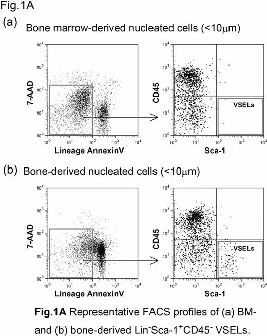
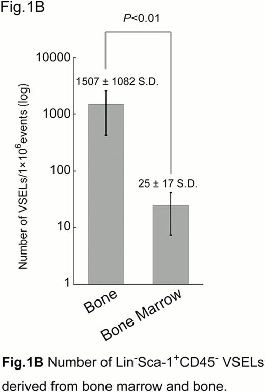
The R1 gate was set on the FSC channel using 4 and 10 μm synthetic beads, based on the predicted very small size of VSELs. The VSELs were isolated from BMNCs and BDNCs by multicolor FACS, as a population of Lin−Sca-1+CD45− cells (Fig. 1A). The incidences of VSELs in the BMNCs and BDNCs were 0.001% and 0.1%, respectively. Therefore, the enzymatic treatment of bone tissues yielded about 100 times the efficiency for the isolation of VSELs (Fig. 1B). The bone-derived (BD) VSELs were small (< 5 μm) and possessed a relatively large nucleus surrounded by a narrow rim of cytoplasm. They expressed CD133, but not PDGFRα. However, they weakly expressed CXCR4. The gene expression profiles were analyzed using real time quantitative PCR (RQ-PCR) to evaluate the expression of ES cell markers (Oct4, Nanog, Rex1, Dppa3), HSC (KSL) markers (c-kit, Tal1, GATA2), and MSC markers (Nestin, Ang1, CXCL12, VE-Cadherin). Unexpectedly, BD VSELs expressed high levels of Nestin and Cadherin. However, they expressed weak levels of Oct4 and Nanog. The gene expression profile of the BD VSELs was clearly distinct from the well-defined populations of ES cells, KSL cells, and MSCs. Interestingly, the number of these BD VSELs significantly increased after the induction of liver injury by carbon tetrachloride administration. They were then most likely mobilized into the peripheral blood (PB). G-CSF did mobilize KSL cells into PB, as previously reported. However, G-CSF did not mobilize the BD VSELs. The effects of sRANKL on the mobilization of BD VSELs were examined in vivo. Interestingly, the number of BD VSELs significantly increased 2–3 days after the administration of sRANKL. However, the number of VSELs in PB did not increase. These results suggest that BD VSELs actively proliferated after liver injury and bone resorption.
The present data suggest that the majority of the Lin−Sca-1+CD45− cells reside in the bone tissue. BD VSELs resemble BM-derived VSELs. However, a RQ-PCR analysis revealed that the gene expression profile of BD VSELs was different from those of the previously reported BM-derived VSELs. Further studies will therefore be required to elucidate their stem cell characteristics and the potential relationship between BD VSELs and BM-derived VSELs.
No relevant conflicts of interest to declare.
Author notes
Asterisk with author names denotes non-ASH members.


This feature is available to Subscribers Only
Sign In or Create an Account Close Modal