Abstract
Abstract 1318
Diffuse large B-cell lymphoma (DLBCL) is the most frequently aggressive Non-Hodgkin's lymphoma (NHL). DLBCL first line therapy consists of R-CHOP regimen, which combines classical chemotherapeutical drugs and corticosteroids with Rituximab. Rituximab is a chimeric anti-CD20 monoclonal antibody which targets the CD20 surface antigen expressed on both normal and malignant B cells. Rituximab triggers antibody- and complement-dependent cytotoxicity, growth inhibition and apoptosis. Rituximab inhibits the expression of the anti-apoptotic proteins Bcl-2/Bcl-XL, down-modulating different survival pathways. Clusterin (CLU) is an ubiquitary protein involved in many physiological and pathological processes such as apoptosis and cancer. CLU has different transcriptional isoforms, which share exons from 2 to 9 but have a unique exon 1 and at least two protein forms: The cytoplasmatic precursor of about 60 kDa is secreted outside the cell after post-traslational modifications (sCLU), whereas a truncated protein (50–55 kDa) is produced only in particular conditions and localizes in the nucleus (nCLU). CLU expression is disregulated in almost all cancers and its role has been deeply investigated in solid tumors. Only limited information is currently available about its function in lymphomas.
Toledo, a DLBCL-derived cell line and normal B lymphocytes were chosen as experimental models. B lymphocytes were purified and immuno-magnetically sorted from peripheral blood of 4 healthy volunteers. Toledo and B lymphocytes were treated with 10 or 100 μg/ml Rituximab or doxorubicin (from 0.05 to 1 μM) for 48 hours. Phenotype analysis was performed by flow cytometry; cell viability was assessed by Trypan blue exclusion; apoptosis was evaluated by Annexin V/propidium iodide. CLU expression and localization were evaluated by real time PCR, Western Blot (WB) and immunocytochemistry (ICC). Primers for the real time PCR were design in order to distinguish the unique exon 1 of each CLU transcriptional isoforms or exons common to all variants (CLU3-4).
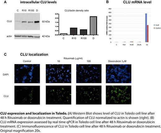
Rituximab treatment doesn't significantly inhibit Toledo proliferation and does not induce apoptosis despite the fact that these cells are CD20+. This suggests that Toledo are resistant to Rituximab. As expected, doxorubicin causes a marked growth inhibition by necrosis and apoptosis. Toledo cells have a very low basal level of the 60 kDa CLU protein form. Both Rituximab and doxorubicin up-regulate the 60 kDa form with 1 μM doxorubicin causing the most dramatic increase. The analysis of transcriptional isoforms by real-time PCR shows that CLU mRNA is almost undetectable in Toledo. CLU transcriptional variant 2 (CLU2) and regions common to all CLU transcripts (CLU3-4) are significantly up-regulated only after 1 μM doxorubicin but not after Rituximab. Fluorescence microscopy clearly shows a cytoplasmic localization of CLU under normal growth conditions. Interestingly, the treatment with both Rituximab and doxorubicin causes the appearance of a nuclear staining. This fact might be consistent with the onset of a process of cell death. 10 and 100 μg/ml Rituximab inhibits B lymphocytes cell survival in a dose-dependent manner (39.7% and 44% respectively after 48 hours). Interestingly, B lymphocytes clearly express high basal levels of the 60 kDa CLU and produce an additional form of about 55 kDa only after Rituximab but not after doxorubicin. Furthermore Rituximab-treated B lymphocytes display a Bcl-2 protein decrease while Toledo cells show high expression of Bcl-2 that doesn't seem to change in response to Rituximab.
Toledo are resistant to Rituximab up to 100 μg/ml and express very low levels of CLU under normal growth conditions. 60 kDa CLU is partially up-regulated and Bcl-2 expression is not significantly affected by Rituximab treatment. Doxorubicin strongly up-regulates the 60 kDa CLU isoform. At the transcriptional level CLU2 shows the same expression profile of the 60 kDa protein form. On the contrary, Rituximab changes the CLU profile of responsive B lymphocytes in that leads to the appearance of a 55 kDa form that is not visible after doxorubicin treatment. Rituximab also causes a reduction in the expression of the antiapoptotic protein Bcl-2. The biological role of CLU during the response of peripheral B lymphocytes to Rituximab treatment is yet to be determined and is currently being investigated.
No relevant conflicts of interest to declare.
Author notes
Asterisk with author names denotes non-ASH members.


This feature is available to Subscribers Only
Sign In or Create an Account Close Modal