Abstract
Abstract 1195
In the murine primitive hematopoietic stem cell (HSC) compartment, MPL/thrombopoietin (THPO) signaling plays an important role for the maintenance of adult quiescent HSCs. However, the role of MPL/THPO signaling in the human primitive HSC compartment has not yet been elucidated. We have previously identified very primitive human cord blood (CB)-derived CD34-negative (CD34−) severe combined immunodeficiency (SCID)-repopulating cells (SRCs) using the intra-bone marrow injection (IBMI) method (Blood 2003:101;2924). Recently, we developed a high-resolution purification method for the primitive CD34− SRCs using 18 lineage (Lin)-specific antibodies (Exp Hematol 2011:39:203). In this study, we investigated the functional significance of the MPL expression in the human primitive CD34− SRCs (HSCs).
First, we sorted 18Lin−CD34+/−MPL+/−cells by FACS. Thereafter, these four fractions of cells were analyzed for their hematopoietic colony-forming capacities (CFCs), the maintenance/production of CD34+ cells in cocultures with human bone marrow-derived mesenchymal stromal cells (DP MSCs) (Blood 2010:116:1575). Finally, these four fractions of cells were transplanted by IBMI into NOD/Shi-scid/IL-2R ƒÁcnull (NOG) mice to investigate their long-term (LT) repopulating capacities. We performed primary, secondary, and tertiary transplantations for up to 18 months.
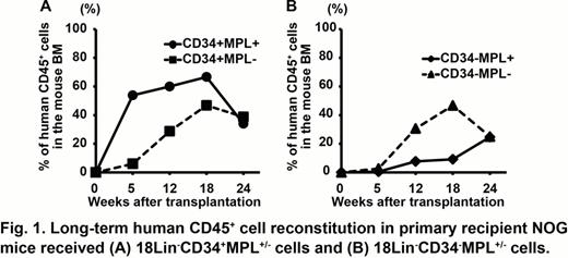
The CFCs of highly purified 18Lin−CD34−cells were quite unique, since they yielded mainly erythroid-bursts (BFU-E) and erythro/megakaryocytes-containing mixed colonies (CFU-EM). Interestingly, they showed very weak myeloid CFCs. Moreover, 200 18Lin−CD34−MPL+cells yielded about 150 colonies, consisting of 20% BFU-E, 20% colony-forming unit-megakaryocytes, and 60% CFU-EM in the presence of 10% platelet-poor plasma, THPO, IL-3, and erythropoietin. The phenotypic and functional characteristics of the 18Lin−CD34+/−MPL+/−cells were further investigated by cocultures with DP MSCs. After 7 days of coculturing the 18Lin−CD34+MPL+/−cells, the total number of cells was observed to expand by about 500 times, 40 % of which consisted of CD34+ cells. On the other hand, the 18Lin−CD34−MPL+/−cells expanded only by about 100 times, 10 to 20% of which consisted of CD34+ cells. Interestingly, the lineage differentiation potentials of the observed 18Lin−CD34+/−MPL+/−cells were significantly different. The percentages of myeloid cells generated from the 18Lin−CD34+MPL+/−cells were significantly greater than those of the 18Lin−CD34−MPL+/−cells. On the contrary, 18Lin−CD34−MPL+cells generated significantly higher percentages of CD41+ cells compared to the other fractions. These observations suggested that the differentiation potentials of 18Lin−CD34+/−MPL+/− cells were different and that these four cell populations contained distinct classes of hematopoietic progenitors as well as HSCs. We next investigated the SRC activities of the 18Lin−CD34+/−MPL+/−cells using NOG mice. In the primary recipient mice, all 18 mice (9 for each cell type) received CD34+MPL+/−SRCs were highly repopulated with human CD45+ cells. On the other hand, 11/13 mice receiving CD34−MPL+SRCs and 12/12 mice receiving CD34−MPL−SRCs showed human cell repopulation. Interestingly, these CD34+/−MPL+/−SRCs showed different repopulation kinetics. The CD34+MPL+ SRCs showed a rapid and sustained repopulating pattern (Fig.1-A). In contrast, the repopulation rates in the mice receiving CD34+MPL− and CD34−MPL+/− SRCs gradually increased, and thereafter reached a high level of repopulation at 18–24 weeks after the transplantation (Fig.1-A, B). All of the primary recipient mice that received CD34+MPL+/− SRCs showed a secondary repopulating capacity. However, only the mice that received CD34+MPL− SRCs showed a tertiary repopulating capacity. Very interestingly, the primary recipient mice that received CD34−MPL− SRCs showed a distinct secondary repopulating capacity. Tertiary transplantation experiments in these mice are now underway in our laboratory.
These results clearly indicated that both the CD34+/− SRCs not expressing MPL sustained a LT (> 1 year) human cell repopulation in NOG mice. Therefore, these findings suggest that the functional significance of the MPL expression in the human primitive HSCs is different from that in murine primitive HSCs.
No relevant conflicts of interest to declare.
Author notes
Asterisk with author names denotes non-ASH members.


This feature is available to Subscribers Only
Sign In or Create an Account Close Modal