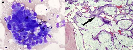
A 70-year-old man was being evaluated for back pain. Two years earlier he was found to have a monoclonal IgA λ protein (0.3 g/dL). Past history included long-standing history of ulcerative colitis with a partial colectomy and ileostomy 31 years earlier. A magnetic resonance imaging scan showed numerous destructive vertebral lesions. His complete blood count was normal except for a minimal normocytic normochromic anemia (hemoglobin 12.9 g/dL). Peripheral blood smear showed moderate anisopoikilocytosis without teardrop forms. Bone marrow biopsy and touch prep smears showed cohesive clusters of tumor cells (panel A). The marrow was replaced by mucin with occasional cellular groupings of colonic type glandular structures (panel B arrow). Immunostains were positive for cytokeratins as well as Cdx2, suggesting gastrointestinal origin, and a diagnosis of metastatic adenocarcinoma was made. Sigmoidoscopy revealed a large friable mass in the distal rectum and the biopsy showed invasive adenocarcinoma.
The clinical suspicion in this case was of myeloma, based on the radiologic findings and the previously known monoclonal protein. Colorectal cancer was unexpected, especially with the previous prophylactic partial colectomy. The bone marrow examination and endoscopy confirmed the metastatic adenocarcinoma.
For additional images, visit the ASH IMAGE BANK, a reference and teaching tool that is continually updated with new atlas and case study images. For more information visit http://imagebank.hematology.org.


This feature is available to Subscribers Only
Sign In or Create an Account Close Modal