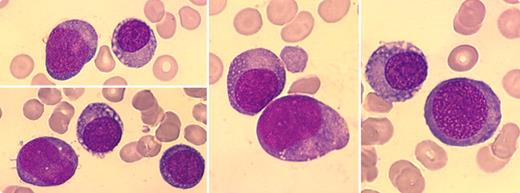
A 60-year-old man presented with an IgG κ monoclonal paraprotein (50 g/L) and pancytopenia (hemoglobin 77 g/L, white blood count 1.1 × 10⁹/L, platelets 75 × 109/L). The bone marrow was hypocellular, lacking in normal hematopoiesis, but contained plasma cells. A diagnosis of multiple myeloma was made and the patient was treated with vincristine, doxorubicin, and dexamethasone, followed by an autologous stem cell transplant. The paraprotein disappeared after treatment. Three years later, the patient became pancytopenic once again. Bone marrow aspirate showed hypocellularity with 95% of the cells appearing with variable size, with round, often eccentric nuclei, chromatin of variable density, abundant basophilic cytoplasm, and occasional fine vacuoles (see figures). The morphology was compatible with early plasma cells and/or erythroid precursors. The continued absence of the paraprotein suggested remission of myeloma or nonsecretory plasma cells. Flow cytometric immunophenotyping showed low expression of CD45 and CD36, glycophorin positivity, and CD138 negativity that suggested erythroid lineage and a diagnosis of pure erythroid leukemia. Multiple karyotypic abnormalities were present. The patient rapidly deteriorated and died shortly after.
Cases of multiple myeloma can evolve into leukemia. In most of these instances, acute nonlymphocytic leukemia or myelodysplasia occurs. Pure erythroid leukemia is an uncommon disease and its evolution from multiple myeloma is distinctly unusual. In this case, the morphologic similarity of plasma/erythroid cells was elucidated by immunophenotyping.
For additional images, visit the ASH IMAGE BANK, a reference and teaching tool that is continually updated with new atlas and case study images. For more information visit http://imagebank.hematology.org.


This feature is available to Subscribers Only
Sign In or Create an Account Close Modal