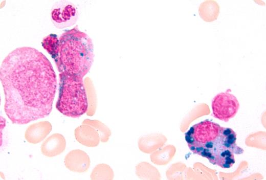
A 70-year-old man with multiple myeloma had been treated with chemotherapy and an autologous stem cell transplant. He had been in remission for 3 years. For 2 years, he had had persistent absolute neutropenia (< 1000/μL), anemia (hemoglobin < 85 g/L), and thrombocytopenia (platelets < 30 000/μL), requiring growth factors and red cell and platelet transfusions every 2-3 weeks. Bone marrow evaluation to investigate the cause for refractory cytopenia showed trilineage dyspoiesis consistent with development of therapy-related myelodysplasia; Prussian blue stain demonstrated increased iron stores, ringed sideroblasts (10%-15%), and plasma cells with hemosiderin iron (shown). Laboratory tests showed high serum iron and markedly increased ferritin. Serum levels of vitamin B12, folate, copper, and ceruloplasmin were within the normal range. The patient was prone to infections due to neutropenia and died from complications of bacterial sepsis.
Iron accumulation in plasma cells is rare, but it has been reported in individuals with alcoholism, myeloma and other malignancies, megaloblastic anemia, other causes of iron overload, including in patients with frequent transfusions, and in copper deficiency. The mechanism for iron accumulation in plasma cell cytoplasm (or cytoplasmic structures) is uncertain.
Many Blood Work images are provided by the ASH IMAGE BANK, a reference and teaching tool that is continually updated with new atlas images and images of case studies. For more information or to contribute to the Image Bank, visit http://imagebank.hematology.org.


This feature is available to Subscribers Only
Sign In or Create an Account Close Modal