

Abstract
Abstract 4736
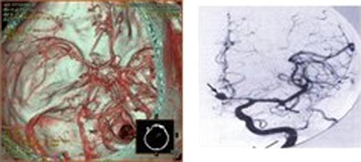
Fusarium is a saprophytic organism that is found widely distributed in soil, subterranean and aerial plants, plant debris and other organic substrates. The organism can cause local tissue infections in immunocompetent patients such as onychomycosis, bone and joint infections, or sinusitis. Since the first case of disseminated Fusarium was described, the incidence of disseminated disease has increased significantly, particularly affecting those immunocompromised with hematological malignancies. We report here a disseminated Fusarium infection with skin manifestations and cerebral aneurysm bleeding in a patient of acute monocytic leukemia. A 49 year old female with acute monocytic leukemia (M5) underwent three courses of therapy with FlAG(cytarabine puls Fludarabine) in second relapse. At the time of hospital admission, she was receiving G-CSF for severe granulocytopenia post-intensification with FLAG, and had also received broad-spectrum antibiotics and fluconazole prophylaxis during previous episodes of febrile neutropenia. Despite treatment with liposomal amphotericin B for five days, She presented persistent fevers, he developed, nonpruritic erythematous Skin Eruption or bleb on her right shin, right shoulder (Figure 1), scalp and foot. The lesions developed central necrosis after four days. The Exudatin in erythematous skin bleb on foot culture was positive for Fusarium solani(Figure 2), which was also confirmed on two different skin lesional drainage culture. Although the patient was treated with liposomal amphotericin B, the clinical symptoms worsened. After liposomal amphotericin B was replaced with voriconazole (VRCZ), Recovery was observed after voriconazole introduction and resolution of neutropenia. Thrombocyte count increased up to 50×109. she suddenly lost consciousness duing lunch. Brain CT scan revealed the hemorrhage in brain. Magnetic Resonance Imaging(MRI) and CTA revealed rupture of brain aneurysm(Figure 3 A,B). Cerebral aneurysm caused by infections differ from congenital brain aneurysm, which often occur the junctions where these arteries come together and form weak spots. The clinical presentation, diagnosis, prognosis and therapeutic options of fusariosis in immunocompromised patients are briefly discussed.

 Close modal
Figure 1:

Skin Eruption
Figure 2:

The Exudatin in erythematous skin bleb culture was positive for Fusarium solani
Figure 3:

Magnetic Resonance Imaging(MRI) and CTA revealed rupture of brain aneurysm(Figure 3 A,B).
Disclosures:
No relevant conflicts of interest to declare.

This feature is available to Subscribers Only
Sign In or Create an Account Close Modal