Abstract
Abstract 3429
Idiopathic PRCA and secondary PRCA associated with thymoma and large granular lymphocyte leukemia are major subtypes of adult-onset chronic PRCA. We have previously shown that these types of PRCA are responsive to immunosuppressive therapy but most patients require long-term maintenance immunosuppressive treatment. These results suggest that acquired chronic PRCA is an autoimmune disorder mediated by T lymphocytes and pathogenic T cell clones may be persistently present during remission. We have previously made an interesting observation that a thymoma-associated PRCA patient had an increase of Vd1 gd T cells in blood. We have also reported that recipients of allogeneic hematopoietic stem cell grafts had an oligoclonal expansion of Vd1 gd T cells and that Vd1 gd T clones had cytotoxicity against autologous EBV-transformed B cell line. Thus, gd T cell repertoires may be altered in PRCA patients in response to certain antigens.
In order to clarify the role for gd T cells in the pathogenesis of chronic acquired PRCA, we have examined the gd T cell receptor repertoire in acquired chronic PRCA patients.
Nineteen PRCA (8 idiopathic, 6 thymoma, 3 LGL-leukemia and 2 SLE) and 107 healthy volunteer donors were included in the study. This study was approved by the Institutional Review Board at Akita University and conducted in accordance with the Declaration of Helsinki. Blood lymphocyte subsets were analyzed by flow cytometry. Clonality of T cells was determined by complementarity-determining region 3 (CDR3) size distribution analysis and junctional sequence was determined by subcloning of PCR products and DNA sequencing. In some experiments, purified gd T cells from PRCA patients were co-cultured with allogeneic erythroid progenitor cells derived from CD34-positive cells in vitro in order to learn whether patient's gd T cells would exert cytotoxic or growth-inhibitory effect on erythroid progenitor cells.
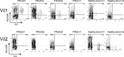
The absolute numbers of ab T cells and gd T cells were normal in patients with PRCA, but there were an increase of Vd1 gd T cells and a decrease of Vd2 T cells (Table 1). More than 50% of Vd1 T cells from PRCA patients expressed HLA-DR, while 20 to 30% of those from healthy individuals expressed HLA-DR (Fig. 1). CDR3 size spectratyping revealed that CDR3 size distribution patterns were skewed in 9 out of 13 PRCA patients examined, although skewed CDR3 size distribution patterns were also observed in 7 out of 10 healthy individuals. In order to determine whether a particular Vd1-Jd rearrangement size was selected in PRCA patients, we performed statistical analysis comparing the CDR3 size distribution of 115 Vd1 TCR clones obtained by subcloning of PCR products in 7 PRCA patients versus 7 controls. No significant difference was found between the two groups (p=0.795 by Mann-Whitney test). Moreover, no apparent consensus amino acid motifs were identified in PRCA patients. Although the T cell clone carrying the -YWGIR- sequence in the CDR3d region was detected in 3 PRCA patients, the T cell clone carrying the -YWGIR- sequence was also detected in one healthy donor. Purified gdT lymphocytes from idiopathic PRCA neither showed an inhibitory effect on proliferation nor cytotoxicity against erythroid progenitor cells in vitro.
gd T cell repertoires in PRCA and aplastic anemia
| T cell . | Healthy donors (N=107) . | PRCA (N=19) . | Aplastic anemia (N=16) . | p value: healthy vs. PRCA . |
|---|---|---|---|---|
| TCRab (/ml) | 1063.9 ± 33.9 | 1256.6 ± 173.9 | 853.5 ± 157.9 | 1.000 |
| TCRgd (/ml) | 68.3 ± 4.2 | 142.2 ± 43.5 | 27.0 ± 4.4 | 1.000 |
| Vd1 (/ml) | 15.6 ± 1.1 | 94.0 ± 30.7 | 6.6 ± 1.5 | 0.000 |
| Vd2 (/ml) | 42.9 ± 3.5 | 15.7 ± 5.4 | 18.0 ± 4.3 | 0.000 |
| Vd2/Vd1 ratio | 4.7 ± 0.6 | 0.3 ± 0.1 | 5.1 ± 1.5 | 0.000 |
Data are expressed as the mean ± SE.
Expression of HLA-DR on expanded Vd1 T cells in PRCA
Adjusted p value was calculated by Kruskal-Wallis ANOVA test.
Expansion of Vd1 T cells and depletion of Vd2 T cells are unique features for chronic acquired PRCA. Expansion of Vd1 T cells does not seem to be the consequence of CDR3-dependent selection. Depletion of Vd2 T cells may be the result of chronic stimulation, because our previous study has revealed that the numbers of Vd2 T cells show an age-dependent decrease and Vd2 T cells are susceptible to activation-induced cell death (Int J Hematol, in press). Failure to demonstrate the cytotoxicity of gd T cells from a PRCA patient against erythroid progenitor cells suggests that expanded gd T cells are not effector T cells.
No relevant conflicts of interest to declare.
Author notes
Asterisk with author names denotes non-ASH members.


This feature is available to Subscribers Only
Sign In or Create an Account Close Modal