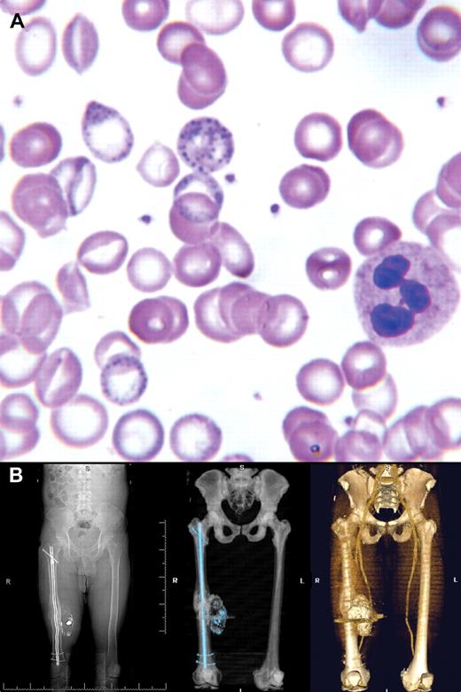
A 44-year-old male presented with chronic abdominal pain that prompted multiple previous hospitalizations with negative assessments. The patient had a past history of moderate alcohol consumption but none in the past 20 years. There was no occupational exposure. Laboratory tests showed microcytic anemia (hemoglobin 6.6 mg/dL, mean corpuscular volume 66.8 fL) with normal iron studies and basophilic stippling on the peripheral smear (panel A). Further history disclosed a retained bullet fragment in his right thigh 2 years previously. Radiographic views of his right thigh affirmed several metallic bullet fragments and posttraumatic soft tissue calcification and ossification. Computed tomography with 3-D reconstruction (panel B) showed a large calcified mass of the thigh, consistent with a complex hematoma.
Lead levels were as high as 306 μg/dL. The patient was started on intramuscular EDTA for chelation. The bullet fragments were surgically removed without complications and the patient was discharged on oral dimercaptosuccinic acid for 2 weeks. Currently, 3 years later, he is asymptomatic with a normal complete blood count (hemoglobin 14.4 g/dL, mean corpuscular volume 86.4 fL), undetectable lead levels, and subsequent peripheral smears that no longer demonstrate any basophilic stippling.
Basophilic stippling was the clue to the underlying diagnosis. Several reports have already described lead toxicity from retained bullets. (The orthopedic aspects of this case were previously reported in Dougherty PJ, van Holsbeeck M, Mayer TG, Garcia AJ, Najibi S. Lead toxicity associated with a gunshot-induced femoral fracture. A case report. J Bone Joint Surg Am. 2009;91(8):2002-2008.)
Many Blood Work images are provided by the ASH IMAGE BANK, a reference and teaching tool that is continually updated with new atlas images and images of case studies. For more information or to contribute to the Image Bank, visit http://imagebank.hematology.org.


This feature is available to Subscribers Only
Sign In or Create an Account Close Modal