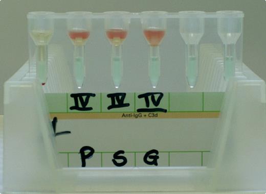
An 84-year-old man with Waldenström macroglobulinemia was seen for routine check-up. Laboratory evaluation revealed hemoglobin 72.0 g/L, white blood cell count 1.5 × 10⁹/L, and platelet count 113 × 109/L. A blood sample was sent to the transfusion center for type and screen.
In antibody screening that utilizes the gel column technique, test erythrocytes and patient's serum usually form a mixture immediately after automated sampling as shown with sample G (a healthy individual). However, test erythrocytes mixed neither with the patient's plasma (sample P) nor with his serum (sample S). Instead, a distinct cellular layer rested on the patient's probes after sampling. This finding raised the suspicion of an increased serum and plasma viscosity that prevented the spreading of test erythrocytes.
This laboratory test led to a diagnosis of hyperviscosity that was confirmed fundoscopically and with the finding of an immunoglobulin M level of 101 g/L. After the patient was treated with plasmapheresis, subsequent plasma and serum samples that were tested for antibody screening no longer showed this abnormality.
Usually clinical features of hyperviscosity initiate the diagnosis. In this case, an unusual finding in antibody screening was subsequently affirmed clinically as hyperviscosity.
Many Blood Work images are provided by the ASH IMAGE BANK, a reference and teaching tool that is continually updated with new atlas images and images of case studies. For more information or to contribute to the Image Bank, visit www.ashimagebank.org.


This feature is available to Subscribers Only
Sign In or Create an Account Close Modal