Abstract
In allogeneic hematopoietic cell transplantation (HSCT), donor T lymphocytes mediate the graft-versus-leukemia (GVL) effect, but induce graft-versus-host disease (GVHD). Suicide gene therapy—that is, the genetic induction of a conditional suicide phenotype into donor T cells—allows dissociating the GVL effect from GVHD. Genetic modification with retroviral vectors after CD3 activation reduces T-cell alloreactivity. We recently found that alloreactivity is maintained when CD28 costimulation, IL-7, and IL-15 are added. Herein, we used the minor histocompatibility (mH) antigens HA-1 and H-Y as model alloantigens to directly explore the antileukemia efficacy of human T cells modified with the prototypic suicide gene herpes simplex virus thymidine kinase (tk) after activation with different stimuli. Only in the case of CD28 costimulation, IL-7, and IL-15, the repertoire of tk+ T cells contained HA-1– and H-Y–specific CD8+ cytotoxic T cells (CTL) precursors. Thymidine kinase–positive HA-1– and H-Y–specific CTLs were capable of self-renewal and differentiation into potent antileukemia effectors in vitro, and in vivo in a humanized mouse model. Self-renewal and differentiation coincided with IL-7 receptor expression. These results pave the way to the clinical investigation of T cells modified with a suicide gene after CD28 costimulation, IL-7, and IL-15 for a safe and effective GVL effect.
Introduction
Adoptive T-cell therapy of cancer, that is, the transfer of tumor-reactive T lymphocytes for cancer treatment, is curative in the case of virus-associated lymphoproliferative disorders1 and can induce long-term remission in patients with metastatic melanoma.2,3 Allogeneic hematopoietic stem cell transplantation (HSCT) is a form of adoptive T-cell therapy of cancer,4 in which donor alloreactive T cells play a crucial therapeutic graft-versus-leukemia (GVL) and a putative graft-versus-tumor effect.5 The widespread exploitation of HSCT for cancer treatment is, however, limited by the frequent occurrence of graft-versus-host disease (GVHD), caused by the attack of donor alloreactive T cells against the recipient's healthy tissues.
The implementation of a suicide gene in donor T lymphocytes is a promising strategy for the operational dissociation of the GVL effect from GVHD.6 A suicide gene encodes for a factor that confers sensitivity to a nontoxic prodrug, enabling the selective elimination of expressing cells on administration of the compound. In patients treated with donor T cells modified ex vivo with a retroviral vector (RV) to express the herpes simplex virus thymidine kinase (tk) suicide gene, the time-wise administration of the prodrug ganciclovir (GCV) blocked GVHD progression both in the HLA-identical7 and in the HLA-mismatched HSCT setting,8 while sparing a significant GVL effect.
Despite these encouraging results, the overall efficiency of suicide gene therapy in HSCT is limited because gene-modified human T lymphocytes produced with current clinical protocols are less alloreactive than unmodified cells.9 This is confirmed by the observation that patients treated with tk+ donor T cells experience less GVHD than expected. Given the close clinical association of GVHD with the GVL effect, it is postulated that suicide gene therapy would greatly benefit from the preservation of T-cell alloreactivity. We and others showed that the phenomenon of reduced alloreactivity of gene-modified T cells is not due to the integration of the transgene itself, but rather to the overall effect of the different steps of in vitro T-cell manipulation required for genetic modification with RV.10,11 Genetic modification of T cells with RV requires proliferation, which is classically induced by the activation with anti-CD3 monoclonal antibodies (mAbs) and culturing with high-dose IL-2, and results in T-cell differentiation.12
There is growing evidence that alloreactivity may segregate at certain stages of T-cell differentiation. On antigen encounter, naive T lymphocytes (TNA) proliferate and differentiate into effector and memory cells.13 The pool of human memory T cells is functionally heterogeneous. The expression of a lymph node addressin, such as CCR7 or L-selectin, identifies central memory T cells (TCM), a subset of CD45RA− cells with high proliferative potential, but with poor effector function. Conversely, effector memory T cells (TEM), identified as CD45RA−/CCR7− or L-selectin− cells, are cells with immediate effector function but with limited proliferative potential. Signal strength possibly controls the transition of TCM to TEM cells, thus ensuring the plasticity of the adaptive immune response in vivo.14 Earlier studies in mouse models showed that, as opposed to TNA cells, the adoptive transfer of CD4+ memory T cells did not cause GVHD.15,16 Later, it was demonstrated that the adoptive transfer of a subset of CD8+ memory T cells, possibly coinciding with human TCM cells, can both induce GVHD and mediate the GVL effect.17
RV-mediated transduction after CD3 activation and culturing with high-dose IL-2 enriches for TEM cells. CD28 costimulation through anti-CD3 and anti-CD28 mAbs coupled to cell-sized paramagnetic beads (CD3/CD28-beads) decreases the need for exogenous IL-2 and leads to gene-modified TCM cells.12 In a xenograft model of GVHD using NOD/scid mice, tk+ human T cells generated after CD28 costimulation with beads and culturing with low-dose IL-2 were more potent than those generated after CD3 activation and culturing with high-dose IL-2, but less than unmodified T cells.12 In a fully humanized model of GVHD based on the grafting of HLA-mismatched skin onto NOD/scid mice, we recently showed that tk+ T cells generated after CD28 costimulation with beads and culturing with a combination of IL-7 and IL-15 are as alloreactive as unmodified cells.18
In the HLA-matched setting, which makes up the majority of HSCT, GVHD and the GVL effect are mediated by donor T cells directed against minor histocompatibility (mH) antigens present in the host but absent in the donor.19 Minor H antigens are allopeptides derived from polymorphic intracellular proteins presented and recognized in the context of HLA. In HLA-matched, mH antigen–mismatched HSCT, the association between leukemia clearance and in vivo emergence of high-avidity cytotoxic T lymphocytes (CTLs) specific for the mH antigens HA-1 and HA-2 suggests a key role for mH antigens in the GVL effect.20 Moreover, we recently demonstrated that targeting HA-1 with CTLs is effective in controlling human leukemia21 and solid tumors22 in NOD/scid mice.
All the studies reported so far anticipated the GVL potential of suicide gene–modified human T lymphocytes using GVHD as a surrogate marker. In the current study, we aimed at directly exploring the antileukemia efficacy of T cells modified with the prototypic suicide gene tk using the mH antigens HA-1 and H-Y as model alloantigens.
Methods
Genetic modification of human T lymphocytes
After informed consent, buffy coats or leukapheresis products were obtained from HLA-A2pos healthy blood donors negative for the mH antigen HA-1 (n = 3, HA1-tk #1, HA1-tk #2, and HA1-tk #3) or for the mH antigen H-Y (n = 4, HY-tk #1, HY-tk #2, HY-tk #3, HY-tk #4). HA-1 negativity was defined as homozygosity for the nonimmunogenic R isoform by molecular typing (R/R). PBMCs were isolated by density-gradient centrifugation (Fresenius), seeded in 6-well plates at 1 × 10⁶ cells/mL in IMDM (GIBCO-BRL) supplemented with antibiotics, glutamine (GIBCO-BRL), and 10% heat-inactivated FCS (BioWhittaker), and activated with anti-CD3 mAb (OKT3, 30 ng/mL; Ortho Biotech,) or with CD3/CD28-beads (ClinExVivo CD3/CD28; Invitrogen) at a 3:1 bead/T-cell ratio. After CD3 activation, T cells were cultured with recombinant human IL-2 at 600 IU/mL (Chiron). After CD3/CD28-bead activation, T cells were cultured with IL-2 at 200 IU/mL, with IL-7 at 5 ng/mL (PeproTech), or with a combination of IL-7 and IL-15 at 5 ng/mL (PeproTech). At days 2 and 3, T cells were resuspended in the SFCMM-323 or in the Mut2 RV supernatant (Molmed SpA) and centrifuged at 2400 rpm for 2 hours at 32°C (spinoculation). The Mut2 RV has been modified to avoid alternative splicing leading to loss of function.24 After spinoculation, CD3/CD28-beads were removed and T cells cultured for an additional 7-10 days in the presence of cytokines.
Flow cytometry
Transduction efficiency, memory T-cell phenotype, intracellular cytokine production, enumeration and sorting of antigen-specific T cells with HLA-A2/peptide tetramers, annexin V/PI binding, and human chimerism in mouse blood were evaluated by flow cytometry. The following mAbs were used: FITC–conjugated anti–human CD45RA, CD27, and IFN-γ, PE–conjugated anti–human CD8, nerve growth factor receptor (NGFR), CCR7, CD28, CD127 (IL-7Rα), and IL-2, peridinin chlorophyll-a protein (PerCP)–conjugated anti–mouse CD45 (Ly5.1), and allophycocyanin (APC)–conjugated mAbs to human CD3 and CD8. All mAbs were purchased from BD and FITC-conjugated annexin V from Bender MedSystems. APC- or PE-conjugated HLA-A2 tetramers were produced in-house and bound to the following peptides: human HA-1 allelic isoform H (VLHDDLLEA), human H-Y (FIDSYICQV), CMV pp65 (NLVPMVATV), EBV BMLF-1 (GLCTLSVAML). All samples were run on a FACSCalibur flow cytometer (BD) and analyzed with CellQuest software version 5.1 (Becton Dickinson).
Generation and expansion of human mH antigen-specific CTLs
DCs were obtained by immunomagnetic bead selection (CD14 microbeads, Miltenyi Biotec) and culture of monocytes in RPMI medium (GIBCO-BRL), supplemented with 10% FCS, human recombinant GM-CSF (Peprotech) at 800 IU/mL and IL-4 (Peprotech) at 1 μg/mL. After 5 days, lipopolysaccharide (Sigma-Aldrich) was added at 1 μg/mL for 18 hours. At day 6, DCs were pulsed with the human HA-1 allelic isoform H (VLHDDLLEA), the human H-Y (FIDSYICQV), the CMV pp65 (NLVPMVATV), or the EBV BMLF-1 (GLCTLSVAML) peptide at and cocultured with T cells at 10:1 T-cell/DC ratio. After sorting by flow cytometry, mH antigen-specific CTLs were cultured with irradiated autologous PBMCs (30 Gy), PHA (GIBCO-BRL) at 2 ng/mL, and IL-2 at 100 IU/mL.
Functional characterization of human mH antigen-specific CTLs
In vitro cytotoxicity was evaluated in a standard 51Cr release assay described elsewhere.21 Specific lysis was calculated as follows: (experimental release − spontaneous release) / (maximal release − spontaneous release) × 100. HLA-A2pos lymphoblastoid cell lines (LCL) or HLA-A2pos leukemic cells were used as targets. Leukemic cells were derived either from patients after informed consent or from the ALL-CM leukemic cell line. HA-1 positivity was defined as heterozygosity (H/R) or homozygosity (H/H) for the immunogenic H isoform. LCL or leukemic cells of male origin were assumed to be H-Ypos. The ALL-CM cell line is a HLA-A2pos/HA-1pos/H-Ypos cell line derived from a CML patient suffering from a Philadelphia chromosome–positive lymphoid blast crisis. An HLA-A2–restricted HA-1–specific cytotoxic T lymphocyte clone (1.7) and an HLA-A2–restricted H-Y–specific cytotoxic T lymphocyte clone (21-17) were used as positive controls and, in some cases, as reference. When used as reference, the cytotoxicity of mH antigen-specific CTLs was calculated by dividing the specific lysis of the mH antigen-specific CTLs against the relevant mH antigen-expressing LCL with that of the respective mH antigen-specific cytotoxic T lymphocyte clone at the same 20:1 ratio. Minor Hag-independent survival was evaluated by plating mH antigen-specific CTLs at 1 × 10⁶/mL in complete IMDM supplemented or not with IL-7 at 5 ng/mL. At different time points, apoptotic cells were enumerated by flow cytometry after annexin V/PI staining. Mean telomere length was determined by quantitative in situ hybridization with flow-cytometry (flow FISH) using a FITC-conjugated telomere probe (Dako) and the 1301 subline of the human T lymphoblastic leukemia cell line CEM as a telomere length control. Relative telomere length was derived after normalization for each donor as follows: mean telomere length of population of interest / mean telomere length of PBL from the same donor. Minor Hag-specific cytokine production was evaluated by intracytoplasmatic staining after coincubation of mH antigen-specific CTLs with the relevant mH antigen-expressing cells at 1:1 ratio for 6 hours at 37°C. The functionality of the tk suicide gene machinery was evaluated by measuring the 3Hthymidine incorporation of mH antigen-specific CTLs in response to autologous feeder and PHA at increasing concentrations of the prodrug GCV. Inhibition of proliferation was calculated as follows: 1 − (incorporation in the presence of GCV / incorporation in the absence of GCV) × 100.
In vivo evaluation of the antileukemia efficacy of human mH antigen-specific CTLs
Antileukemia efficacy was evaluated in a humanized model for adoptive T-cell therapy described elsewhere.21 Briefly, after approval from the Leiden University Medical Center review board on animal studies, 6- to 8 week-old NOD/scid female mice were treated with a rat anti–mouse IL-2/IL-15Rβ mAbs (1 mg/mouse intraperitoneally, TMβ-1, kindly provided by Prof Toshiyuki Tanaka, Tokyo Metropolitan Institute of Medical Sciences, Japan). The day after, 107 ALL-CM cells were inoculated via the tail vein. After 3 days, 2 × 10⁷ antigen-specific CTLs were infused through the same route. During the first week from infusion, mice were given rhIL-2 intraperitoneally (2 × 10⁴ IU/mouse daily). ALL-CM chimerism was monitored by weekly bleeding through the tail vein. After osmotic lysis, flow cytometry was used to measure the relative percentage of human cells over total nucleated cells. Cytotoxic T lymphocyte chimerism was calculated as follows: human CD3+ cells / (human CD3+ + mouse CD45+ cells) × 100. ALL-CM chimerism was calculated as follows: human CD45+/CD3− cells / (human CD45+/CD3− + mouse CD45+ cells) × log100. When ALL-CM chimerism was > 70%, mice had to be killed for ethical reasons.
Statistical analysis
Paired data were analyzed with a 2-tailed Student t test, unpaired data with a 2-tailed Mann-Whitney U test, and 2-variable data with linear regression. Leukemia-free survival in NOD/scid mice was analyzed with a generalized model based on linear regression (Prism; GraphPad).
Results
RV-mediated tk transduction after CD28 costimulation and culturing with IL-7/IL-15 preserves human mH antigen-specific cytotoxic T lymphocyte alloreactivity in vitro
In vitro stimulation of T lymphocytes from HLA-A2pos/HA-1neg healthy donors with autologous DCs loaded with HLA-A2-restricted HA-1 peptides and IL-2 induces substantial numbers of HA-1–specific CTLs that can be isolated with HLA-A2HA-1 tetramers.25 This protocol was used to identify the requirements for the preservation of gene-modified HA-1–specific cytotoxic T lymphocyte alloreactivity. Hereto, after RV-mediated tk transduction in different conditions, T cells from HLA-A2pos/HA-1neg donors were stimulated with HA-1 peptide-loaded DCs. Unmodified T cells from the same individuals were stimulated in parallel as control. The different conditions for transduction included the activation with an anti-CD3 mAb and culturing with high-dose IL-2 (OKT3), according to our current clinical protocol,8 or the activation with CD3/CD28-beads and culturing with either low-dose IL-2 (B + IL-2), with IL-7 (B + IL-7) or with a combination of IL-7 and IL-15 (B + IL-7/IL-15). IL-15 alone was not used because it promotes the differentiation into TEM cells.26 A clinical-grade RV vector, which encodes for the tk suicide gene along with a truncated form of NGFR, was used in all conditions. As expected, transduction efficiency, final tk+ T-cell yields and CD4/CD8 were higher in the case of previous activation with CD3/CD28-beads,18 regardless of the cytokine used for culturing, than with OKT3 (Table 1).
RV-mediated tk transduction and induction of human mH antigen-specific CTLs
| Condition* . | Transduction,† (%) . | Yields,‡ ×106 . | CD4/CD8,§ n . | HLA-A2HA-1 tet+,¶ % . | HLA-A2H-Y tet+,¶ % . |
|---|---|---|---|---|---|
| PBL | — | 10 | 2.1 ± 0.3 | 2.4 (2.1-3.5) | 2.7 (1.4-3.7) |
| OKT3 | 18.4 ± 4.6 | 23 ± 6.3‖ | 1.1 ± 0.2‖ | 0.7 (0.4-1.2) | 0.5 (0.2-0.9) |
| B + IL-2 | 41.1 ± 8.1‖ | 65 ± 11.3‖ | 1.8 ± 0.2 | 1.0 (0.4-1.4) | 0.8 (0.3-1.2) |
| B + IL-7 | 36.7 ± 7.5‖ | 47.3 ± 6.4‖ | 2.2 ± 0.2 | 0.3 (0.1-0.5) | 0.7 (0.5-0.9) |
| B + IL-7/IL-15 | 40.1 ± 9.6‖ | 70.1 ± 11.3‖ | 1.8 ± 0.2 | 3.1 (2.8-4.1) | 2.4 (2.1-4.7) |
Conditions for transduction: OKT3, anti-CD3 mAb, and IL-2; B + IL-2, CD3/CD28-beads and IL-2; B + IL-7, CD3/CD28-beads and IL-7; B + IL-7/IL-15, CD3/CD28-beads, IL-7, and IL-15
Transduction efficiency
NGFR+ T-cell yields after transduction
CD4/CD8 ratio on NGFR+ cells
HLA-A2HA-1 or HLA-A2H-Y tetramer binding on NGFR+/CD8+ T cells
indicates P < .05
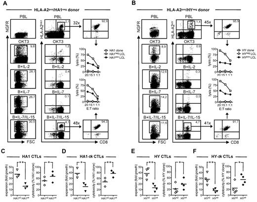
In all donors tested (n = 3), the only tk condition from which it was possible to induce HLA-A2HA-1 tetramer-binding CD8+ T lymphocytes with efficiency comparable to unmodified T cells was B + IL-7/IL-15 (Figure 1A and Table 1). After isolation by flow cytometry and polyclonal stimulation with phytohemagglutinin, tk+ HLA-A2HA-1 tetramer-binding CD8+ T cells from the B + IL-7/IL-15 condition expanded and were cytotoxic against targets naturally expressing HA-1 (Figure 1A). To exclude that these findings were restricted to the mH antigen HA-1, similar experiments were carried out with T cells from HLA-A2pos/H-Yneg (female) donors (n = 4), using H-Y as an alternative model alloantigen. Again, the only tk condition in which the efficiency of H-Y–specific cytotoxic T lymphocyte induction was comparable to unmodified T cells was B + IL-7/IL-15 (Figure 1B and Table 1). In summary, these results show that RV-mediated tk transduction after CD28 costimulation with beads and culturing with IL-7/IL-15 preserves mH antigen-specific cytotoxic T lymphocyte alloreactivity in vitro. Consequently, all subsequent experiments were performed with tk+ T cells generated with B + IL-7/IL-15.
Expansion and cytotoxicity of human tk+ mH antigen-specific CTLs. (A) The transduction efficiency (left panels) of peripheral blood lymphocytes (PBLs) from HLA-A2pos/HA-1neg donors either after activation with an anti-CD3 mAb (OKT3) and culture with IL-2 or after activation with CD3/CD28-beads and culture with IL-2 (B + IL-2), IL-7 (B + IL-7) or a combination of IL-7 and IL-15 (B + IL-7/IL-15) was evaluated by flow cytometry and is shown as the percentage of T cells positive for the NGFR marker gene (y-axis) over the FSC (x-axis). Flow cytometry was also used to measure (middle panels) and to isolate (right panels) CD8+ T cells (x-axis) binding the HLA-A2HA-1 tetramer (y-axis). In the case of RV-mediated transduction, HLA-A2HA-1 tetramer-binding CD8+ T cells were gated for NGFR expression. Inset fold numbers indicate the expansion rate after PHA stimulation. Cytotoxicity is expressed as the percentage of lysis (y-axis) at different effector-to-target ratios (x-axis) against an HLA-A2pos/HA-1neg lymphoblastoid cell line (HA1neg LCL, ◊) or an HLA-A2pos/HA1pos LCL (HA1pos LCL, ♦). The cytotoxicity of an HA-1–specific cytotoxic T lymphocyte clone (HA1 clone) against the HA1pos LCL is shown as reference (□). Maximal background cytotoxicity of the HA1 clone against the HA1neg LCL was always < 5% (not shown). Results from 1 donor representative of n = 3 are shown. (B) The same experimental procedure was applied to HLA-A2neg/H-Yneg (female) donors using appropriate reagents and control cell lines. Results from 1 donor representative of n = 4 are shown. (C) The expansion of HA-1–specific CTLs (HA1 CTLs, left panel) generated after either 2 rounds (HA1×2, ▿) or 4 rounds of antigenic stimulation (HA1×4, ▾) is expressed as fold growth. The cytotoxicity of HA1 CTLs (right panel) generated after either HA1×2 (◊) or HA1×4 (♦) is expressed as a relative percentage over that of the HA1 clone (see “Functional characterization of human mH antigen-specific CTLs”). Each symbol represents the result from a single donor. (D) The expansion (left panel) and cytotoxicity (right panel) of HA-1–specific CTLs generated from B + IL-7/IL-15 tk+ T cells (HA1-tk CTLs) after either HA1×2 or HA1×4 are shown. Each symbol represents the result from a single donor. (E-F) Experiments were repeated with H-Y–specific CTLs generated from PBLs (HY CTLs) or from B + IL-7/IL-15 tk+ cells (HY-tk CTLs) of HLA-A2neg/H-Yneg donors after either 2 (HYx2) or 4 (HYx4) rounds of antigenic stimulation, using appropriate reagents and control cell lines. Expansion (left panels) and cytotoxicity (right panels) are shown. Each symbol represents the result from a single donor. Results from a paired t test statistical analysis are shown (*P < .05, **P < .01).
Expansion and cytotoxicity of human tk+ mH antigen-specific CTLs. (A) The transduction efficiency (left panels) of peripheral blood lymphocytes (PBLs) from HLA-A2pos/HA-1neg donors either after activation with an anti-CD3 mAb (OKT3) and culture with IL-2 or after activation with CD3/CD28-beads and culture with IL-2 (B + IL-2), IL-7 (B + IL-7) or a combination of IL-7 and IL-15 (B + IL-7/IL-15) was evaluated by flow cytometry and is shown as the percentage of T cells positive for the NGFR marker gene (y-axis) over the FSC (x-axis). Flow cytometry was also used to measure (middle panels) and to isolate (right panels) CD8+ T cells (x-axis) binding the HLA-A2HA-1 tetramer (y-axis). In the case of RV-mediated transduction, HLA-A2HA-1 tetramer-binding CD8+ T cells were gated for NGFR expression. Inset fold numbers indicate the expansion rate after PHA stimulation. Cytotoxicity is expressed as the percentage of lysis (y-axis) at different effector-to-target ratios (x-axis) against an HLA-A2pos/HA-1neg lymphoblastoid cell line (HA1neg LCL, ◊) or an HLA-A2pos/HA1pos LCL (HA1pos LCL, ♦). The cytotoxicity of an HA-1–specific cytotoxic T lymphocyte clone (HA1 clone) against the HA1pos LCL is shown as reference (□). Maximal background cytotoxicity of the HA1 clone against the HA1neg LCL was always < 5% (not shown). Results from 1 donor representative of n = 3 are shown. (B) The same experimental procedure was applied to HLA-A2neg/H-Yneg (female) donors using appropriate reagents and control cell lines. Results from 1 donor representative of n = 4 are shown. (C) The expansion of HA-1–specific CTLs (HA1 CTLs, left panel) generated after either 2 rounds (HA1×2, ▿) or 4 rounds of antigenic stimulation (HA1×4, ▾) is expressed as fold growth. The cytotoxicity of HA1 CTLs (right panel) generated after either HA1×2 (◊) or HA1×4 (♦) is expressed as a relative percentage over that of the HA1 clone (see “Functional characterization of human mH antigen-specific CTLs”). Each symbol represents the result from a single donor. (D) The expansion (left panel) and cytotoxicity (right panel) of HA-1–specific CTLs generated from B + IL-7/IL-15 tk+ T cells (HA1-tk CTLs) after either HA1×2 or HA1×4 are shown. Each symbol represents the result from a single donor. (E-F) Experiments were repeated with H-Y–specific CTLs generated from PBLs (HY CTLs) or from B + IL-7/IL-15 tk+ cells (HY-tk CTLs) of HLA-A2neg/H-Yneg donors after either 2 (HYx2) or 4 (HYx4) rounds of antigenic stimulation, using appropriate reagents and control cell lines. Expansion (left panels) and cytotoxicity (right panels) are shown. Each symbol represents the result from a single donor. Results from a paired t test statistical analysis are shown (*P < .05, **P < .01).
Antigen-specific enrichment of human tk+ mH antigen-specific CTLs in vitro associates with loss of proliferative potential and acquisition of effector function
With the aim to generate large numbers of CTLs for further studies, after induction, mH antigen-specific tk+ CTLs were exposed to additional rounds of antigenic stimulation. Similar to previous studies,27 this led to a significant enrichment of CD8+ T cells binding the relevant tetramer. Namely, tk+ HLA-A2HA-1 tetramer-binding CD8+ T cells increased from mean 3.1% (range 2.8-4.1) after 2 rounds of HA-1–specific stimulation to mean 7.2% (range 5.0-8.2) after 4 rounds (P < .05), and tk+ HLA-A2H-Y tetramer-binding CD8+ T cells increased from mean 2.4% (range 2.1-4.7) after 2 rounds of H-Y–specific stimulation to mean 6.1% (range 4.8-7.5) after 4 rounds (P < .01). Both tk+ HLA-A2HA-1 and HLA-A2H-Y tetramer-binding CD8+ T cells isolated by flow cytometry after 4 rounds of stimulation expanded less than cells isolated after 2 rounds, but displayed a higher degree of cytotoxicity against the relevant mH antigen (Figure 1D,F). The phenomenon was also evident in the case mH antigen-specific tetramer-binding CD8+ T cells generated from unmodified T cells (Figure 1C,E). Altogether, these results indicate that antigen-specific enrichment of tk+ mH antigen-specific CTLs in vitro associates with loss of proliferative potential and acquisition of effector function.
Loss of proliferative potential of human tk+ mH antigen-specific CTLs in vitro associates with loss of functional IL-7 receptor expression
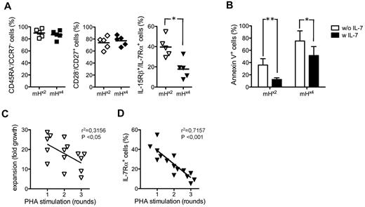
Because the loss of proliferative potential could be related to a progression in T-cell differentiation, before or after antigen-specific enrichment, the phenotype of mH antigen-specific tk+ CTLs was compared. Before or after the procedure, tk+ mH antigen-specific CTLs were homogenously negative for CD45RA and CCR7, that is, they belonged to the putative TEM cell subset (Figure 2A). Moreover, they were indifferently positive for the costimulatory receptor CD27, but negative for CD28 (Figure 2A). However, whereas before enrichment tk+ mH antigen-specific CTLs coexpressed the α subunit of the IL-7 receptor (IL-7Rα) and the β subunit of the receptor for IL-2/IL-15 (IL-15Rβ), after enrichment there was a selective depletion of cells expressing the α subunit of the receptor for IL-7 (IL-7Rα; Figure 2A).
Differentiation phenotype and IL-7 responsiveness of human tk+ mH antigen-specific CTLs. (A) The differentiation phenotype of tk+ mH antigen-specific CTLs after either 2 rounds (mH×2) or 4 rounds (mH×4) of antigenic stimulation was evaluated by flow cytometry and is expressed as the percentage of CD45RA−/CCR7− (left panel), of CD28−/CD27+ (middle panel), and of IL-15Rβ+/IL-7Rα+ cells (right panel). Each symbol represents the result from a single donor of a group of n = 2 HLA-A2pos/HA-1neg and n = 3 HLA-A2pos/H-Yneg donors. Results from a t test paired statistical analysis are shown (*P < .05). (B) The percentage of either mH×2 or mH×4 tk+ mH antigen-specific CTLs binding annexin V after culture in the absence of antigen and in the presence (■) or in the absence (□) of exogenous IL-7 was evaluated by flow cytometry. Means ± SD of the data from n = 2 HLA-A2pos/HA-1neg and n = 2 HLA-A2pos/H-Yneg donors and results from a paired t test statistical analysis are shown (*P < .05, **P < .01). (C) The expansion of mHx2tk+ mH antigen-specific CTLs after multiple, weekly rounds of stimulation with PHA (x-axis) is expressed as fold growth (y-axis). (D) The percentage of IL-7Rα+ cells (y-axis) was evaluated by flow cytometry before each round of PHA stimulation (x-axis). Results from linear regression analysis are shown (exact P values).
Differentiation phenotype and IL-7 responsiveness of human tk+ mH antigen-specific CTLs. (A) The differentiation phenotype of tk+ mH antigen-specific CTLs after either 2 rounds (mH×2) or 4 rounds (mH×4) of antigenic stimulation was evaluated by flow cytometry and is expressed as the percentage of CD45RA−/CCR7− (left panel), of CD28−/CD27+ (middle panel), and of IL-15Rβ+/IL-7Rα+ cells (right panel). Each symbol represents the result from a single donor of a group of n = 2 HLA-A2pos/HA-1neg and n = 3 HLA-A2pos/H-Yneg donors. Results from a t test paired statistical analysis are shown (*P < .05). (B) The percentage of either mH×2 or mH×4 tk+ mH antigen-specific CTLs binding annexin V after culture in the absence of antigen and in the presence (■) or in the absence (□) of exogenous IL-7 was evaluated by flow cytometry. Means ± SD of the data from n = 2 HLA-A2pos/HA-1neg and n = 2 HLA-A2pos/H-Yneg donors and results from a paired t test statistical analysis are shown (*P < .05, **P < .01). (C) The expansion of mHx2tk+ mH antigen-specific CTLs after multiple, weekly rounds of stimulation with PHA (x-axis) is expressed as fold growth (y-axis). (D) The percentage of IL-7Rα+ cells (y-axis) was evaluated by flow cytometry before each round of PHA stimulation (x-axis). Results from linear regression analysis are shown (exact P values).
Elaborating on the assumption that the difference in IL-7Rα expression had a functional relevance, the effect of exogenous IL-7 on the ability of tk+ mH antigen-specific CTLs isolated before or after antigen-specific enrichment to survive on antigen deprivation was compared in vitro. After 1 week of culture in the absence of antigen, the majority of tk+ mH antigen-specific CTLs isolated after enrichment was in apoptosis, as demonstrated by annexin V staining, whereas a considerable fraction of CTLs isolated before enrichment was still viable (Figure 2B). The addition of IL-7 almost completely rescued tk+ mH antigen-specific CTLs isolated before enrichment, but failed in the case of CTLs isolated after enrichment (Figure 2B).
With the hypothesis that the loss of proliferative potential associated with the loss of IL-7 receptor expression, before antigen-specific enrichment, tk+ mH antigen-specific CTLs were isolated, exposed to weekly rounds of polyclonal stimulation, and analyzed for IL-7Rα expression. The expansion rate (Figure 2C) as well as the proportion of tk+ mH antigen-specific CTLs expressing IL-7Rα (Figure 2D) progressively decreased with the rounds of stimulation.
IL-7 receptor expression identifies human tk+ mH antigen-specific CTLs capable of self-renewal and differentiation into effectors in vitro
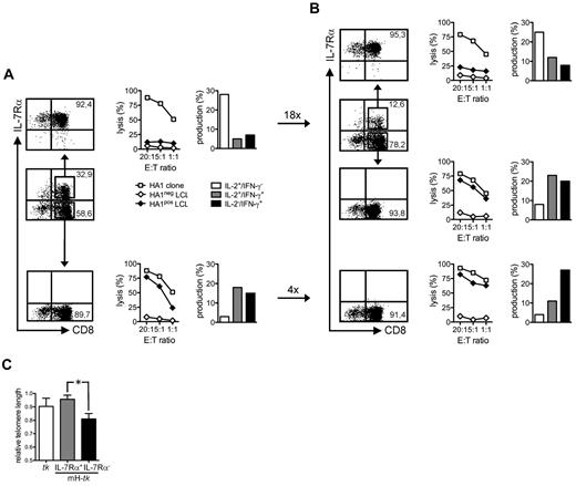
To confirm that the loss of IL-7 receptor expression was linked to the acquisition of effector function, experiments based on sequential sorting were performed. Before antigen-specific enrichment, tk+ mH antigen-specific CTLs were sorted for IL-7Rα expression and analyzed for cytotoxicity and cytokine production. Interestingly, whereas IL-7Rα+ cells were poorly cytotoxic and mainly produced IL-2, IL-7Rα− cells were highly cytotoxic and essentially produced IFN-γ (Figure 3A and Table 2). On polyclonal stimulation, IL-7Rα+ cells expanded and gave rise to a mixed progeny of IL-7Rα+ and IL-7Rα− cells (Figure 3B and Table 2). In contrast, IL-7Rα− cells did not expand substantially and homogenously lacked IL-7Rα expression (Figure 3B and Table 2). After expansion, tk+ mH antigen-specific CTLs were re-sorted for IL-7Rα expression and functional testing revealed the same differences in cytotoxicity and cytokine production between IL-7Rα+ and IL-7Rα− cells. Interestingly, flow cytometric analysis of mean telomere length of tk+ mH antigen-specific CTLs after the first sorting revealed that IL-7Rα+ cells had longer telomeres than IL-7Rα− cells (Figure 3C), implying that the loss of proliferative potential associated with IL-7 receptor loss coincided with telomere erosion.
Self-renewal and differentiation of human tk+ mH antigen-specific CTLs based on IL-7Rα expression. (A) HA1-specific CTLs generated after 2 rounds of antigenic stimulation were sorted for IL-7Rα expression by flow cytometry (left panels). The percentage of CD8+ T cells (x-axis) positive for the IL-7Rα (y-axis) is shown before and after sorting (arrows). Cytotoxicity (middle panels) is expressed as the percentage of lysis (y-axis) at different effector-to-target ratios (x-axis) against an HLA-A2pos/HA1neg lymphoblastoid cell line (HA1neg LCL, ◊) or an HLA-A2pos/HA1pos LCL (HA1pos LCL, ♦). The cytotoxicity of a HA1-specific cytotoxic T lymphocyte clone (HA1 clone) against the HA1pos LCL is shown as reference (□). Maximal background cytotoxicity of the HA1 clone against the HA1neg LCL was always < 5% (not shown). Cytokine production (right panels) was evaluated by flow cytometry after antigenic stimulation (see “Generation, expansion and functional characterization of human mH antigen-specific CTLs”) and is expressed as the percentage of cells with the following profile of intracytoplasmic staining: IFN-γ−/IL-2+ (□), IFN-γ+/IL-2+ (▩), IFN-γ+/IL-2− (■). Inset fold numbers indicate the expansion rate of sorted populations after PHA stimulation. (B) IL-7Rα expression (left panels), cytotoxicity (middle panels), and cytokine production (right panels) of the progeny of IL-7Rα+ HA-1–specific CTLs cells before and after re-sorting (arrows). After re-sorting, cells were re-stimulated with PHA. Fold growth of IL-7Rα+ cells from IL-7Rα+ cells was 10.4, fold growth of IL-7Rα− cells from IL-7Rα+ cells was 4.7, and fold growth of IL-7Rα− from IL-7Rα− cells was 2.3. Results from the representative donor HA1-tk #1 are shown. (C) Relative telomere length (y-axis) of tk+ T cells generated with CD3/CD28-beads and IL-7/IL-15 (tk, □) and of tk+ mH antigen-specific CTLs (mH-tk) sorted as IL-7Rα+ (▩) or IL-7Rα− (■) was measured by flow FISH. Means ± SD of relative telomere length from n = 2 HLA-A2pos/HA-1neg and n = 1 HLA-A2pos/H-Yneg donors and results from a paired t test statistical analysis are shown (*P < .05).
Self-renewal and differentiation of human tk+ mH antigen-specific CTLs based on IL-7Rα expression. (A) HA1-specific CTLs generated after 2 rounds of antigenic stimulation were sorted for IL-7Rα expression by flow cytometry (left panels). The percentage of CD8+ T cells (x-axis) positive for the IL-7Rα (y-axis) is shown before and after sorting (arrows). Cytotoxicity (middle panels) is expressed as the percentage of lysis (y-axis) at different effector-to-target ratios (x-axis) against an HLA-A2pos/HA1neg lymphoblastoid cell line (HA1neg LCL, ◊) or an HLA-A2pos/HA1pos LCL (HA1pos LCL, ♦). The cytotoxicity of a HA1-specific cytotoxic T lymphocyte clone (HA1 clone) against the HA1pos LCL is shown as reference (□). Maximal background cytotoxicity of the HA1 clone against the HA1neg LCL was always < 5% (not shown). Cytokine production (right panels) was evaluated by flow cytometry after antigenic stimulation (see “Generation, expansion and functional characterization of human mH antigen-specific CTLs”) and is expressed as the percentage of cells with the following profile of intracytoplasmic staining: IFN-γ−/IL-2+ (□), IFN-γ+/IL-2+ (▩), IFN-γ+/IL-2− (■). Inset fold numbers indicate the expansion rate of sorted populations after PHA stimulation. (B) IL-7Rα expression (left panels), cytotoxicity (middle panels), and cytokine production (right panels) of the progeny of IL-7Rα+ HA-1–specific CTLs cells before and after re-sorting (arrows). After re-sorting, cells were re-stimulated with PHA. Fold growth of IL-7Rα+ cells from IL-7Rα+ cells was 10.4, fold growth of IL-7Rα− cells from IL-7Rα+ cells was 4.7, and fold growth of IL-7Rα− from IL-7Rα− cells was 2.3. Results from the representative donor HA1-tk #1 are shown. (C) Relative telomere length (y-axis) of tk+ T cells generated with CD3/CD28-beads and IL-7/IL-15 (tk, □) and of tk+ mH antigen-specific CTLs (mH-tk) sorted as IL-7Rα+ (▩) or IL-7Rα− (■) was measured by flow FISH. Means ± SD of relative telomere length from n = 2 HLA-A2pos/HA-1neg and n = 1 HLA-A2pos/H-Yneg donors and results from a paired t test statistical analysis are shown (*P < .05).
Cytotoxicity and expansion of human tk+ mH antigen-specific CTLs after IL-7Rα sequential sorting
| CTLs* . | IL-7Rα+,† % . | Sorting . | IL-7Rα+,† % . | Re-sorting . | ||||
|---|---|---|---|---|---|---|---|---|
| IL-7Rα,‡ % . | Cytotoxicity,§ % . | Expansion,¶ fold . | IL-7Rα,‡ % . | Cytotoxicity,§ % . | Expansion,¶ fold . | |||
| HA1-tk #1 | 32.9 | 92.4pos | 12.7 | 17.6 | 12.6 | 95.3pos | 21.2 | 10.4 |
| < 1neg | 65.7 | 4.1 | < 1neg | 78.5 | 4.7 | |||
| HA1-tk #2 | 54.6 | 86.7pos | 16.8 | 24.2 | 10.9 | 91.7pos | 11.5 | 14.5 |
| 2.3neg | 54.2 | 4.6 | 1.8neg | 84.5 | — | |||
| HY-tk #1 | 21.7 | 81.8pos | 21.2 | 35.1 | 15.4 | 89.6pos | 22.6 | 12.3 |
| 5.6neg | 74.0 | 8.7 | 1.5neg | 81.2 | 3.5 | |||
Thymidine kinase–positive CTLs used for sorting: HA1-tk #1, specific for HA-1 and induced from an HLA-A2pos/HA-1neg donor; HA1-tk #2, specific for HA-1 and induced from a second HLA-A2pos/HA-1neg donor; HY-tk #1, specific for H-Y and induced from an HLA-A2pos/H-Yneg (female) donor
Thymidine kinase–positive mH antigen-specific CTLs expressing IL-7Rα
Thymidine kinase–positive mH antigen-specific CTLs expressing IL-7Rα in the positive (pos) and in the negative (neg) fraction after IL-7Rα sorting
Cytotoxicity of IL-7Rα+ and IL-7Rα−tk+ mH antigen-specific CTLs against an LCL expressing the relevant mH antigen at a 20:1 ratio
Expansion of IL-7Rα+ and IL-7Rα−tk+ mH antigen-specific CTLs
In summary, these results suggest that IL-7 receptor expression identifies tk+ mH antigen-specific CTLs capable of self-renewal and differentiation into effectors in vitro.
Human tk+ mH antigen-specific CTLs are cytotoxic against leukemia and sensitive to suicide induction by prodrug administration in vitro
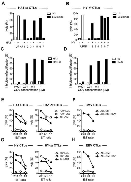
Evidently, demonstrating the antileukemia efficacy of tk+ mH antigen-specific CTLs would underscore the clinical relevance of our findings. For this purpose, tk+ mH antigen-specific CTLs were evaluated for cytotoxicity against a panel of HLA-A2pos leukemic cells derived from patients (supplemental Table 1, available on the Blood Web site; see the Supplemental Materials link at the top of the online article). Thymidine kinase–positive HA-1–specific CTLs were cytotoxic against HA-1pos leukemic cells derived from patients with different types of leukemia, but not against HA-1neg leukemic cells (Figure 4A and supplemental Figure 1A). By analogy, tk+ H-Y-specific CTLs were cytotoxic against leukemic cells of male origin (H-Ypos) but not against those of female origin (H-Yneg; Figure 4B and supplemental Figure 1B).
Cytotoxicity of tk+ mH antigen-specific CTLs against leukemia and suicide induction by GCV administration in vitro. (A) The cytotoxicity of tk+ HA-1–specific CTLs (HA1-tk CTLs) generated from an HLA-A2pos/HA-1neg representative donor (HA1-tk #1) was evaluated against different HLA-A2pos targets, including lymphoblastoid cell lines (LCL, □) and patient-derived leukemic cells typed for HA-1 (negative if R/R, positive if H/R or HH) and is expressed as the percentage of lysis at a 20:1 effector-to-target ratio. (B) The cytotoxicity of tk+ H-Y-specific CTLs (HY-tk CTLs) generated from an HLA-A2pos/H-Yneg representative (female) donor (HY-tk #1) was evaluated against different HLA-A2pos targets, including lymphoblastoid cell lines (LCL, □) and leukemic cells derived from female (HY negative) or male (HY positive) patients and is expressed as the percentage of lysis at a 20:1 effector-to-target ratio. (C) Suicide induction on HA1 and HA1-tk CTLs at increasing concentrations of GCV (x-axis) is expressed as GCV sensitivity (y-axis, see “Functional characterization of human mH antigen-specific CTLs”). Results from the HA1-tk #1 donor are shown. (D) Suicide induction was also assayed on HY and HY-tk CTLs. Results from the donor HY-tk #1 donor are shown. (E) The cytotoxicity of HA1-specific CTLs generated either from PBLs (HA1 CTLs) or from tk+ T cells (HA1-tk CTLs) of the HA1-tk #1 donor is expressed as the percentage of lysis (y-axis) at different effector-to-target ratios (x-axis) against an HLA-A2pos/HA1neg lymphoblastoid cell line (HA1neg LCL, ◊), an HLA-A2pos/HA1pos LCL (HA1pos LCL, □), or ALL-CM leukemic cells (♦), which are HLA-A2pos/HA-1pos/H-Ypos. (F) The cytotoxicity of CMV-specific CTLs (CMV CTLs) generated from PBLs of the HA1-tk #1 donor is expressed as the percentage of lysis (y-axis) at different effector-to-target ratios (x-axis) against ALL-CM leukemic cells loaded (♦) or not (◊) with the CMV peptide used for generation. (G) H-Y–specific CTLs were generated from PBLs (HY CTLs) and from tk+ T cells (HY-tk CTLs) of the HY-tk #1 donor and characterized for cytotoxicity against leukemia using appropriate reagents and control cell lines. (H) EBV-specific CTLs (EBV CTLs) were generated from PBLs of the HY-tk #1 donor and characterized.
Cytotoxicity of tk+ mH antigen-specific CTLs against leukemia and suicide induction by GCV administration in vitro. (A) The cytotoxicity of tk+ HA-1–specific CTLs (HA1-tk CTLs) generated from an HLA-A2pos/HA-1neg representative donor (HA1-tk #1) was evaluated against different HLA-A2pos targets, including lymphoblastoid cell lines (LCL, □) and patient-derived leukemic cells typed for HA-1 (negative if R/R, positive if H/R or HH) and is expressed as the percentage of lysis at a 20:1 effector-to-target ratio. (B) The cytotoxicity of tk+ H-Y-specific CTLs (HY-tk CTLs) generated from an HLA-A2pos/H-Yneg representative (female) donor (HY-tk #1) was evaluated against different HLA-A2pos targets, including lymphoblastoid cell lines (LCL, □) and leukemic cells derived from female (HY negative) or male (HY positive) patients and is expressed as the percentage of lysis at a 20:1 effector-to-target ratio. (C) Suicide induction on HA1 and HA1-tk CTLs at increasing concentrations of GCV (x-axis) is expressed as GCV sensitivity (y-axis, see “Functional characterization of human mH antigen-specific CTLs”). Results from the HA1-tk #1 donor are shown. (D) Suicide induction was also assayed on HY and HY-tk CTLs. Results from the donor HY-tk #1 donor are shown. (E) The cytotoxicity of HA1-specific CTLs generated either from PBLs (HA1 CTLs) or from tk+ T cells (HA1-tk CTLs) of the HA1-tk #1 donor is expressed as the percentage of lysis (y-axis) at different effector-to-target ratios (x-axis) against an HLA-A2pos/HA1neg lymphoblastoid cell line (HA1neg LCL, ◊), an HLA-A2pos/HA1pos LCL (HA1pos LCL, □), or ALL-CM leukemic cells (♦), which are HLA-A2pos/HA-1pos/H-Ypos. (F) The cytotoxicity of CMV-specific CTLs (CMV CTLs) generated from PBLs of the HA1-tk #1 donor is expressed as the percentage of lysis (y-axis) at different effector-to-target ratios (x-axis) against ALL-CM leukemic cells loaded (♦) or not (◊) with the CMV peptide used for generation. (G) H-Y–specific CTLs were generated from PBLs (HY CTLs) and from tk+ T cells (HY-tk CTLs) of the HY-tk #1 donor and characterized for cytotoxicity against leukemia using appropriate reagents and control cell lines. (H) EBV-specific CTLs (EBV CTLs) were generated from PBLs of the HY-tk #1 donor and characterized.
After demonstration of the antileukemia efficacy of tk+ mH antigen-specific CTLs in vitro, the functionality of the suicide gene machinery was investigated. As expected, exposing tk+ but not unmodified mH antigen-specific CTLs to increasing concentrations of the prodrug GCV caused a dose-dependent inhibition of cell proliferation (Figure 4C-D).
Thymidine kinase–positive mH antigen-specific CTLs display substantial antileukemia efficacy in vivo
In the final part of the study, we aimed at verifying whether suicide gene modification might decrease the antileukemia efficacy of mHag-specific CTLs in a clinically relevant humanized mouse model.21 To this aim, different populations of CTLs were generated from 2 of the HLApos donors previously analyzed. The 2 donors were chosen as representative for the HA-1 and for the H-Y antigen, respectively. In the case of the HA-1neg donor, tk+ or unmodified HA-1–specific CTLs and control unmodified CMV-specific CTLs were generated after induction with 2 rounds of antigenic stimulation, isolation, and expansion by polyclonal stimulation. Interestingly, tk+ HA-1–specific CTLs could be generated as efficiently as unmodified HA-1–specific CTLs, confirming that previous RV-mediated tk transduction with B + IL-7/IL-15 was not interfering with the potential for secondary expansion (not shown). Expanded tk+ and unmodified HA-1–specific CTLs were highly and equally cytotoxic against ALL-CM leukemic cells (Figure 4E), whereas CMV-specific CTLs were not, unless leukemic cells were pulsed with the CMV peptide used for generation (Figure 4F). Using the same procedure, tk+ and unmodified H-Y–specific CTLs were also generated from the H-Yneg representative (female) donor and were cytotoxic against ALL-CM leukemic cells, which were of male origin (H-Ypos; Figure 4G). As expected, unmodified EBV-specific CTLs generated from the same donor as control did not recognize ALL-CM leukemic cells, unless these were pulsed with the peptide used for generation (Figure 4H).
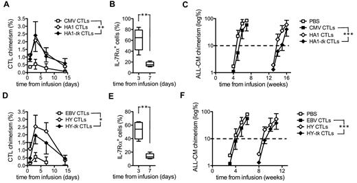
NOD/scid mice were inoculated with 107 ALL-CM leukemic cells and, 3 days later, infused either with 2 × 10⁷tk+ or with unmodified HA-1–specific CTLs. Control mice received saline or 2 × 10⁷ CMV-specific human CTLs. HA-1–specific CTLs were detected in the peripheral circulation of all mice (Figure 5A). Circulating tk+ and unmodified HA-1–specific CTLs peaked at day 3 and persisted up to day 14. CMV-specific CTLs also peaked at day 3, but from day 7 on they were barely detectable (Figure 5A). Interestingly, despite before infusion only 18.3% of tk+ HA1-specific CTLs expressed IL-7Rα, the majority of peaking circulating CTLs were IL-7Rα+ (mean 65.1%, range 33%-84%; Figure 5B). This proportion declined over time, possibly indicating ongoing differentiation on antigen-encounter in vivo. Compared with CMV-specific CTLs, tk+ and unmodified HA-1–specific CTLs equally and significantly prolonged leukemia-free survival (approximately 9 weeks; Figure 5C, P = .001). In another experiment, after leukemia inoculum, mice were infused either with tk+ or with unmodified H-Y–specific CTLs. In vivo persistence of tk+ H-Y–specific CTLs and kinetics of IL-7 receptor expression was similar as in the case of tk+ HA-1–specific CTLs (Figure 5D-E; IL-7Rα+ cells before infusion: 15.5%, on peaking circulating CTLs: mean 52%, range 36%-65%). Compared with the respective control group infused with EBV-specific CTLs, tk+ H-Y–specific CTLs also significantly prolonged leukemia-free survival, albeit to a slightly lower extent than HA-1–specific CTLs (approximately 5 weeks; Figure 5C, P = .003).
In vivo persistence and antileukemia efficacy of mH antigen-specific tk+ CTLs in NOD/scid mice. (A) At different time points (x-axis) after ALL-CM leukemia inoculum and later infusion of unmodified CMV-specific CTLs (CMV CTLs, ■), unmodified (HA1 CTLs, ◊), or tk+ HA-1–specific CTLs generated from the HA1-tk #1 donor, cytotoxic T lymphocyte chimerism (y-axis, see “In vivo evaluation of the antileukemia efficacy of human mH antigen-specific CTLs”) was evaluated in the peripheral blood of individual NOD/scid mice by flow cytometry. Control mice were infused with PBS (□). Means ± SD of the data from n = 5 mice/group and results from an unpaired Mann-Whitney U test statistical analysis are shown (**P < .01 for cytotoxic T lymphocyte chimerism at day 3). (B) At days 3 and 7 after infusion, the percentage of circulating tk+ HA-1–specific CTLs expressing IL-7Rα (y-axis) was evaluated by flow cytometry. (C) At different time points (x-axis), mice were also individually monitored for ALL-CM chimerism (y-axis). Mean log percentages of circulating leukemic cells from n = 5 mice/group ± SD (y-axis) are shown (***P < .005). (D-F) Cytotoxic T lymphocyte chimerism, IL-7Rα expression, and ALL-CM chimerism were also monitored in mice inoculated with leukemia and later infused with unmodified EBV-specific CTLs (EBV CTLs, ■), unmodified (HY CTLs, ◊), or tk+ HY-specific CTLs generated from the HY-tk #1 donor (*P < .05 for cytotoxic T lymphocyte chimerism at day 3; **P < .01; ***P < .005).
In vivo persistence and antileukemia efficacy of mH antigen-specific tk+ CTLs in NOD/scid mice. (A) At different time points (x-axis) after ALL-CM leukemia inoculum and later infusion of unmodified CMV-specific CTLs (CMV CTLs, ■), unmodified (HA1 CTLs, ◊), or tk+ HA-1–specific CTLs generated from the HA1-tk #1 donor, cytotoxic T lymphocyte chimerism (y-axis, see “In vivo evaluation of the antileukemia efficacy of human mH antigen-specific CTLs”) was evaluated in the peripheral blood of individual NOD/scid mice by flow cytometry. Control mice were infused with PBS (□). Means ± SD of the data from n = 5 mice/group and results from an unpaired Mann-Whitney U test statistical analysis are shown (**P < .01 for cytotoxic T lymphocyte chimerism at day 3). (B) At days 3 and 7 after infusion, the percentage of circulating tk+ HA-1–specific CTLs expressing IL-7Rα (y-axis) was evaluated by flow cytometry. (C) At different time points (x-axis), mice were also individually monitored for ALL-CM chimerism (y-axis). Mean log percentages of circulating leukemic cells from n = 5 mice/group ± SD (y-axis) are shown (***P < .005). (D-F) Cytotoxic T lymphocyte chimerism, IL-7Rα expression, and ALL-CM chimerism were also monitored in mice inoculated with leukemia and later infused with unmodified EBV-specific CTLs (EBV CTLs, ■), unmodified (HY CTLs, ◊), or tk+ HY-specific CTLs generated from the HY-tk #1 donor (*P < .05 for cytotoxic T lymphocyte chimerism at day 3; **P < .01; ***P < .005).
Discussion
In this study, we demonstrate that CD28 costimulation through CD3/CD28-beads and culturing with a combination of IL-7 and IL-15 are necessary and sufficient for the RV-mediated tk transduction of human T lymphocytes retaining mH antigen-specific cytotoxic T lymphocyte alloreactivity. Moreover, we provide direct proof-of-concept of the in vitro and in vivo antileukemia efficacy of mH antigen-specific CTLs derived from tk+ T cells. Interestingly, during the process of in vitro expansion, we observed that tk+ mH antigen-specific CTLs progressively lost proliferative potential and reciprocally acquired effector function. Within the bulk population, we identified a subset of tk+ mH antigen-specific CTLs characterized by the highest proliferative potential and by the expression of the IL-7 receptor. This subset could self-renew and give rise to a progeny of effectors, that is, properties of putative memory stem T cells.
At present, the reason why tk+ human T lymphocytes generated after CD28 costimulation with beads and culturing with IL-7/IL-15, but not with the other combinations tested, retain mH antigen-specific cytotoxic T lymphocyte alloreactivity is unknown. The different cytokines belonging to the γ-chain family apparently have distinct roles in T-cell responses. IL-2 and IL-15, for example, are potent T-cell growth factors that share the IL-2/IL-15 receptor β. As opposed to IL-2, IL-15 does not prime T cells for activation-induced cell death28 and strongly contrasts regulatory T cells.29 Importantly, IL-7 is far less potent than IL-2 and IL-15 at inducing T-cell proliferation, but appears to be critically involved in memory T-cell survival.30 The preservation of mH antigen-reactivity may be explained by a combined action of the 2 cytokines, which could specifically promote the proliferation and the survival of suicide gene–modified precursors for mH antigen-specific CTLs.
The progressive loss of proliferative potential and the reciprocal acquisition of effector function on in vitro expansion of tumor-reactive CD8+ T lymphocytes has been already observed in mouse models of adoptive T-therapy of melanoma, and has been shown to associate with the transition from TCM to TEM cells.31 We recently described that CD28 costimulation with beads before RV-mediated tk transduction enriches for human TCM cells, regardless of the cytokine used for culture.18 Rather unexpectedly, despite their predominant TEM phenotype, tk+ mH antigen-specific CTLs could undergo multiple rounds of expansion in vitro, apparently without losing antileukemia efficacy after subsequent transfer in vivo. This is in agreement with the observation in a primate model that virus-specific CD8+ TEM clones generated from TCM, but not from TEM cells, maintain the functional characteristics of the population of origin.32
Differently from the linear model of T-cell memory, which assumes a continuous transition among TNA, memory T cells, and effector T cells, the stem cell model proposes that antigen encounter determines the early generation of a long-lived putative memory stem T cell characterized by the ability to self-renew and differentiate into memory T cells and effectors.33 In a mouse model of GVHD, CD8+ T cells primed in vitro for a short period of time with host antigens in the presence of IL-15 were able to persist after adoptive transfer throughout the disease course and to give rise to different T-cell types, including effectors.34 In analogy, after antigenic stimulation, we found that a subpopulation of tk+ mH antigen-specific CTLs, identified by the expression of the IL-7 receptor, was characterized by the highest proliferative potential and by the capability to self-renew and to differentiate into potent antileukemia effectors. These characteristics match with those of memory stem T cells. Signaling through Wnt has recently been shown to be involved in the generation of CD8+ memory stem T cells in mice.35 It would be interesting in the future to verify whether Wnt signaling also plays a role in the generation of gene-modified memory stem T cells in humans.
Our observation of shorter telomeres in cells losing IL-7 receptor expression suggests that the differentiation of tk+ mH antigen-specific CTLs into full effectors is unidirectional, as erosion of telomeres in nonmalignant cells is thought to be irreversible (discussed in Zhang et al36 ). Conversely, the higher proliferative potential of tk+ mH antigen-specific CTLs maintaining IL-7 receptor expression could explain their selective enrichment early after infusion into NOD/scid mice. Data in the literature support our conclusions. For example, in vivo persistence and antitumor efficacy of melanoma-reactive human CD8+ T cells associate with both increased IL-7 receptor expression37 and longer telomeres.38,39 Moreover, replicative senescence of EBV-specific human CTLs correlates with irreversible IL-7 receptor deficiency, which can be counterfeited by the genetic insertion of the IL-7 receptor.40 The demonstration of an association between IL-7 receptor expression and memory stem T-cell properties of tk+ mH antigen-specific CTLs bears important consequences not only to suicide gene therapy applied to HSCT, but clearly also to the general field of adoptive T-cell therapy. Currently, different improved protocols for adoptive T-cell therapy propose multiparametric sorting before ex vivo manipulation, including sorting for TNA before genetic redirection41 or TCM before antigenic stimulation.42 In alternative, our results indicate the feasibility and the efficacy of a reverse approach, in which starting from a heterogeneous population gene-modified and/or antigen-specific T cells with high proliferative potential are first generated and then sorted according a single, broadly applicable surface biomarker, namely IL-7Rα. Moreover, our finding of selective in vivo persistence of IL-7Rα+ T cells after adoptive transfer of unselected T cell provides evidence for the opportunity of boosting ex vivo generated T cells by in vivo administration of recombinant IL-7.43
The basic concept of suicide gene therapy applied to HSCT is that, by infusing polyclonal donor T lymphocytes modified with a suicide gene, it is possible to reconstitute the patient with the whole T-cell repertoire of a healthy donor, including T-cell specificities for viral, tumor, and alloantigens, while having the possibility to switch off GVHD. Current clinical protocols for RV-mediated transduction critically reduce alloreactivity and the antileukemia efficacy of suicide gene–modified T cells.9 The final specific aim of this study was to verify whether suicide gene modification following CD28 costimulation with beads and culturing with IL-7/IL-15 might decrease the antileukemia efficacy of T cells in vivo. This crucial issue was approached in a clinically relevant humanized mouse model of adoptive T-cell therapy of leukemia, in which targeting a single mH antigen with CTLs significantly prolongs disease-free survival.21,44 In this setting, we found that single mH antigen-specific CTLs, either HA-1 or H-Y, derived from tk+ T cells were as effective as those from unmodified T cells, therefore indicating that genetic modification did not interfere with antileukemia efficacy.
In summary, the results of the present study indicate that a protocol including CD28 costimulation with beads and culturing with a combination of IL-7 and IL-15 preserves the antileukemia efficacy of suicide gene–modified T cells, possibly through the direct targeting of a population of precursors for alloreactive memory stem T cells. A phase 1/2 clinical trial of suicide gene therapy implementing tk+ T cells generated with this protocol is currently in preparation for the treatment of leukemia patients in the context of HSCT and will be instrumental for the further investigation of IL-7 receptor expression as a biomarker of sustained, yet controllable, cytotoxic T lymphocyte alloreactivity in vivo.
The online version of this article contains a data supplement.
The publication costs of this article were defrayed in part by page charge payment. Therefore, and solely to indicate this fact, this article is hereby marked “advertisement” in accordance with 18 USC section 1734.
Acknowledgments
We thank Anna Mondino for helpful discussions, Massimo Bernardi for help with leukemia classification, and Zuzana Stachová-Rychnavská and Tamara Koudijs for technical assistance.
A.B. was supported by the European Union (Individual Marie Curie Fellowship, SGT for GVL). L.H. was supported by the Dutch Cancer Society (Koningin Wilhelmina Fonds, project code UL2006-3482). The study has been supported in part by the Netherlands Organization for Scientific Research (NOW; Den Haag, The Netherlands) and by the Italian Association for Cancer Research (AIRC).
Authorship
Contribution: A.B. designed and performed research, analyzed and interpreted the data, performed statistical analysis, and wrote the manuscript; L.H. designed research, analyzed and interpreted the data, and wrote the manuscript; Z.A. performed research and collected the data; B.N. and M.R. provided vital new reagents; S.K. and S.M. designed research; K.F., F.C., and C. Bordignon analyzed and interpreted the data; and C. Bonini and E.G. designed research, analyzed and interpreted the data, and wrote the manuscript.
Conflict-of-interest disclosure: M.R. and C. Bordignon are employees of Molmed S.p.A., a company whose potential product was studied in the present work. The remaining authors declare no competing financial interests.
Correspondence: Dr C. Bonini, Experimental Hematology Unit, Division of Regenerative Medicine, Stem Cells and Gene Therapy S. Raffaele Scientific Institute, Via Olgettina, 58, 20132 Milano, Italy; e-mail bonini.chiara@hsr.it.
References
Author notes
A.B. and L.H. contributed equally to this study.

This feature is available to Subscribers Only
Sign In or Create an Account Close Modal