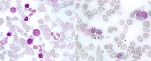
An 84-year-old woman with a long-standing history of progressive exertional dyspnea and fatigue was assessed for concomitant anemia. She also had coronary artery disease, chronic obstructive pulmonary disease, hypertension, and gout. Her hemoglobin was 109 g/L with a mean corpuscular volume of 76.1 fL and red blood cell distribution width (RDW) at 19.4%. White cell count including differential and platelets were within normal limits. Hypochromia and microcytosis were noted on the peripheral smear as well as an occasional hypogranular granulocyte. The serum ferritin was 18 ng/L. A bone marrow examination showed no iron and mild erythroid dysplasia (left). An extensive work-up for blood loss was negative.
Iron infusions were administered monthly on 3 occasions with correction of microcytosis, but with a persistence of normocytic anemia, and a further elevation of RDW to 26%. The peripheral smear showed more dysplasia of the granulocytes. Seven months after the initial bone marrow, a repeat examination revealed the presence of abundant iron stores, prominent ring sideroblasts (right), and multilineage dysplastic changes.
This case highlights an example wherein iron deficiency masked the appearance of underlying ring sideroblasts in a patient with evolving myelodysplasia.
Many Blood Work images are provided by the ASH IMAGE BANK, a reference and teaching tool that is continually updated with new atlas images and images of case studies. For more information or to contribute to the Image Bank, visit http://imagebank.hematology.org.


This feature is available to Subscribers Only
Sign In or Create an Account Close Modal