Abstract
Donor lymphocyte infusion is an alternative treatment for Epstein-Barr virus (EBV)–associated lymphoproliferative disorders (LPDs) but with risk of graft-versus-host diseases (GVHDs). According to the fetal-maternal microchimerism tolerance, we assumed that maternal lymphocyte infusion may be effective without causing GVHD. In 54 cases when a child required cytotherapy or hematopoietic stem cell transplantation, we studied the mother for child-mother microchimerism with use of insertion-deletion polymorphisms as allogeneic markers and a combination of nested polymerase chain reaction (PCR) and real-time quantitative PCR. Thirteen mothers were child-microchimerism–positive at the ratio of 10−5-10−3. Among them, 5 children had non–transplant-associated, EBV+ T-cell LPD. In these 5 cases, high doses of human leukocyte antigen–haploidentical maternal peripheral blood mononuclear cells (> 108/kg/infusion) were infused 1-4 times. Symptoms of all 5 patients improved between 3 and 10 days after the infusion; thereafter, 3 cases showed complete remission for 6-18 months without further therapy and 2 had partial remission. During the period of observation, none developed obvious GVHD. By quantitative PCR, in some patients maternal cells were found to be eliminated or decreased after infusions, indicating existence of host-versus-graft reaction. We suggest that high doses of mother's lymphocyte infusion may be an effective and safe treatment for non–transplant-associated EBV+ T-cell LPD.
Introduction
Greater than 90% of humans are infected with the Epstein-Barr virus (EBV), and the infection persists for life. Most persons have a chronic asymptomatic infection with EBV, but the virus has been associated with several malignancies and can infect B cells, T cells, natural killer cells, and epithelial cells. Patients with iatrogenic, congenital, or acquired immunodeficiency are at increased risk of EBV-associated lymphomas, which are in nearly all instances of B-cell lineage. In patients without known immunodeficiency, chronic active EBV (CAEBV) disease has been defined as a systemic EBV-positive lymphoproliferative disease (LPD) characterized by fever, lymphadenopathy, and splenomegaly that develops after primary virus infection.1 The subtype of T-cell origin CAEBV has a strong racial predisposition, with most cases occurring in Asians and some cases in Native American populations in the Western hemisphere from Mexico, Peru, and Central America. It is rare in whites and African Americans.2 In the 2008 World Health Organization (WHO) classification, the systemic EBV-positive T-cell LPD of childhood (a clonal T-cell LPD) has been classified into the group of malignant lymphomas.3 In China, several therapies such as antiviral agents (acyclovir, ganciclovir), immunomodulators (interferon-γ), chemotherapy (etoposide, dexamethasone) have been tried for EBV-positive T-cell LPD of childhood without satisfactory curative effect. Alternative approaches that used adoptive immunotherapies sometimes offer attractive results in some patients. EBV may be the target for immunotherapy because the disease is usually associated with CAEBV.4,5 In fact, the use of donor lymphocyte infusion (DLI) has significantly improved the outcome of malignant EBV-LPD especially among patients with posttransplantation lymphoproliferative disorder, of whom most are associated with EBV infection.6,7 The rationale for DLI may be that EBV-seropositive persons contain EBV-specific T-cell precursors in their body, so that DLI facilitates to restore the immune responses to EBV, or that donor's lymphocytes can recognize and eliminate malignant tumor cells.8,9 Transfusion of unmanipulated allogeneic T cells had significantly improved the outcome of malignant EBV-associated lymphomas.10-12 However, donor lymphocytes also contain many alloreactive T cells that induce graft-versus-host disease (GVHD).13 The risk of developing acute GVHD correlates with the dose of donor's T cells as well as donor-recipient human leukocyte antigen (HLA) compatibility.5 Donor's T cells < 107 cells/kg usually do not induce GVHD, but they also do not have significant antitumor activity.14 Acute GVHD is found in ∼ 10% of patients receiving T cells 107/kg, in 20%-30% of patients receiving T cells 3-5 × 10⁷/kg, and in ≥ 50% of patients receiving T cells ≥ 108/kg.15 However, there is a statistically strong relationship between the development of GVHD and the induction of favorable responses. Collins et al16 had reported that 42 of the 45 complete responders (93%) developed acute GVHD, and 36 of the 41 assessable complete responders (88%) developed chronic GVHD.15 Although DLI has been confirmed to be effective in EBV-LPD, the obstacle to this therapy is GVHD due to HLA incompatibility between donor and patient. Subsequently, the use of DLI for the treatment of EBV-LPD is still rare to date.
We have known that a small number of maternal blood cells exist in the newborn's blood, and, in turn, blood cells derived from the offspring can be detected in the mother's blood long after labor.17 This phenomenon is referred to as fetal-maternal microchimerism, suggesting the presence of immunologic tolerance between the mother and her offspring. Recent experimental evidences show the association between microchimerism and acquired immune hyporesponsiveness, which is useful for solid-organ transplantation and hematopoietic stem cell transplantation (HSCT).18,19 The long-term child-maternal microchimerism as a form of acquired child-maternal tolerance was first proposed by Tokita et al.20 They reported the dramatic regression of thymic carcinoma in a mother who received an infusion of peripheral blood stem cells from her HLA-haploidentical daughter without any signs of GVHD. Microchimerism of donor's alleles in the recipient were detected before and long after the infusion, prompting them to speculate that the mother had been tolerant to the inherited paternal antigens (IPAs); thus, the infused effector lymphocytes from her daughter survived longer in the face of the maternal immune system. Soon thereafter, Ochiai et al21 reported a case of non–T cell–depleted mother-to-child HSCT from his microchimeric mother. Although the mother and son had serologic mismatches at HLA-A, -B, and -DR in the graft-versus-host direction, acute GVHD was restricted to skin and rapidly improved after standard therapy. This approach was soon found to be feasible by several groups.22,23 The favorable role of fetal-maternal immunologic tolerance in allogeneic HSCT was recently shown. Maternal stem cell donation for HSCT was better than paternal donation, based on the results of a nationwide HSCT survey conducted in Japan.24 van Rood et al25 also showed that the recipients of non–T cell–depleted maternal transplants had a significantly lower incidence of chronic GVHD than the recipients of paternal transplants in haploidentical 1- or 2-antigen–mismatched transplantations. These observations also support the hypothesis that the microchimeric mother may be hyporesponsive to IPAs of her offspring.
We assumed that lymphocyte infusion from a HLA-haploidentical mother would have a better effect to non–transplantation-associated EBV-LPD of her offspring. The prerequisite for this therapy is the presence of microchimerism to IPAs in mother's blood long after the labor and the resultant mother-child immunologic tolerance. The level of child-maternal microchimerism is low. Detection of child-maternal microchimerism requires methods with higher sensitivity.26,27 So we developed some methods that use allogeneic markers to raise the sensitivity. We found some mothers had child-maternal microchimerism certainly. Five patients who had non–transplantation-associated EBV-positive T-cell LPD were chosen to be treated with infusion of high doses of their IPA-microchimerism–positive, HLA-haploidentical mother's lymphocytes. During the observation period, none of them developed obvious GVHD.
Methods
Patients and donors
Fifty-four pairs of patients with acute leukemia or lymphoma 5-31 years of age and their mothers as the donors for cytotherapy or HSCT were examined for HLA typing with the use of the HLA sequence-based typing technology and microchimerism of IPAs in the mother's blood. Among them, 5 patients in the age range of 6-14 years had their condition diagnosed as EBV-positive T-cell LPD in childhood. Three of them had accepted etoposide, glucocorticoid, or antiviral therapy in the past but without remission (Table 1). Quantification of EBV DNA with the use of real-time quantitative polymerase chain reaction (PCR) in these 5 patients' plasma and titer of anti-EBV antibodies in their mothers' sera was performed. These 5 patients were treated with high doses of DLI from their mothers at the Department of Hematology, Peking University First Hospital, from August 2008 to February 2010. The treatment protocols were approved by the Ethics Committee of Peking University First Hospital, and written consent was obtained from the parents in accordance with the Declaration of Helsinki.
Symptoms and pathologic characteristics of the 5 patients
| Case . | Age, y/sex . | Symptoms . | Biopsy . | Previous therapy . |
|---|---|---|---|---|
| 1 | 10/M | Fever, skin lesion, lymphadenopathy, hepatosplenomegaly | Skin and lymph node biopsies showed diffuse infiltration of atypical lymphocytes. Immunohistochemistry staining showed tumor cells expressing CD3+, CD8+, CD20−, Ki-67+ 60%, CD56−, CD68+, CD30−, Gram-B−. In situ hybridization was EBER+. | Acyclovir for > 6 mo |
| 2 | 6/M | Fever, skin lesion, lymphadenopathy, hepatosplenomegaly | Skin biopsy showed diffuse infiltration of atypical T lymphocytes. Immunohistochemistry staining showed tumor cells expressing CD3+, CD20light+, Gram-B−, CD56−, TIA-1+, CD68+. In situ hybridization was EBER+. | IFN-α for 10 months; prednisone for ∼ 6 mo |
| 3 | 14/M | Fever, lymphadenopathy, hepatosplenomegaly | Liver biopsy showed infiltration of atypical T lymphocytes. Immunohistochemistry staining showed tumor cells expressing CD3+, CD20−, CD2+, CD56light+, CD5+, CD7+, CD8+, CD4light+, Gram-B−, Ki-67+ 10%-30%, CD79a−. In situ hybridization was EBER+. | No specific treatment |
| 4 | 7/M | Fever, parotid gland swelling | Parotid gland biopsy showed infiltration of atypical lymphocytes. Immunohistochemistry staining showed tumor cells expressing CD3+, CD4+, Granzyme B+, CD20−, CD56−, CD8− Ki-67+ 20%. In situ hybridization was EBER+. | No specific treatment |
| 5 | 13/M | Fever, lymphadenopathy, hepatosplenomegaly, rhinopolypus | Rhinopolypus biopsy showed infiltration of atypical lymphocytes. Immunohistochemistry staining showed tumor cells expressing CD3+/−, Granzyme B+, CD20−, CD56+, CD8+/−, Ki-67+ 30%. In situ hybridization was EBER+. | Intermittent antiviral therapy for ∼ 4 y; etoposide and prednisone for ∼ 1 y |
EBER indicates Epstein-Barr virus–encoded early RNAs; TIA-1, T-cell intercellular antigen-1; and IFN-α, interferon-α.
IPA in mother's blood and the survival of mother's lymphocytes in patient's blood
| Case . | Mother's PBMCs infused per infusion, ×108/kg . | IPAs in mother's blood* . | Ratio of NIMAs in patient's blood before the infusion* . | Ratio of NIMAs in patient's blood after the infusion* . |
|---|---|---|---|---|
| 1; 4 infusions in total | 2 | SRY+; | Negative | 4 days after the first infusion: 10−3.9; |
| 1.3 | 10−5.15 by quantification | 8 days after the first infusion: none | ||
| 1.0 | ||||
| 1.1 | ||||
| 2; 4 infusions in total | 6.8 | SRY+; | † | † |
| 5.7 | 10−4.12 by quantification | |||
| 3.6 | ||||
| 4 | ||||
| 3; 2 infusions in total | 1.1 | SRY+; | 10−3.13 | 5 days after the first infusion: 10−2.84; |
| 1.0 | † | 13 months after the first infusion: 10−4.92 | ||
| 4 | 3.5 | SRY+; 10−4.67 by quantification | † | † |
| 5 | 1.7 | SRY+; † | 10−2.33 | 4 days after the first infusion:10−2.46; 42 days after the first infusion:10−2.61 |
Quantification of microchimerism by specific insertion/deletion polymorphism method.
We used 9 insertion/deletion polymorphisms, trying to identify the polymorphism specific for IPAs or NIMAs, but no such insertion/deletion polymorphism was found. Therefore, quantification of microchimerism could not be conducted.
Evaluation of microchimerism of IPAs in mother's blood
The amounts of inherited paternal antigens (IPAs) in mother's blood and noninherited maternal antigens (NIMAs) in patient's are rare, for which the commercially available kit on the basis of short tandem repeat polymorphism clinically is used for chimerism status after stem cell transplantation is unsuitable because of its limited detection capacity (> 1%), and single nucleotide polymorphisms commonly used for allele identification are sometimes nonspecific. So we combined nested PCR and real-time quantitative PCR to amplify several small insertion/deletion polymorphisms for measurement of microchimerism ratio of IPAs in the mother's blood and NIMAs in the patient's blood. We first searched for polymorphisms of 4-6 nucleotide insertion or deletion (InDel) with an appropriate heterozygosity rate for the Chinese population in the human genome database, arbitrarily selected 9 InDel polymorphisms, and used PCR and capillary electrophoresis of the PCR products to find out the InDel polymorphism that was heterozygous to the patient and homozygous to the patient's mother to identify IPA in mother's blood (see supplemental data for details, available on the Blood Web site; see the Supplemental Materials link at the top of the online article).
For male patients, nested PCR for the amplification of the SRY gene on the Y chromosome was used to identify IPAs in mother's blood (see supplemental data for details).
Mother's lymphocyte infusion
Peripheral blood mononuclear cells (PBMCs) from the mother were collected with the use of a blood component apheresis separator and were immediately infused into her child. The cell number infused was < 107 PBMCs/kg for the first time, then increased to > 108 PBMCs/kg for the next infusions.
Evaluation of transfused mother's lymphocytes by determination of NIMAs in patient's blood
We used 9 InDel polymorphisms that have the approriate heterozygosity rate for the Chinese population to select the suitable loci that were homozygous to the patient but heterozygous to the mother as the mother-specific marker to identify NIMAs in patient's blood after the mother's lymphocyte transfusion (supplemental data).
Results
Microchimerism of IPAs in blood samples of 54 mothers
Fifty-four mother-child pairs were evaluated, including the 5 pairs that we treated with the infusion of the mother's lymphocytes in this study. With the use of the InDel polymorphisms as the allogeneic markers, 13 mothers were found to have the microchimerism of IPAs in the ratio of 10−5 to ∼10−3. The microchimerism could not be identified in 8 mother-child pairs because of the same genotypes of the 9 InDel polymorphisms between mother and child. We did not try other InDel polymorphisms for the identification. Therefore, the rate of IPA microchimerism in mothers is ∼ 28%.
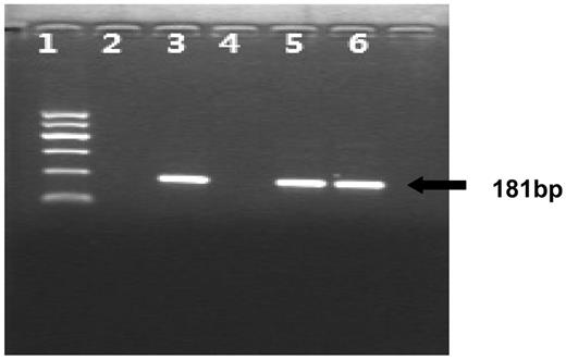
Because the 5 patients with EBV-positive LPD were all boys, the SRY gene in the sex-determining region on the Y chromosome can be used as the offspring's marker. All of these 5 mothers were positive for the SRY gene. Meanwhile, among our 36 son-mother pairs, 13 were positive for the SRY gene; the rate was 36% (Figure 1).
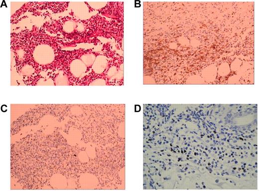
Pathologic features of the skin biopsy from patient 2. (A) Atypical T lymphocyte infiltration (hematoxylin-eosin stain; original magnification 20×10). (B) Immunohistochemistry stain shows tumor cells expressing CD3 (original magnification 20×10). (C) Immunohistochemistry stain shows tumor cells expressing TIA-1 (original magnification 20×10). (D) In situ hybridization of EBV-RNA.
Pathologic features of the skin biopsy from patient 2. (A) Atypical T lymphocyte infiltration (hematoxylin-eosin stain; original magnification 20×10). (B) Immunohistochemistry stain shows tumor cells expressing CD3 (original magnification 20×10). (C) Immunohistochemistry stain shows tumor cells expressing TIA-1 (original magnification 20×10). (D) In situ hybridization of EBV-RNA.
Clinical characteristics of the 5 mother-child pairs
The diagnosis of EBV-positive T-cell LPD of the 5 children is based on the 2008 WHO International Working Formulation for Lymphoma Classification.2 All of the 5 children had had fever > 6 months, accompanied with lymphadenopathy, hepatosplenomegaly. Two children showed skin lesion, and one patients had swelling of the parotid gland. Plasma EBV-DNA was positive in all 5 patients, with the titer of > 4 × 10⁴ copies/mL. The diagnosis was confirmed by pathologic examination of tissue biopsies. In situ hybridization showed positive EBV-encoded early RNAs in the tumor cells of the biopsies (Figure 2D; Table 1).
Nested PCR for the identification of the SRY gene in the mothers. (Lane 1) Molecular weight marker; (lane 2) without template (negative control); (lane 3) DNA sample from a male (positive control); (lane 4) DNA sample from a female without pregnancy (negative control); (lane 5) DNA sample from the mother of patient 1; (lane 6) DNA sample from the mother of patient 2.
Nested PCR for the identification of the SRY gene in the mothers. (Lane 1) Molecular weight marker; (lane 2) without template (negative control); (lane 3) DNA sample from a male (positive control); (lane 4) DNA sample from a female without pregnancy (negative control); (lane 5) DNA sample from the mother of patient 1; (lane 6) DNA sample from the mother of patient 2.
All 5 patients' mothers were accepted as the donors because of the presence of child-microchimerism shown as the positive SRY gene and polymorphisms of child-specific alleles (Figure 3). They were healthy and seropositive for EBV. We also examined 4 HLA loci (HLA-A, HLA-B, HLA-Cw, and HLA-DRB1), which are important to the HLA compatibility for HSCT. They had mismatch of 2 (patient no. 1) or 3 (patients no. 2 and no. 4) or 4 (patients no. 3 and no. 5) loci (Table 3).
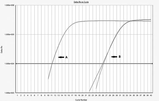
Detection of child microchimerism by QT-PCR. (A) The mother's allele, or major allele, has low cycle number corresponding to its comparatively high DNA concentration. (B) The minor allele or microchimeric cell population has a much higher cycle number because the minor DNA exists at a much lower concentration than the major allele. The method is to calculate the change in cycle threshold (ΔCT) to show the ratio of the minor population to the major population.
Detection of child microchimerism by QT-PCR. (A) The mother's allele, or major allele, has low cycle number corresponding to its comparatively high DNA concentration. (B) The minor allele or microchimeric cell population has a much higher cycle number because the minor DNA exists at a much lower concentration than the major allele. The method is to calculate the change in cycle threshold (ΔCT) to show the ratio of the minor population to the major population.
HLA compatibility of the 5 child-mother pairs
| Child-mother pairs . | HLA-A locus . | HLA-B locus . | HLA-DRB1 locus . | HLA-Cw locus . | ||||
|---|---|---|---|---|---|---|---|---|
| Case 1 | ||||||||
| Patient | 0207 | 0207 | 4601 | 4601 | 0901 | 14BCAD* | 0102 | — |
| Mother | 0207 | 1101 | 4601 | 4601 | 0901 | 12DUKV† | 0102 | — |
| Case 2 | ||||||||
| Patient | 0301 | 0207 | 4001 | 4601 | 1202 | 0803 | 0702 | 0102 |
| Mother | 0301 | 3308 | 4001 | 5401 | 1202 | 0405 | 0702 | 0102 |
| Case 3 | ||||||||
| Patient | 3201 | 1101 | 4403 | 5101 | 0701 | 1602 | 0401 | 1402 |
| Mother | 3201 | 2402 | 4403 | 4002 | 0701 | 1403 | 0401 | 0304 |
| Case 4 | ||||||||
| Patient | 3101 | 0201 | 1518 | 5101 | 1403 | 1501 | 0704 | 1402 |
| Mother | 3101 | 1101 | 1518 | 1502 | 1403 | 1501 | 0704 | 0801 |
| Case 5 | ||||||||
| Patient | 2402 | 3303 | 5101 | 4403 | 1602 | 0701 | 1402 | 0701 |
| Mother | 2402 | 1101 | 5101 | 1301 | 1602 | 1602 | 1402 | 0304 |
14BCAD>DRB1*1401/1454.
12DUKV>DRB1*1201/1206/1210/1217.
Complete clinical remission in 3 cases and partial clinical remission in 2 cases after of mother's lymphocyte infusion
We collected PBMCs from mothers and infused them ino the patients. At the beginning, the number of cells infused was < 107 PBMCs/kg per infusion without any side effects. We then increase the number of cells to > 108 PBMCs/kg per infusion. The infusions were performed for 1-4 times for a patient (Table 2). Clinical improvement was achieved in all of the patients between 3 and 10 days after the initial infusions, with remarkable reduction of lymph nodes, liver, and spleen. Plasma EBV-DNA was undetectable or reduced remarkably in all of the patients. Clinical remission maintained for 6-18 months without further therapy except 2 patients who had recurrence of lymphadenopathy and fever ∼ 2 months after the first infusion. One of them had received high doses of his mother's lymphocytes 4 times but could not get complete remission; he did not continue the therapy. The other patient had prepared for HSCT (Table 4).
Clinical improvement and the change of EBV-DNA copies in the patients
| Case . | Clinical outcomes . | Plasma EBV-DNA copies/mL in patients' before infusion . | EBV-DNA copies in patients' 15-30 d after the first infusion . |
|---|---|---|---|
| 1 | Lymph nodes in his neck became smaller after 3-5 days. Skin rashes decreased, and the temperature was back to normal. Two months after the first infusion, the fever and lymphadenopathy recurred; he then received another 3 infusions from his mother. Lymph nodes became smaller within a week after each infusion, but intermittent fever and lymphadenopathy happened 1-2 months after each infusion. He only got a partial remission and did not continue the therapy. | − | − |
| 2 | His rashes and lymphadenopathy improved in a week after the first infusion. Low-grade intermittent fever lasted for 2 months. He received another 3 infusions from his mother, the fever stopped, rashes disappeared, and no lymphadenopathy occurred. After 17 months, he has achieved complete remission | 6.0 | − |
| 3 | Abnormal liver function disappeared. Spleen became unpalpable. Ultrasonography showed reduction of liver and spleen sizes. Low-grade intermittent fever lasted for 3 months. Thirteen months later, he remained afebrile. He had a complete remission. | 8.0 | − |
| 4 | Parotid gland became smaller a week after the infusion; intermittent fever lasted for ∼ 1-2 months. Ten months after the infusion, he remained afebrile with good health and impalpable parotid gland. | 2.0 | No data |
| 5 | The temperature returned to normal 3 days after the infusion, and the ultrasonography showed reduction of liver and spleen sizes within 2 weeks. Two months later, the fever and hepatosplenomegaly recurred. He then prepared for HSCT. | 6.7 | − |
– indicates EBV-DNA was undetectable; and no data, no test had been performed.
Survival of mother's lymphocytes in child's blood
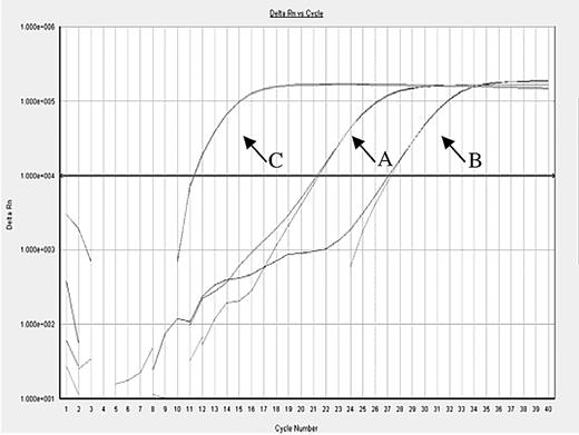
Appropriate InDel polymorphisms to examine the amount of NIMA in child's blood before and after the infusion could only be found in 3 patients (Table 2). In patient no.1, the ratio of mother's cells was undetectable before infusion and increased only to 10−3.9 4 days after the first infusion but was undetectable 8 days after the infusion. In patient no.3, the ratio was 10−3.13 before the infusion, increased only to 10−2.54, and then decreased to 10−4.92 after 13 months (Figure 4). In patient no.5, the ratio was 10−2.33 before the infusion and 10−2.61 42 days after the infusion.
Monitor the infused mother's lymphocyte by QT-PCR in patient 3. (A) The mother's allele before lymphocyte infusion. (B) The mother's allele 13 months after lymphocyte infusion. (C) The child's allele or the major allele.
Monitor the infused mother's lymphocyte by QT-PCR in patient 3. (A) The mother's allele before lymphocyte infusion. (B) The mother's allele 13 months after lymphocyte infusion. (C) The child's allele or the major allele.
Discussion
EBV is a successful member of the herpesvirus family, infecting > 90% of the world's adult population. Like all herpesviruses, EBV is able to persist in the host for life, but in most healthy carriers the virus causes no disease.4 This is because a delicate balance is maintained between the host immune system, which limits production of virus particles, and the virus, which persists and is successfully transmitted in the face of host antiviral immunity. Disruption of this balance, resulting from primary or acquired immunodeficiency, may lead to the development of EBV-associated disease.
Because of the WHO 2008 International Working Formulation for Lymphoma Classification,3 all 5 conditions were diagnosed as EBV-positive T-cell LPD in childhood. Among them, 2 patients had accepted etoposide or glucocorticoid in the past, some had accepted antiviral agents (acyclovir) or immunomodulators (interferon-α) for 6 months or 4 years without satisfactory curative effect. It is difficult for those patients with these treatments. We had treated some patients with EBV-LPD with cytotoxic T-lymphocytes in the past, mostly in patients with posttransplantation lymphoproliferation diseases, some of them had encouraging results.28-30 But in some other patients with EBV-LPD patients, because they lack expression of immunodominant viral antigens and because many tumor-associated target antigens are functionally weak stimulators of the immune response, it had proved harder to exploit the equivalent promise of infused T lymphocytes, and lack of T helper (CD4+) cell function in vivo may lead to lack of in vivo expansion.12,31,32 So our overall response rate in these kinds of patients has still been low. Many researchers had shown that DLI could significantly improve the outcome of EBV-LPD,5-7 but the main problem was GVHD. So in the past, we chose to use a HLA-identical donor. The existence of child-mother microchimerism suggests the presence of immunologic tolerance between a mother and her offspring. We assumed that a mother's lymphocyte infusion could benefit EBV-LPD without obvious GVHD.
We confirmed that the child's microchimerism would persist in the mother's blood long after labor. To confirm if child-mother microchimerism is a common phenomenon, a highly sensitive assay is needed to detect microchimerism. The level of fetal-maternal microchimerism was low (a fraction concentration of 10−5 to 10−4)17 ; analysis of microchimerism requires highly sensitive assays able to detect specific minor populations in the context of a high background of allogeneic cells. Therefore, an allogeneic marker unique to the chimeric cell population and an analytically sensitive assay are needed. The usual method, such as short tandem repeat PCR to detect microchimerism after HSCT, can only reach the sensitivity of 10−2.33 Nested PCR of SRY is highly sensitive with the detection level of 1 male cell among 106 female cells in these mothers; however, it cannot be used for quantitation.34 InDel polymorphisms are common in the population.35 IPAs can be detected in mother's blood when the child is heterozygous for both alleles of InDel polymorphisms while the mother is homozygous for one of them.36 We set up the assay of combining nested PCR with real-time quantitative PCR to improve the sensitivity to a level of 10−6 to 10−5 while quantifying the microchimerism. Among the 54 child-mother pairs we had chosen for detecting microchimerism, 13 mothers were child-microchimerism–positive with the level of ∼ 10−5 to 10−3.
We experimentally treated 5 patients who had EBV-positive T-cell LPD. All these patients' mothers had their child's microchimerism to a level of 10−5 to 10−4, which indicated that these mothers would tolerate their child's IPAs. These patients were infused with large doses of mother's lymphocyte > 1 time. Clinical improvement was achieved in all of the patients between 3 and 10 days after the first infusion; the body temperature was back to normal with remarkable reduction of lymph nodes, liver, and spleen sizes. Plasma EBV DNA became undetectable or reduced remarkably in all of the patients. The infusions were effective, and recurrence of lymphadenopathy and fever was found in 2 patients 2 months after the first infusion. One of them had received high doses of his mother's lymphocytes several times, but could not get complete remission. During the observation period, none of the patients developed obvious GVHD. These 5 mothers were HLA-haploidentical. So the question is whether the existence of microchimerism benefits the GVHD.
To study how the mother's lymphocyte survived in the child's blood and how these allogenic lymphocytes developed antitumor effect, we used InDel polymorphism loci to detect the mother's cell in child's blood. It can only be done when the child is homozygous and the mother is heterozygous. Because only 3 of the child-mother pairs got suitable InDel loci for such detection, we had monitored the quantitation of mother's cells in these 3 children's body. One patient had no mother's microchimerism before the infusion, the maternal-microchimerism existed < 1 week after the infusion. Another 2 patient had their mothers' microchimerism before the infusion, one of them had decreased maternal-microchimerism after infusion, whereas the other had no obvious change. Our research showed that maybe the mother's lymphocyte could help the patients to restore the immune responses to EBV-associated tumor but not kill tumor cells directly. Meanwhile, our results also showed that large doses of haploidentical donor lymphocytes would arouse host-versus-graft reaction in immunocompetent patients. The mechanism is still unclear.
Our research showed that high doses of mother's lymphocyte infusion may be an effective and safe treatment for EBV-positive T-cell LPD of childhood.
The online version of this article contains a data supplement.
The publication costs of this article were defrayed in part by page charge payment. Therefore, and solely to indicate this fact, this article is hereby marked “advertisement” in accordance with 18 USC section 1734.
Acknowledgments
We thank Ying Zhang of the Hematology Laboratory of Peking University First Hospital for research coordination, Dr Dingfang Bu from the Center Lab of Peking University First Hospital for expert technical assistance, and Dr Peng Cai and his staff of Beijing Daopei Hospital for assistance in donor lymphocyte preparation and quality assurance.
This work was supported by the International Cooperative Medical Research Project from National Ministry of Science and Technology of China (2006 DFB 31430).
Authorship
Contribution: Q.W. was a coprincipal investigator on the clinical trial, cared for some of the patients, performed the therapy and molecular biology studies, and wrote this manuscript; H.L. developed the clinical trial and performed molecular biology studies; X. Zhang participated in the clinical study and cared for the patients; Q.L. carried out some molecular biology studies; X. Zhou and Y.X. performed pathologic studies; C.T. contributed to laboratory studies and mother's lymphocyte preparation; and P.Z. proposed this therapy, organized the treatment and related experiments, and reviewed this manuscript.
Conflict-of-interest disclosure: The authors declare no competing financial interests.
Correspondence: Ping Zhu, Department of Hematology of Peking University First Hospital, No. 8 Xishiku St, Xicheng District, Beijing, China 100034; e-mail: zhuping@bjmu.edu.cn.


This feature is available to Subscribers Only
Sign In or Create an Account Close Modal