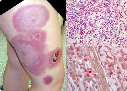
A 12-year-old boy with Fanconi anemia presented with a 3-week history of painless, circular, bluish skin lesions, up to 15 cm in diameter, most prominently seen on the lateral portions of his right upper leg (see figure, top left). He was afebrile with anemia (Hb 90 g/L) and thrombocytopenia (platelets 45 × 103/μL), but a normal leukocyte count. There were no blasts in the peripheral blood smear.
A skin biopsy showed an infiltration with myelomonocytic, POX-positive and NACE-positive blast cells, compatible with acute myelogenous leukemia (AML), French-American-British (FAB) subtype M4 (see figure, right). The bone marrow was hypocellular without evidence of acute leukemia. Treatment resulted in a complete remission, but the patient died with disseminated adenovirus following an allogeneic bone marrow transplantation.
Bone marrow failure and acute leukemia are well-known complications of Fanconi anemia. Extramedullary manifestations of AML may be observed in some subtypes of AML, most commonly FAB M2, but are infrequent in Fanconi anemia.
Many Blood Work images are provided by the ASH IMAGE BANK, a reference and teaching tool that is continually updated with new atlas images and images of case studies. For more information or to contribute to the Image Bank, visit www.ashimagebank.org.


This feature is available to Subscribers Only
Sign In or Create an Account Close Modal