Abstract
Abstract 2935
Chromosomal abnormalities not only demonstrate the clonality of the disease, but also help to predict the outcome of the patients in MDS. While IPSS suggests conventional cytogenetic analysis as a standard method in detecting clonal abnormalities in MDS, FISH is widely used due to its ability to detect minor clones and to quantitate the clonal abnormalities. In this study, we compared the results of conventional cytogenetic analysis and FISH, and investigated their different prognostic impact in MDS.
The study included 129 MDS patients composed of 10 RA, 5 RARS, 15 RCMD, 39 RAEB-1, 33 RAEB-2 and 5 MDS, U (WHO classification, 2001). A standard protocol was used to perform conventional cytogenetic analysis (G-banding). Interphase FISH was performed to detect any abnormalities of chromosomes 5, 7, 8, 20 and 1. The result was analyzed as per the manufacturer¡&hibar;s instructions and recorded according to ISCN (2005) criteria.
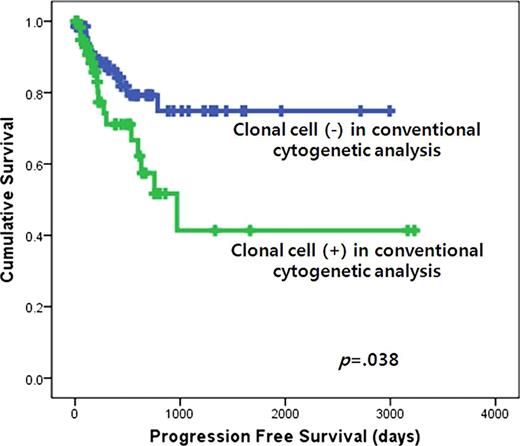
The incidence of −5/5q, −7/7q, +8, −20q and +1q were 13.2%, 14.0%, 19.4%, 7.0% and 7.8%, respectively. Among them, FISH detected occult abnormalities which were not detected in conventional cytogenetic analysis in 1.6%, 3.1%, 5.4%, 1.6% and 5.4%, respectively. Overall, FISH detected occult abnormalities in 18.6% of the patients (24/129), and IPSS grouping was changed in 4.9% (6/129: 1 Low to Int-1; 4 Int-1 to Int-2; 1 Int-2 to High). The abnormalities in −5/5q, −7/7q and +1q were more common with other chromosomal abnormalities, whereas the abnormalities in +8 and −20q were more common as a sole abnormality. The quantity of clonal cells for each chromosome detected in conventional cytogenetic analysis did not correlate with the quantity of clonal cells detected in FISH. The presence of clonal cells either in conventional cytogenetic analysis or in FISH did not correlate with prognostic factors included in IPSS. However, the clonal cell percentage detected in conventional cytogenetic analysis was higher in patients with >5% bone marrow blasts than those with <5% (79.1% vs. 57.9%, p=.013). The clonal cell percentage detected in FISH did not show any relationship with IPSS factors including bone marrow blast percentage. Multivariate analysis showed the presence of clonal cells in conventional cytogenetic analysis were independent prognostic factors in predicting progression free survival into acute leukemia in this patient group (p=.038).
FISH is advantageous in identifying cryptic cytogenetic abnormalities which could be overlooked in conventional cytogenetic analysis, and can change the IPSS risk grouping in MDS patients. The quantity of clonal cells detected in conventional cytogenetic analysis correlates with bone marrow blast percentage, whereas the quantity of clonal cells detected in FISH does not. Blasts possess higher proliferating activity than maturing hematopoietic precursors, thus they are more likely to form metaphase required in conventional cytognetic analysis. In contrast, the FISH results performed on non-dividing, interphase cells might reflect the true quantity of clonality both in blasts and maturing hematopoietic precursors. The relationship between the quantity of clonal cells and bone marrow blast percentage, and the prognostic significance of the presence of clonality suggest the novel diagnostic utility of conventional cytogenetic analysis in MDS.
The presence of clonal cell in conventional cytogenetic analysis is an independent prognostic factor in predicting the progression free survival into acute leukemia in myelodysplastic syndromes.
The presence of clonal cell in conventional cytogenetic analysis is an independent prognostic factor in predicting the progression free survival into acute leukemia in myelodysplastic syndromes.
No relevant conflicts of interest to declare.
Author notes
Asterisk with author names denotes non-ASH members.


This feature is available to Subscribers Only
Sign In or Create an Account Close Modal