To the editor:
Patients suffering from anhidrotic ectodermal dysplasia with immunodeficiency (EDA-ID) fail to activate the canonical nuclear factor-κB (NF-κB) pathway due to mutations in the IKKG (inhibitory κB kinase gamma) gene encoding IKKγ, also known as NEMO (NF-κB essential modulator).1 This results in a combined cellular and humoral immune defect. In a recent study by Luengo-Blanco et al, the authors conclude that NF-κB activity is required for the transcription of the phagocyte nicotinamide adenine dinucleotide phosphate (NADPH) oxidase genes CYBB and NCF1, encoding gp91phox and p47phox, respectively.2 Both proteins are essential components of the NADPH-oxidase complex, and in patients with chronic granulomatous disease (CGD) failure to express these proteins results in a severe immunodeficiency.3 Luengo-Blanco et al base their conclusions on studies with Epstein-Barr virus (EBV)–transformed B-cell lines from patients diagnosed with EDA-ID as well as studies with pharmacologic inhibitors of NF-κB activation in the monocytic cell line U937 and repressor transfected U937 cells. In these model systems, expression of gp91phox and p47phox was severely impaired, resulting in a failure to activate the NADPH oxidase upon stimulation with phorbol 12-myristate 13-acetate (PMA), similar to EBV-transformed B cell lines from CGD patients.2
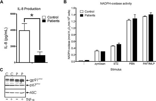
Recently, we had the opportunity to perform functional tests with primary neutrophils from 3 different EDA-ID patients. The patients were 1, 2 and 5 years of age at the time of investigation. The second patient was the brother of the third. All 3 patients had variable degrees of dermatitis and minor peripheral lymphadenopathy since birth, as well as recurrent bacterial infections, and had the pale, sparse hair and conical incisors typical for EDA-ID patients.1 The genetic defects were all identified in the IKBKG gene resulting in a stop codon (p.Q365X) in exon 9 (c.1093C>T) in one patient and a missense mutation (p.Q205P) in exon 5 (c.614A>C) in the other 2 patients. NF-κB activation was severely impaired in all 3 patients, as demonstrated in Figure 1A by a significantly reduced production of interleukin-8 (IL-8), when determined by ELISA after overnight stimulation of the isolated neutrophils with lipopolysaccharide (LPS, 10 ng/mL) and lipid binding protein (LBP, 50 ng/mL). However, in contrast to the results from Luengo-Blanco et al, NADPH-oxidase activity was completely normal in the neutrophils of these EDA-ID patients upon stimulation with various stimuli, as shown in Figure 1B. In addition, expression of both gp91phox and p67phox were found to be relatively normal in the neutrophils of the patients (Figure 1C). This suggests that, at least in primary neutrophils, NEMO-dependent NF-κB activation is not required for the expression and function of the NADPH oxidase. Instead, the recurrent bacterial infections in our EDA-ID patients seem to be due to the poor antibody reactivity against (encapsulated) pathogens (not shown),1 possibly combined with a failure to produce proinflammatory cytokines to recruit phagocytes to the site of infection (Figure 1A).
Neutrophils from EDA-ID patients display normal NADPH-oxidase activity. Neutrophils were isolated from heparinized peripheral blood of patients and controls over isotonic Percoll, as described.4 (A) Cells were cultured overnight in HEPES medium (132 mM NaCl, 20 mM HEPES, 6 mM KCl, 1 mM MgSO4, 1.2 mM K2HPO4, 1 mM CaCl2, 5 mM glucose and 2% (vol/vol) human serum albumin, pH 7.4) in the presence of 10 ng/mL LPS and 50 ng/mL LBP at a concentration of 5 × 10⁶/mL, after which the IL-8 concentration was determined in the supernatant by ELISA, according to the manufacturer's instructions (Sanquin Reagents, Amsterdam, The Netherlands). Data represent the means (± SEM) of 3 independent experiments performed in duplicate. Statistical significance was determined by a Student t test. *P = .0173. (B) NADPH-oxidase activity was assessed as hydrogen peroxide production determined by an Amplex Red kit (Molecular Probes). Neutrophils (106/mL) were stimulated with 1 mg/mL bare or serum-treated zymosan (STZ), 100 ng/mL phorbol myristate acetate (PMA) or PAF + fMLP (formyl-methionyl-leucylphenylalanine; both 1 μM, added simultaneously), in the presence of 0.5 μM Amplex Red and 1 U/mL horseradish peroxidase, as described.5 Fluorescence was measured at 30-second intervals for 20 minutes on a Spectra Fluor Plus spectrophotometer (Tecan, Zürich, Switzerland). Maximal slope of H2O2 release was assessed over a 2-minute interval. Data represent the means (± SEM) of 3 independent experiments performed in duplicate. No significant difference was found between patient and control cells. (C) Expression of gp91phox and p67phox was assessed on Western blot. Freshly isolated neutrophils of a healthy control donor (C) or an EDA-ID patient (P) were stimulated for 2 hours with 10 ng/mL TNF-α (+) or left unstimulated (−), as indicated. Afterward, cell lysates were prepared and analyzed on Western blot for the indicated targets. Cells (1.5 × 10⁶) were loaded in each lane and, in addition, ASC expression is shown as a loading control. Mouse anti-gp91phox (clone 48) was obtained from Sanquin Reagents, rabbit anti-p67phox was obtained from Upstate Biotechnology (Lake Placid, NY), mouse anti-ASC was obtained from MBL International (Woburn, MA). IRDye680 or 800CW conjugated secondary antibodies were obtained from LI-COR Biosciences (Lincoln, NE). The blots were analyzed with an Odyssey Infrared Imager (LI-COR).
Neutrophils from EDA-ID patients display normal NADPH-oxidase activity. Neutrophils were isolated from heparinized peripheral blood of patients and controls over isotonic Percoll, as described.4 (A) Cells were cultured overnight in HEPES medium (132 mM NaCl, 20 mM HEPES, 6 mM KCl, 1 mM MgSO4, 1.2 mM K2HPO4, 1 mM CaCl2, 5 mM glucose and 2% (vol/vol) human serum albumin, pH 7.4) in the presence of 10 ng/mL LPS and 50 ng/mL LBP at a concentration of 5 × 10⁶/mL, after which the IL-8 concentration was determined in the supernatant by ELISA, according to the manufacturer's instructions (Sanquin Reagents, Amsterdam, The Netherlands). Data represent the means (± SEM) of 3 independent experiments performed in duplicate. Statistical significance was determined by a Student t test. *P = .0173. (B) NADPH-oxidase activity was assessed as hydrogen peroxide production determined by an Amplex Red kit (Molecular Probes). Neutrophils (106/mL) were stimulated with 1 mg/mL bare or serum-treated zymosan (STZ), 100 ng/mL phorbol myristate acetate (PMA) or PAF + fMLP (formyl-methionyl-leucylphenylalanine; both 1 μM, added simultaneously), in the presence of 0.5 μM Amplex Red and 1 U/mL horseradish peroxidase, as described.5 Fluorescence was measured at 30-second intervals for 20 minutes on a Spectra Fluor Plus spectrophotometer (Tecan, Zürich, Switzerland). Maximal slope of H2O2 release was assessed over a 2-minute interval. Data represent the means (± SEM) of 3 independent experiments performed in duplicate. No significant difference was found between patient and control cells. (C) Expression of gp91phox and p67phox was assessed on Western blot. Freshly isolated neutrophils of a healthy control donor (C) or an EDA-ID patient (P) were stimulated for 2 hours with 10 ng/mL TNF-α (+) or left unstimulated (−), as indicated. Afterward, cell lysates were prepared and analyzed on Western blot for the indicated targets. Cells (1.5 × 10⁶) were loaded in each lane and, in addition, ASC expression is shown as a loading control. Mouse anti-gp91phox (clone 48) was obtained from Sanquin Reagents, rabbit anti-p67phox was obtained from Upstate Biotechnology (Lake Placid, NY), mouse anti-ASC was obtained from MBL International (Woburn, MA). IRDye680 or 800CW conjugated secondary antibodies were obtained from LI-COR Biosciences (Lincoln, NE). The blots were analyzed with an Odyssey Infrared Imager (LI-COR).
We suggest that the results of Luengo-Blanco et al, which were all obtained from in vitro–cultured model cell lines, can be explained by a failure to differentiate these NF-κB–deficient model cell lines in vitro to fully functional phagocytes or B cells. Primary phagocytes, or at least neutrophils, that differentiate in vivo under different circumstances, do not appear to depend on NF-κB activity for the expression of the NADPH oxidase.
Authorship
Conflict-of-interest-disclosure: The authors declare no competing financial interests.
Correspondence: Bram J. van Raam, Burnham Institute for Medical Research, 10901 North Torrey Pines Rd, La Jolla, CA 92037; e-mail: B.J.vanRaam@gmail.com.


This feature is available to Subscribers Only
Sign In or Create an Account Close Modal