Abstract
Signals mediated by the transforming growth factor-β superfamily of growth factors have been implicated in thymic epithelial cell (TEC) differentiation, homeostasis, and function, but a direct reliance on these signals has not been established. Here we demonstrate that a block in canonical transforming growth factor-β signaling by the loss of Smad4 expression in TECs leads to qualitative changes in TEC function and a progressively disorganized thymic microenvironment. Moreover, the number of thymus resident early T-lineage progenitors is severely reduced in the absence of Smad4 expression in TECs and directly correlates with extensive thymic and peripheral lymphopenia. Our observations hence place Smad4 within the signaling events in TECs that determine total thymus cellularity by controlling the number of early T-lineage progenitors.
Introduction
The thymus provides a specialized environment adept to attract lymphoid precursor cells and to control their survival, expansion, differentiation, and selection to functionally mature T cells, which are ultimately exported to peripheral lymphoid tissues. Within this microenvironment, thymic epithelial cells (TECs) constitute the most abundant stromal component. TECs are arranged both in the cortex and the medulla as a 3-dimensional scaffold and can be subdivided into distinct subpopulations according to functional, structural, and specific antigenic features.1,2 Committed by embryonic day (E) 10.5 to a thymic fate, TEC precursors bud from the ventral endodermal lining of the third pharyngeal pouch and form a separate primordium by E11.5 to E12.5.
The necessary steps in the homing process of hematopoietic precursor cells to the postnatal thymus are initiated in blood vessels at the corticomedullary junction and involve not only the expression of different adhesion molecules on endothelial cells but also the presence of chemokines. Specifically, Ccl21 and Ccl25 have been implicated for this thymic colonization as they are expressed both in the combined fetal thymus/parathyroid anlage and in the postnatal thymus.3–8 Maintenance and proliferation of the most immature lymphoid cells within the thymus microenvironment are regulated by interleukin-7 (IL-7) and stem cell factor (SCF), which use distinct but synergizing signaling pathways to control early stages of T-cell development.9–11
The earliest intrathymic T-lymphoid precursors are known as early T-lineage progenitors (ETPs), display a high CD117 (c-kit) cell surface expression, do not express several lineage-specific markers (designated lineage-negative, Lin−), and are found within the CD44+CD25− subpopulation of immature CD4−CD8− (so-called double-negative [DN]) thymocytes. The developmental potential of ETPs to give rise to B cells, natural killer cells, macrophages, and dendritic cells in culture is progressively lost during further differentiation along the αβ T-cell receptor (TCR) lineage.12,13 The sequential maturation of DN thymocytes (DN1: CD44+CD25−; > DN2: CD44+CD25+; > DN3: CD44−CD25+; > DN4: CD44−CD25−) leads to cells that simultaneously express the coreceptors CD4 and CD8 and are designated double-positive (DP) thymocytes. After the surface expression of a complete αβTCR/CD3 complex and contact with thymic stromal cells, DP cells are subject to positive and negative selection. This process results in the generation of single-positive (SP; CD4+CD8− or CD4−CD8+) thymocytes that are self-tolerant but responsive to foreign antigens.14
Members of the transforming growth factor-β (TGF-β) superfamily of signaling molecules, including TGF-β, bone morphogenetic proteins (BMPs), and activins, regulate essential cellular functions during organ development, such as proliferation, differentiation, apoptosis, morphogenesis, and tissue remodeling.15–17 A direct role for signaling by TGF-β superfamily members has been extensively demonstrated for thymic T-cell maturation,18 although an analogous involvement in TEC differentiation and function remains largely unknown.
The binding of TGF-β superfamily members to their corresponding type II receptor induces the formation of a heterodimeric receptor by recruiting a transmembrane type I receptor chain to the complex. Type II receptor chains are constitutively active serine/threonine kinase receptors that activate the type I receptors through phosphorylation. Activated type I receptor chains propagate signals downstream via phosphorylation of specific receptor-regulated Smad proteins (R-Smads), whereby phosphorylated R-Smads form heteromeric complexes with a single common partner, designated Smad4 (or co-Smad). These complexes translocate to the nucleus where they participate in cooperation with DNA-binding transcription factors and coactivators in the regulation of target gene transcription. Additional proteins either modify ligand binding to receptors (eg, noggin) or alter the cytoplasmic signal transduction by competing for phosphorylated R-Smads (eg, Smad7). The TGF-β family of cytokines and corresponding receptors encompass at least 29 ligands, 5 type II and 7 type I receptor chains, reflecting the striking complexity of this pathway.19–22
To test whether the canonical TGF-β signaling pathway plays a role in TEC development and function, we generated mice that lacked functional Smad4 in TECs. Here we demonstrate that disruption of the canonical TGF-β signaling pathway in TECs results in minor changes of TEC numbers but causes a progressive structural disorganization of the thymic microenvironment. Furthermore, this targeted lack of Smad4 expression results in a dramatic reduction of ETP numbers in the thymus, leading to a proportional decrease in total thymocyte cellularity. These changes in the lymphoid compartment of the thymus, which occur before the gross structural alterations changes in the thymic microenvironment, result in a significant peripheral T-cell lymphopenia.
Methods
Mice
Mice homozygous for LoxP flanked Smad4 exon 9 (Smad4co/co)23 were bred to transgenic mice expressing Cre recombinase under the control of Foxn1 regulatory elements. The F1 generation was interbred to obtain Smad4co/co;Foxn1Cre mice and Smad4co/co control mice. Unless noted otherwise, mice were used between 6 and 10 weeks of age. For developmental staging, the day of the vaginal plug was designated as E0.5. Mice were housed at the Center for Biomedicine's animal facility in accordance with institutional review board approval from the University of Basel and the Cantonal Veterinary Office.
Cell isolation
To obtain TECs, the thymi were sequentially digested 3 times for 15 minutes at 37°C with Hank balanced salt solution containing collagenase D (1 mg/mL; Roche Diagnostics, Basel, Switzerland) and DNase I (2 μg/mL; Roche Diagnostics). After each step, the thymic organoids were gently resuspended, allowed to settle, and the supernatant was collected. During the last step, remaining organoids were vigorously resuspended until a homogeneous cell suspension was achieved. To enrich TECs for cell sorting, the first supernatant was discarded. For TEC quantification, all cell supernatants were collected, counted, and stained. Total TEC numbers were calculated by adding the numbers from all 3 digestion steps. Figure S1 (available on the Blood website; see the Supplemental Materials link at the top of the online article) shows comparable TEC viability isolated from Smad4co/co and Smad4co/co;Foxn1Cre mice. To obtain thymocytes, the thymi were placed between a 40-μm gauze mesh and gently squeezed with bent forceps until all cells were in suspension.
Flow cytometry
Antibodies against CD3ϵ, CD4, CD8, CD44, CD45, CD25, TCRβ, Sca-1, major histocompatibility complex (MHC) II (I-Ab), and c-kit were obtained from eBioscience (San Diego, CA). For lineage staining, biotinylated antibodies against CD3ϵ, CD4, CD8, TCRβ, TCRγδ, NK1.1, DX5, CD11b, CD11c, GR1, Ter119, and CD19 were used followed by phycoerythrin (PE)–Cy7-conjugated streptavidin (all eBioscience). For ETP quantification, CD25 was included in the lineage cocktail. Anti-EpCAM mAb (G8.8) was purified and biotinylated in our laboratory using standard procedures. Flow cytometry was performed on a FACSCalibur and analyzed using Cellquest Pro software (BD Biosciences, San Jose, CA). Cells were sorted on a FACSAria (BD Biosciences), and sort purity routinely was more than or equal to 95%.
BrdU labeling and detection
For the quantification of DN proliferation, mice were injected intraperitoneally 4 hours and 0.5 hours before analysis with 1 mg 5-bromo-2-deoxyuridine(BrdU; Sigma-Aldrich, St Louis, MO) in phosphate-buffered saline (PBS). For the assessment of intrathymic transition kinetics, mice were injected twice intraperitoneally with 1 mg BrdU within 4 hours, and BrdU incorporation was analyzed 4 days later in SP thymocytes. Single-cell suspensions were stained, resuspended in 0.5 mL ice-cold 0.15 M NaCl, and fixed by the dropwise addition of 1.2 mL ice-cold 95% ethanol for 30 minutes on ice. After washing with PBS, the cells were resuspended in PBS containing 1% paraformaldehyde and 0.01% Tween for 30 minutes at room temperature. After washing with PBS, the samples were treated with DNase I (5 μg/mL in 0.15 M NaCl, 4.2 mM MgCl2) for 10 minutes at room temperature. Subsequently, cells were resuspended in PBS containing 5% fetal calf serum and 0.5% Tween, and 10 μL anti-BrdU mAb (BD Biosciences) was added for 30 minutes at room temperature. After a final wash step, cells were analyzed by flow cytometry.
Immunohistochemistry
Thymus lobes were frozen in optimal cutting temperature (OCT) compound (Medite Medizintechnik, Burgdorf, Germany), and sections (8 μm) were blocked in PBS/5% goat serum and stained with a combination of 4′,6-diamidino-2′-phenylindole (DAPI; Sigma-Aldrich) and antibodies against cytokeratin (CK) 5 (Covance; Labforce, Nunningen, Switzerland), CK18 (Progen, Heidelberg, Germany), ERTR7 (provided by Willem van Ewijk, Utrecht, The Netherlands), and Aire (provided by Hamish Scott, Parkville, Australia). Antirabbit Alexa 488, antirat Alexa 555, and streptavidin-Cy5 were used as secondary reagents (Invitrogen, Carlsbad, CA). All images were captured at room temperature in Hydromount (National Diagnostics, Atlanta, GA) on a Zeiss LSM 510 Meta Laser Scanning Confocal Microscope system (10×/0.5 NA, 20×/0.5 NA, and 40×/0.5 NA objectives; Carl Zeiss, Jena, Germany). Overlays of stainings were colored by computer-assisted management of confocal microscopy data using Zeiss LSM 510 software, version 3.2. Hematoxylin-and-eosin–stained sections were viewed on a Nikon Eclipse E600 microscope system with a Nikon 4×/0.13 NA Plan Fluor objective, captured with a Nikon DXM 1200F camera, and processed with Nikon ACT-1 software (Nikon Instruments Europe, Amstelveen, The Netherlands).
RNA isolation and quantitative reverse-transcribed polymerase chain reaction analysis
Five experiments were performed to analyze gene transcription in TECs derived from Smad4co/co and Smad4co/co;Foxn1Cre mice. To obtain sufficient TEC numbers for RNA isolation, thymi from mice of both sexes were and different ages were pooled (experiment 1: 7 female + 7 male Smad4co/co and 9 female + 7 male Smad4co/co;Foxn1Cre mice 4-8 weeks of age; experiment 2: 5 female + 3 male Smad4co/co and 8 female + 4 male Smad4co/co;Foxn1Cre mice 4-6 weeks of age; experiment 3: 4 female + 5 male Smad4co/co and 6 female + 6 male Smad4co/co;Foxn1Cre mice 4-5 weeks of age; experiment 4: 7 female + 4 male Smad4co/co and 8 female + 7 male Smad4co/co;Foxn1Cre mice 4-9 weeks of age; experiment 5: 2 female Smad4co/co and 4 female Smad4co/co;Foxn1Cre mice 5 weeks of age), digested, and TECs were sorted based on MHC class II and CD45 expression. Total RNA was isolated using the RNeasy Micro kit (QIAGEN, Basel, Switzerland), and Oligo (dT20) or random-N6 primed cDNA was generated with Superscript 3 (Invitrogen) according to standard protocols. Gel pictures were acquired on a Molecular Imager Gel Doc XR system using Quantity One 4.6.2 software (Bio-Rad, Hercules, CA). For quantitative real-time PCR, the SybrGreen-based method was used (SensiMix; Quantace Biolabo, Châtel St Denis, Switzerland). The oligonucleotide sequences used were: Smad4del: TCCCACATTCCTCTTAGTTTTGA and CCAGCTTCTCTGTCCAGGTAGTA; Ccl21: AGCTATGTGCAAACCCTGAGGA and GAAAGCCTTCCGCTACCTTCTT; Ccl25: GTTACCAGCACAGGATCAAAT and GGAAGTAGAATCTCACAGCA; IL7: ATTATGGGTGGTGAGAGCCG and GTTCATTATTCGGGCAATTACTATCA; Scf: AAGGAGATCTGCGGGAATCC and CGGCGACATAGTTGAGGGTTA; Foxn1: GTGGAACTGGAGTCCACG and TGTTGGGCATAGCTCAAGCC; Aire: CCAGTGAGCCCCAGGTTAAC and GACAGCCG-TCACAACAGATGA; glyceraldehyde-3-phosphate dehydrogenase: ACCATGTAGTTGAGGTCAATGAAGG and GGTGAAGGTCGGTGTGAACG. Polymerase chain reaction (PCR) specificity was controlled by analyzing the melting curves and by agarose gel electrophoresis of the PCR products. Amounts of specific mRNA were normalized to glyceraldehyde-3-phosphate dehydrogenase.
Statistical analysis
P values were calculated with the Student t test with Microsoft Excel software (Microsoft, Redmond, WA) using 2-tailed distribution and 2-sample equal variance parameters.
Results
Loss of Smad4 expression in TECs causes thymic hypoplasia
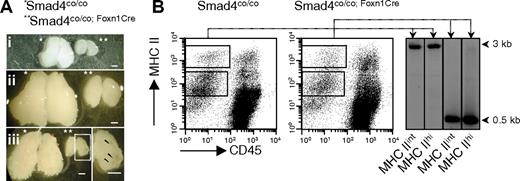
Because a general deficiency in Smad4 results in early embryonic lethality,24,25 mice were generated in which the inactivation of Smad4 was restricted to TECs using tissue-specific conditional gene targeting. For this purpose, mice expressing the Cre recombinase under the control of the Foxn1 locus (Foxn1Cre) were used because Foxn1 transcripts are confined in the thymus to epithelial cells.26 Foxn1Cre mice were crossed to mice homozygous for a conditional Smad4 allele (Smad4co/co), where exon 9 is flanked by loxP sites and recombination generates a null allele.23 Mice with a Smad4 deficiency in TECs (designated Smad4co/co;Foxn1Cre) developed a thymus. However, its size was drastically reduced compared with Smad4co/co littermate controls (Figure 1A). To rule out that the development of a small thymus was not mediated by a population of TECs that did not recombine the Smad4 locus, we analyzed the timing and efficiency of Cre-mediated loss of exon 9. Recombination was first detected in thymic tissue at E11.5 (data not shown). Complete recombination was obtained in purified TECs as early as E16 (Figure S2A). In adult mice, both TECs with an intermediate and a high cell surface expression of MHC class II were devoid of exon 9 (Figure 1B). Hence, inactivation of Smad4 in TECs was efficient during embryonic thymus development and eventually affected practically all TEC populations expressing detectable amounts of MHC class II molecules.
Inactivation of Smad4 in TECs causes thymic hypoplasia. (A) Representative photomicrographs of thymi from neonate (i), 5-week- (ii), and 20-week-old (iii) Smad4co/co (*) and Smad4co/co;Foxn1Cre (**) mouse littermates. Boxed area is shown in higher magnification to point out cysts in aged Smad4co/co;Foxn1Cre mice. Scale bars represent 1 mm. (B) Smad4 exon 9 deletion was verified in adult MHCIIint and MHCIIhi sorted TEC populations. Rectangles in the dot plots indicate sorting gates. Deletion was quantified by genomic PCR, yielding a 3-kb WT fragment and a 0.5-kb recombined fragment.
Inactivation of Smad4 in TECs causes thymic hypoplasia. (A) Representative photomicrographs of thymi from neonate (i), 5-week- (ii), and 20-week-old (iii) Smad4co/co (*) and Smad4co/co;Foxn1Cre (**) mouse littermates. Boxed area is shown in higher magnification to point out cysts in aged Smad4co/co;Foxn1Cre mice. Scale bars represent 1 mm. (B) Smad4 exon 9 deletion was verified in adult MHCIIint and MHCIIhi sorted TEC populations. Rectangles in the dot plots indicate sorting gates. Deletion was quantified by genomic PCR, yielding a 3-kb WT fragment and a 0.5-kb recombined fragment.
Smad4 signaling is required for maintenance of a structured thymic microenvironment
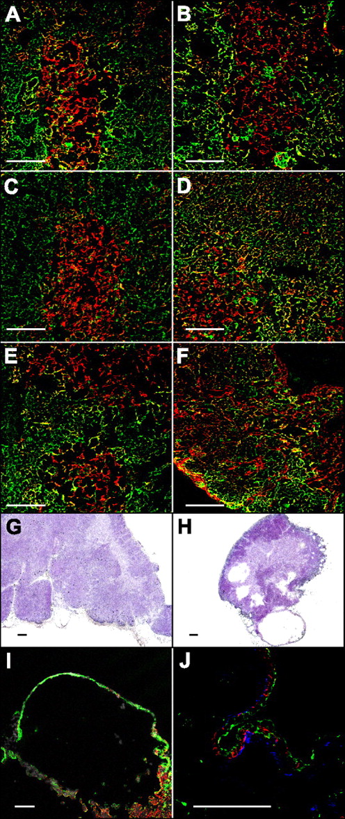
We next examined how the loss of Smad4 function in TECs influenced the cellular composition and organization of the thymic microenvironment. Histologic analysis of the thymus of newborn Smad4co/co;Foxn1Cre mice did not reveal any apparent abnormalities compared with control animals with the exception of an obvious difference in size. A clear separation between cortex and medulla was seen at this age both by hematoxylin and eosin staining (data not shown) and by immunofluorescence analysis using the intracellular markers CK18 (predominantly expressed by cortical TECs) and CK5 (predominantly expressed by medullary TECs; Figure 2A,B). At 6 weeks of age, the demarcation between cortical and medullary TECs (as evidenced by CK18 and CK5 staining) became blurred in Smad4co/co;Foxn1Cre mice compared with newborns of the same genotype or age-matched controls (Figure 2B-D). This was largely the result of a higher number of TECs expressing both cytokeratins concomitantly. Furthermore, an increased number of stromal cells in the Smad4co/co;Foxn1Cre mice expressed ERTR7, a mesenchymal marker identifying fibroblasts (Figure S3A,B).
Disorganized thymic microenvironment in aged Smad4co/co;Foxn1Cre mice. Immunofluorescence analysis of thymi from neonatal (A,B), 6-week- (C,D), and 20-week-old mice (E,F) with a Smad4co/co (A,C,E) or Smad4co/co;Foxn1Cre (B,D,F) genotype stained for cytokeratin 5 (red) and cytokeratin 18 (green). Hematoxylin and eosin staining of thymi from 20-week-old Smad4co/co (G) or Smad4co/co;Foxn1Cre (H) mice. Detailed analysis of a cyst stained either (I) with cytokeratin 5 (green), cytokeratin 18 (red), and DAPI (white), or (J) with cytokeratin 5 (red), cytokeratin 18 (green), and ERTR7 (blue). Scale bars represent 100 μm.
Disorganized thymic microenvironment in aged Smad4co/co;Foxn1Cre mice. Immunofluorescence analysis of thymi from neonatal (A,B), 6-week- (C,D), and 20-week-old mice (E,F) with a Smad4co/co (A,C,E) or Smad4co/co;Foxn1Cre (B,D,F) genotype stained for cytokeratin 5 (red) and cytokeratin 18 (green). Hematoxylin and eosin staining of thymi from 20-week-old Smad4co/co (G) or Smad4co/co;Foxn1Cre (H) mice. Detailed analysis of a cyst stained either (I) with cytokeratin 5 (green), cytokeratin 18 (red), and DAPI (white), or (J) with cytokeratin 5 (red), cytokeratin 18 (green), and ERTR7 (blue). Scale bars represent 100 μm.
At 20 weeks of age, immunofluorescence analysis now showed a highly aberrant expression pattern for the CK18 and CK5 markers compared either with age-matched control mice or mutant mice at 6 weeks of age (Figure 2D-F). In addition, patches devoid of CK18- and CK5-positive TECs were observed, which were filled by stromal cells expressing ERTR7 (Figure S3C). Furthermore, large cysts were visible in the thymi of all Smad4co/co;Foxn1Cre mice analyzed. The cysts were devoid of nucleated cells as evidenced by hematoxylin and eosin and DAPI staining (Figure 2H,I), and the cyst walls were composed of cytokeratin and ERTR7-positive thymic stroma cells, respectively (Figure 2J). Despite the progressive changes in composition and organization of the thymic microenvironment (including the appearance of large cysts and an increase in mesenchymal cells in older Smad4co/co;Foxn1Cre mice), distinct cortical and medullary areas could nonetheless be distinguished in hematoxylin and eosin sections of thymi at 20 weeks of age (Figure 2G,H). Moreover, the differentiation of TECs into Aire+ cells, defining a subset of postmitotic, mature medullary TECs,27 was unaffected (Figure S3D,E), and some small patches with a normal CK18 and CK5 distribution were still present in older Smad4co/co;Foxn1Cre mice (Figure 2F, Figure S3E). Taken together, we conclude that, with ongoing age, Smad4 expression in TECs is required at large for the maintenance of a normal compositional architecture of the thymic microenvironment.
Thymocyte development is unperturbed in Smad4co/co;Foxn1Cre mice
The inactivation of Smad4 in TECs resulted in a significant reduction in total thymic cellularity. A pattern of age-related changes in cellularity could be observed with a typical increase during young adulthood and a decrease after the onset of sexual maturity (Figures 3A, S2B). A careful analysis of the cellular composition revealed that the various thymus cell types were differentially affected by the TEC-targeted loss of Smad4. Whereas the absolute number of thymocytes was greatly diminished in Smad4co/co;Foxn1Cre mice at any time point investigated, absolute TEC cellularity remained normal in these animals, with the exception of young mutant mice where TECs were temporarily decreased by 20% to 50% (Figure 3A). Hence, the TEC frequency in relation to total thymus cellularity was significantly increased in Smad4co/co;Foxn1Cre mice at all ages analyzed except at E14, where both numbers and frequencies of TECs were significantly reduced, which could reflect delayed kinetics in TEC differentiation imposed by the absence of Smad 4, which is normalized at the end of gestation (Figures 3A, S2B). Mixed bone marrow chimeras of wild-type recipients and either Smad4co/co or Smad4co/co;Foxn1Cre donors resulted in equal thymic cellularity, thereby excluding the possibility of an unexpected cell-autonomous effect of the Foxn1Cre transgene in thymocytes (Figure S4).
Smad4co/co;Foxn1Cre mice display normal intrathymic T-cell development but have a reduced peripheral T-cell pool. (A) Thymus cellularity (i), absolute (ii), and relative (iii) TEC numbers of Smad4co/co and Smad4co/co;Foxn1Cre mice at different ages. (B) Relative numbers of different thymic subpopulations defined by CD44 and CD25 expression in the Lin− thymocyte population to detail the DN1-DN4 developmental stages, and CD4, CD8, and CD3/TCR expression to assess DP and SP populations in neonatal (i), 6-week- (ii), and 16-week-old mice (iii). (C) Absolute (i) and relative (ii) numbers of splenic T cells at different ages. For all experiments, a minimum of 3 mice per group were analyzed. Values are mean plus SD. □ represents Smad4co/co; ■, Smad4co/co;Foxn1Cre. *P < .05, **P < .01, ***P < .005, ****P < .001.
Smad4co/co;Foxn1Cre mice display normal intrathymic T-cell development but have a reduced peripheral T-cell pool. (A) Thymus cellularity (i), absolute (ii), and relative (iii) TEC numbers of Smad4co/co and Smad4co/co;Foxn1Cre mice at different ages. (B) Relative numbers of different thymic subpopulations defined by CD44 and CD25 expression in the Lin− thymocyte population to detail the DN1-DN4 developmental stages, and CD4, CD8, and CD3/TCR expression to assess DP and SP populations in neonatal (i), 6-week- (ii), and 16-week-old mice (iii). (C) Absolute (i) and relative (ii) numbers of splenic T cells at different ages. For all experiments, a minimum of 3 mice per group were analyzed. Values are mean plus SD. □ represents Smad4co/co; ■, Smad4co/co;Foxn1Cre. *P < .05, **P < .01, ***P < .005, ****P < .001.
Intrathymic T-cell development was largely normal in Smad4co/co;Foxn1Cre mice analyzed during fetal development (E14 and E18), at birth, and at 6 and 16 weeks of age (Figures 3B, S2C). Although statistically significant differences (P < .05) could be observed intermittently for some subpopulations, they were always minimal (3.1 ± 0.6 vs 4.2 ± 0.3, 6.5 ± 0.3 vs 8.7 ± 0.4 and 2.8 ± 0.3 vs 2.2 ± 0.1 among neonatal DN1, DN2, and CD8 SP populations, respectively; and 5.6 + 1.0 vs 7.0 + 0.4 among DN1 thymocytes from 16-week-old mice). The frequency of thymocytes with a high density of TCR-αβ/CD3 molecules and the expression of the cell surface markers CD69 and CD24 on SP thymocytes was similar for both mouse strains (Figure 3B and data not shown). These results demonstrate that the loss of Smad4 function in TECs did not impact on intrathymic T-cell development, including the positive selection of thymocytes.
The decreased thymus cellularity correlated in Smad4co/co;Foxn1Cre mice with a peripheral T-cell lymphopenia affecting both the relative and absolute numbers of CD4 and CD8 T cells (Figure 3C). As the number of T cells improved with age in Smad4co/co;Foxn1Cre mice but never reached normal values, we tested the homeostatic expansion of T cells in Smad4co/co and Smad4co/co;Foxn1Cre mice. Adoptively transferred WT and TCRtg T cells displayed an increased homeostatic expansion in mutant hosts compared with controls, suggesting the presence of a compensatory mechanism for the reduced thymic output (Figure S5A). In accordance, T cells with a memory phenotype (CD44hi) were more frequent in Smad4co/co;Foxn1Cre mice, a finding reflective of homeostatic expansion (Figure S5B).28 Naive peripheral T cells from Smad4co/co;Foxn1Cre mice displayed normal functionality as revealed by their regular CD3-mediated proliferative response in vitro, by their homeostatic expansion after transfer to lymphocyte-deficient recipient mice, and by their ability to provide B-cell help to a T-dependent antigen (data not shown).
Smad4 deficiency in TECs reduces the number of early T-lineage progenitors
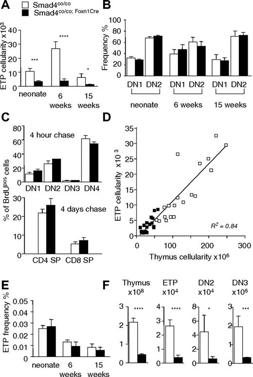
Because intrathymic T-cell maturation was normal but the overall number of thymocytes was decreased, we next determined in Smad4co/co;Foxn1Cre mice the absolute number of ETPs. Defined as Lin−CD25−CD44+Sca-1+c-kithi cells, ETPs were significantly reduced in mutant mice at all time points analyzed (Figure 4A, Figure S2D). However, the developmental potential of ETPs remained unchanged in Smad4co/co;Foxn1Cre mice because the DN1/DN2 relationship was identical in comparison to Smad4co/co controls (Figure 4B). Moreover, BrdU pulse/chase experiments demonstrated identical DN thymocyte proliferation values and a regular transition kinetic regarding the progression to a mature phenotype comparing Smad4co/co;Foxn1Cre and control mice (Figure 4C). Thymic hypoplasia in Smad4co/co;Foxn1Cre mice is therefore linked to low ETP numbers but does not reflect a divergence from regular αβTCR lineage development or a change in thymocyte proliferation during early T-cell maturation.
Reduced numbers of ETPs correlate to thymic hypoplasia in Smad4co/co;Foxn1Cre mice. (A) Absolute number of thymic ETPs (Lin−CD25−CD44+Sca-1+c-kithi cells) in mice of different ages. (B) Analysis of developmental progression from the DN1 to the DN2 stage in Lin−c-kithi thymic progenitors. (C) Quantification of proliferation in DN thymocytes 4 hours after BrdU administration and determination of intrathymic transition kinetics 4 days after the BrdU pulse in SP thymocytes. DN1 and DN2 populations were identified as c-kithi CD25neg and c-kithi CD25pos, respectively. (D) Linear correlation of ETP numbers with total thymic cellularity in mice ranging in age from 1.5 to 15 weeks. (E) Relative numbers of thymic ETPs in mice of different ages. (F) Total thymus cellularity and cell numbers of ETP, DN2, and DN3 populations in 6-week-old mice. DN2 cells were identified as c-kithi CD44+CD25+. A minimum of 3 mice per group were analyzed at each time point. Values are mean plus SD. □ represents Smad4co/co; ■, Smad4co/co;Foxn1Cre. *P < .05, **P < .01, ***P < .005, ****P < .001.
Reduced numbers of ETPs correlate to thymic hypoplasia in Smad4co/co;Foxn1Cre mice. (A) Absolute number of thymic ETPs (Lin−CD25−CD44+Sca-1+c-kithi cells) in mice of different ages. (B) Analysis of developmental progression from the DN1 to the DN2 stage in Lin−c-kithi thymic progenitors. (C) Quantification of proliferation in DN thymocytes 4 hours after BrdU administration and determination of intrathymic transition kinetics 4 days after the BrdU pulse in SP thymocytes. DN1 and DN2 populations were identified as c-kithi CD25neg and c-kithi CD25pos, respectively. (D) Linear correlation of ETP numbers with total thymic cellularity in mice ranging in age from 1.5 to 15 weeks. (E) Relative numbers of thymic ETPs in mice of different ages. (F) Total thymus cellularity and cell numbers of ETP, DN2, and DN3 populations in 6-week-old mice. DN2 cells were identified as c-kithi CD44+CD25+. A minimum of 3 mice per group were analyzed at each time point. Values are mean plus SD. □ represents Smad4co/co; ■, Smad4co/co;Foxn1Cre. *P < .05, **P < .01, ***P < .005, ****P < .001.
Plotting ETP numbers calculated in individual Smad4co/co;Foxn1Cre and Smad4co/co mice against their total thymus cellularity established a linear relationship (Figure 4D). Emphasizing such an interrelationship, the frequency of ETPs in relation to total thymus cellularity was identical in E18 embryos, neonatal, and adult mice of both genotypes (Figures 4E, S2D). Thymus cellularity has previously been correlated with the size of the DN3 compartment.6,29 The absolute cell number of DN3 thymocytes was as proportionately reduced in Smad4co/co;Foxn1Cre mice compared with DN2 cells and ETPs, extending the aforementioned correlation to cells with an even more immature phenotype (Figure 4F). These data hence demonstrate that the size of the thymic lymphoid compartment correlated with the number of ETPs.
Altered chemokine transcription by Smad4-deficient TECs
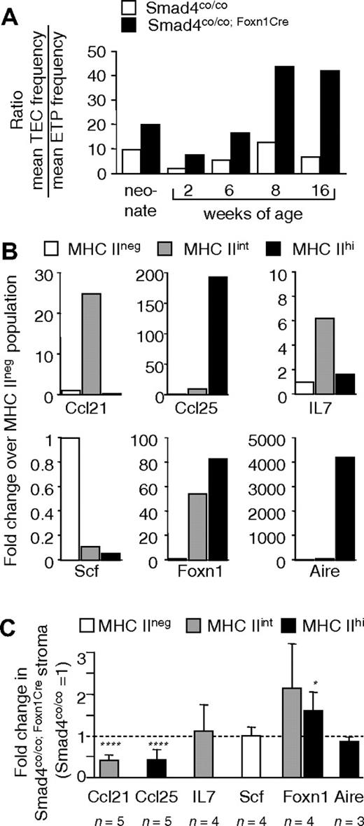
Thymopoietic activity is dependent on and maintained by the homing of hematopoietic progenitors. To interrogate the role of Smad4-mediated signaling in TECs for the number of ETPs, we determined the mean TEC/ETP ratio for both Smad4co/co and Smad4co/co;Foxn1Cre mice at different ages (Figures 5A, S2E). Smad4co/co;Foxn1Cre mice showed on average a 3.5-fold (±1.4-fold) higher TEC/ETP ratio, suggesting that the observed disparity in ETP numbers was the result of qualitative rather than quantitative differences in the microenvironment composed of Smad4-deficient TECs. Therefore, we next investigated the frequency of hematopoietic precursors in blood and bone marrow, whether a Smad4 deficiency in TECs limited the capacity to attract these cells and whether intrathymic T-cell precursors were limited in their survival in a microenvironment composed of Smad4-deficient TECs. The relative number of progenitors from bone marrow and peripheral blood defined as Lin−Sca-1+c-kithi30 or CD62L+ cells within this Lin−Sca-1+c-kithi population, an efficient subpopulation of T-lineage progenitors,31 was comparable for Smad4co/co;Foxn1Cre and control mice. These results excluded an extrathymic cause for the lower numbers of ETPs observed in Smad4co/co;Foxn1Cre mice (Figure S6A,B). Therefore, we analyzed factors important for the settling of these precursors into the thymus and for the promotion of their intrathymic survival and expansion. Using quantitative PCR, Scf was most prominently transcribed in CD45neg stromal cells lacking MHC class II expression (ie, nonepithelial stroma) from control mice, whereas Ccl21 and IL-7 transcripts were abundantly detected in CD45neg stromal cells that transcribe Foxn1 but not Aire and that express intermediate amounts of MHC class II (Figure 5B). In contrast, Ccl25 transcripts were most abundantly recovered from the population of MHC class IIhi cells, which include medullary Aire+ TECs. Thymic stromal cells from Smad4co/co;Foxn1Cre mice expressed similar amounts of IL-7, Scf, and Aire transcripts and slightly elevated amounts of Foxn1 mRNA compared with controls (Figure 5C). However, Smad4-deficient TECs, ie, MHC class IIint and MHC class IIhi stromal cells, expressed significantly decreased amounts of Ccl21 and Ccl25 transcripts but in a pattern identical to that of controls. These results suggest a specific role of the coactivator Smad4 for Ccl21 and Ccl25 transcription in TECs.
Qualitative changes in gene transcription in Smad4-deficient TECs. (A) Relationship between the average TEC and ETP frequencies in thymi from Smad4co/co and Smad4co/co;Foxn1Cre mice at different ages. Three to 5 mice per group were used at each time point. (B) Fold change of various transcripts in sorted MHCIIint ( ) and MHCIIhi (■) TEC populations from WT mice compared with sorted MHCIIneg stroma (□). (C) Fold change of various transcripts in TEC subpopulations (
 and ■) and in nonepithelial CD45neg stroma (□) from Smad4co/co;Foxn1Cre mice compared with Smad4co/co mice. Values are means of 3 to 5 independent experiments. Bars represent SD. *P < .05, **P < .01, ***P < .005, ****P < .001.
Qualitative changes in gene transcription in Smad4-deficient TECs. (A) Relationship between the average TEC and ETP frequencies in thymi from Smad4co/co and Smad4co/co;Foxn1Cre mice at different ages. Three to 5 mice per group were used at each time point. (B) Fold change of various transcripts in sorted MHCIIint ( ) and MHCIIhi (■) TEC populations from WT mice compared with sorted MHCIIneg stroma (□). (C) Fold change of various transcripts in TEC subpopulations (
 and ■) and in nonepithelial CD45neg stroma (□) from Smad4co/co;Foxn1Cre mice compared with Smad4co/co mice. Values are means of 3 to 5 independent experiments. Bars represent SD. *P < .05, **P < .01, ***P < .005, ****P < .001.
Discussion
The TEC-restricted loss of Smad4 expression results in a progressive alteration of the thymus stroma and a qualitative change in the thymic microenvironment, precluding the normal recruitment of ETPs. However, the thymic microenvironment maintains its capacity to support regular thymopoiesis from the most immature stage to full maturation and complete functional competence. Smad4 is therefore indispensable to create a microenvironment capable of efficiently attracting T-cell precursors but is not required for TEC cellularity and differentiation into different subpopulations.
A striking finding in Smad4co/co;Foxn1Cre mice is the progressive change in the thymic stromal architecture leading to a loss of the boundary between cortex and medulla, an increased proportion of fibroblasts, and the formation of cysts. As similar changes are also observed in very old, wild-type mice,32,33 it is tempting to speculate that a deficiency of Smad4 expression in TECs accelerates thymic involution. The molecular mechanisms operational in the physiologic process of thymic involution remain largely unknown but are likely to affect the thymic stromal compartment, which in turn might be influenced by the number of resident thymocytes.32,34
Two opposing explanations may account for the fact that TEC cellularity and competence to support T-cell development are unaffected by the loss of TEC-targeted Smad4 expression: signaling via TGF-β superfamily molecules may not be essential for these aspects of TEC biology or, alternatively, Smad4-independent pathways account for the transduction of signals initiated by TGF-β superfamily molecules.19,35 With TGF-βs, BMPs, activins, and some of their receptors expressed by thymic stromal cells,36–39 the former explanation seems unlikely. Moreover, a link between disrupting TGF-β superfamily signaling and changes in TEC biology has previously been suggested because the overexpression of the inhibitor Smad7 in a subpopulation of TECs resulting in a block in TGF-β and BMP signaling causes thymocyte apoptosis arguably resulting from alterations in TEC differentiation.40 However, Smad7 affects yet other functions independent of TGF-β/BMP signaling, such as activation of the JNK signaling pathway41,42 and degradation of β-catenin,43 which may explain the phenotypic differences between Smad7 transgenic and Smad4co/co;Foxn1Cre mice. In contrast, a block in thymic BMP signaling resulting from the overexpression of the BMP antagonist Noggin by TECs results in an ectopically located, dysplastic thymus.44 Given the experimental conditions used, BMP signaling is inhibited in both epithelial and mesenchymal cells of the developing thymus anlage, which probably explains the more severe phenotype observed in these animals compared with Smad4co/co;Foxn1Cre mice. In keeping with our results, the few thymocytes present in the Noggin transgenic mice developed with normal kinetics and displayed a wild-type phenotype, suggesting that an early thymic block in BMP signaling affected primarily the development of the stromal compartment. Because TEC cellularity and the capacity to support thymocyte development are unaffected in Smad4co/co;Foxn1Cre mice but compromised in animals with other deficiencies in signaling via TGF-β superfamily molecules,40,44 we conclude that Smad4-dependent pathways do not regulate these aspects of thymus biology.
Smad4 is, however, required for the competence of the thymic microenvironment to attract blood-borne ETPs. It has previously been noted that interactions of the chemokines Ccl21 and Ccl25 with their respective ligands, CCR7 and CCR9 control the homing of T cell precursors to the fetal and adult thymus.3–8 Although adult thymus size is normal in CCR9-deficient and CCR7/CCR9-deficient mice, a careful analysis in CCR9-deficient mice revealed a significant reduction of ETP numbers.5,6 It is conceivable that a combined decrease in the transcripts for these 2 chemokines at the site of precursor entry limit the number of immigrating ETPs in Smad4co/co;Foxn1Cre mice. Once having gained access to the thymic microenvironment, proliferation and differentiation of these precursors to mature T cells appear unperturbed despite the absence of Smad4 in TECs.
How Smad4 regulates the transcription of Ccl21 in MHCIIint TECs and Ccl25 in MHChi TECs is currently unknown. Our preliminary analyses have identified Smad4 binding sites45,46 in the promoter sequences of Ccl21 and Ccl25. Cooperation of Smad4 with caudal-related homeobox (Cdx) transcription factors could play a role here not least because Smad4 can bind to Cdx proteins, which are involved in the transcriptional regulation of Ccl25.47,48 Indeed, Cdx-1 is expressed in TECs (data not shown). Because Cdx-1 is also a Wnt target gene, it is the interaction of Smad4 with the closely related transcription factors Lef1 and TCF that provides a molecular node for such a crosstalk between the TGF-β and the Wnt signaling pathways.49,50
Nonhematopoietic stromal cells are thought to generate a limited number of physical niches that provide signals for survival, temporary retention, and subsequent release of early immature thymocytes for further differentiation.1 Although little is presently known regarding the cellular and molecular features of these niches, our data suggest that the lack of Smad4-mediated signaling in TECs disturbs the collective competence of these niches to attract/accommodate ETPs, independent from the changes in thymic architecture observed in older Smad4co/co;Foxn1Cre mice, whereas their capability to direct early thymocyte development appears to be unaffected. Two mutually nonexclusive models may account for the thymic hypocellularity observed in Smad4co/co;Foxn1Cre mice. A first model would suggest that the number of niches is significantly decreased as a consequence of a lack of Smad4-mediated signaling in TECs. Because there is no measure to quantify niches, a numerical decrease of such sites can presently not be tested. Nonetheless, this explanation appears to be less probable because the overall cellularity of TECs was only mildly affected in younger and normal in older mice despite the marked decrease of thymocytes. A second model would alternatively propose that the lack of Smad4-mediated signaling in TECs results in a functional impairment of such niches. However, such a microenvironmental deficiency probably doesn't compromise the expansion and differentiation of ETP to later developmental stages because there is a normal frequency of the different thymocyte subpopulations and normal proliferative kinetics in fetal and adult Smad4co/co;Foxn1Cre.
Our results contrast recent observations correlating total thymus cellularity with that of the DN3 compartment. In CCR9−/− mice, ETP and DN2 compartments were significantly reduced compared with control animals, whereas the DN3 cell numbers were unaffected, suggesting that CCR9−/− DN3 cells can compensate for a reduced thymus settling capacity.6 However, an equivalent compensatory mechanism is not evident in Smad4co/co;Foxn1Cre mice. This discrepancy could be explained by the assumption that wild-type DN3 thymocytes differ from their CCR9−/− counterparts in respect to their proliferative conduct or, alternatively, Smad4-deficient TECs do not support this proliferative expansion. Likewise, experiments in mice with variable T-lineage deficiencies using repopulation assays with wild-type bone marrow revealed a critical role for the DN3 compartment in determining total thymus size.29 Here, the size of the preexisting host DN3 compartment directly correlated with the extent of thymus reconstitution by engrafted donor cells. However, these experiments do not allow direct conclusions regarding the overall contribution of donor ETPs to thymus size because they encountered competition with host cells all along until the DN3 stage. Because we quantified the separate thymocyte subpopulations in the absence of cellular competition inherent to transfer models and without genetic alterations targeted to the lymphoid lineage, our results specify that the overall cellularity of the thymus is directly determined by the number of ETPs, although a size-limiting mechanism may be operational only at the DN3 stage.
Because the loss of Smad4 expression affects signaling in a manner not usually targeted by the overexpression of single inhibitory molecules or by the loss of specific ligands or their receptors, our results identify in a tissue-specific fashion the global role of canonical TGF-β superfamily signaling for TEC biology. Detailed understanding of the molecular mechanisms responsible for seeding hematopoietic precursors to TEC niches may lead to novel strategies to enhance thymic reconstitution in lymphopenic individuals having undergone immunoablative therapies.
The online version of this article contains a data supplement.
The publication costs of this article were defrayed in part by page charge payment. Therefore, and solely to indicate this fact, this article is hereby marked “advertisement” in accordance with 18 USC section 1734.
Acknowledgments
The authors thank Thomas Boulay, Katrin Hafen, and Annick Peter for expert technical help, Angelika Offinger and Rodrigo Recinos for excellent animal care, and Ed Palmer, Willem van Ewijk, and Hamish Scott for providing reagents.
This work was supported by the Swiss National Science Foundation (grant 3100-68310.02, G.A.H.; and grant 3235-062696.00, L.T.J.), the European Community 6th Framework Programme Euro-Thymide Integrated Project (G.A.H.), the National Institutes of Health (Besthesda, MD; grant ROI-A1057477-01, G.A.H.), and the Roche Research Foundation (M.H.-H.).
National Institutes of Health
Authorship
Contribution: L.T.J. designed and performed experiments and assisted in writing the paper; T.B. designed and performed experiments and wrote the paper; M.P.K., S.Z., and M.H.-H. designed and performed experiments; C.-X.D. provided reagents; and G.A.H. designed experiments and wrote the paper.
Conflict-of-interest disclosure: The authors declare no competing financial interests.
Correspondence: Georg A. Holländer, Department of Biomedicine, Laboratory of Pediatric Immunology, Mattenstrasse 28, 4058 Basel, Switzerland; e-mail: georg-a.hollaender@unibas.ch.
References
Author notes
*L.T.J. and T.B. contributed equally to this study.


This feature is available to Subscribers Only
Sign In or Create an Account Close Modal