Abstract
Vascular endothelial–cadherin (VE-cad) is localized to adherens junctions at endothelial cell borders and forms a complex with α-, β-, γ-, and p120-catenins (p120). We previously showed that the VE-cad complex disassociates to form short-lived “gaps” during leukocyte transendothelial migration (TEM); however, whether these gaps are required for leukocyte TEM is not clear. Recently p120 has been shown to control VE-cad surface expression through endocytosis. We hypothesized that p120 regulates VE-cad surface expression, which would in turn have functional consequences for leukocyte transmigration. Here we show that endothelial cells transduced with an adenovirus expressing p120GFP fusion protein significantly increase VE-cad expression. Moreover, endothelial junctions with high p120GFP expression largely prevent VE-cad gap formation and neutrophil leukocyte TEM; if TEM occurs, the length of time required is prolonged. We find no evidence that VE-cad endocytosis plays a role in VE-cad gap formation and instead show that this process is regulated by changes in VE-cad phosphorylation. In fact, a nonphosphorylatable VE-cad mutant prevented TEM. In summary, our studies provide compelling evidence that VE-cad gap formation is required for leukocyte transmigration and identify p120 as a critical intracellular mediator of this process through its regulation of VE-cad expression at junctions.
Introduction
Vascular endothelial-cadherin (VE-cad) is a transmembrane protein expressed in the vascular endothelium1 that participates in endothelial barrier function, angiogenesis, signaling, and endothelial cell survival (reviewed in Dejana et al2 ). Surface-expressed VE-cad localizes to cell-cell junctions and associates with α-catenin, β-catenin, plakoglobin (γ-catenin), and p120-catenin (p120) through its cytoplasmic tail, and with the actin cytoskeleton3,4 in combination with vinculin and α-actinin, which is thought to be critical for VE-cad adhesive interactions (reviewed in Vestweber5 ).
p120 is a substrate for Src family kinases and other receptor tyrosine kinases6,7 and regulates cadherin-dependent adhesion positively and negatively, depending on the cell system under study (reviewed in Alemà and Salvatore8 ). p120 associates with the juxtamembrane cytoplasmic region of VE-cad,9 and this is crucial to maintain cadherin surface expression.10 Overexpression of VE-cad mutants that competed for p120 binding, or siRNA knockdown of p120 in endothelium, resulted in dramatically decreased surface-expressed VE-cad and concomitant increased VE-cad degradation by an endocytic pathway. In contrast, overexpression of wild-type p120 augments VE-cad surface expression and diminishes its endocytosis.11 The precise mechanism(s) by which p120 controls the turnover and endocytosis of junctional VE-cad is not completely understood,8,11 but it is conclusive that cytosolic levels of p120 regulate VE-cad surface expression in endothelial cells, and the level of E-cadherin in epithelial cells.12
The idea that VE-cad acts as a gatekeeper for passage of leukocytes has been proposed previously.13-15 The passage of leukocytes through the endothelium at cell-cell junctions coincided with selective and reversible loss of VE-cad and cytosolic β- and γ-catenin staining. Subsequent analyses revealed that small transient gaps in the VE-cad complex occurred selectively at locations of leukocyte transendothelial migration (TEM).16,17 We confirmed and extended these observations by live cell fluorescence imaging of a VE-cad–GFP fusion protein in endothelium, revealing that de novo “gaps” or enlargement of small preexisting gaps in VE-cad staining accompanied TEM, and these resealed once leukocytes completed TEM.18 However, the function of VE-cad gaps in leukocyte transmigration is not clear and the intracellular molecules that regulate these gaps have not been identified. This, together with the newly described role for p120 in cadherin turnover prompted us to test the hypothesis that cytosolic p120 regulated the formation of VE-cad gaps during leukocyte TEM and had functional consequences for leukocyte diapedesis. Here we demonstrate that overexpression of p120GFP fusion protein in large vessel (human umbilical vein endothelial cells [HUVECs]) and microvascular (microvascular endothelial cells [MVECs]) endothelium dramatically increases VE-cad expression at cell-cell junctions and significantly reduces neutrophil and mononuclear leukocyte transmigration. The level of inhibition of transmigration correlates with the staining intensity of p120GFP. In MVECs, where VE-cad internalization is highly regulated by p120 and is the site of TEM in vivo, we do not detect VE-cad internalization during TEM by live cell imaging or by sensitive biochemical approaches. Instead, our data indicate that p120 can directly modulate leukocyte passage at cell-cell junctions by regulating VE-cad displacement at the sites of leukocyte TEM. The molecular mechanisms for initiating VE-cad gap formation during TEM are related to phosphorylation events. Indeed, recent studies found that VE-cadherin is phosphorylated at the p120-binding site, among others, during human THP-1 adhesion and ICAM-1 engagement in HUVEC or T-lymphoblast adhesion in mouse and rat endothelial cell lines.19,20 We extend these findings and show that p120GFP overexpression results in decreased tyrosine phosphorylation of VE-cad and that overexpression of nonphosphorylatable tyrosine VE-cad mutant at the p120-binding site results in decreased leukocyte TEM and gap formation. Together our data demonstrate that VE-cad redistribution is critical for leukocyte transmigration and is likely regulated, in part, by p120-mediated effects on VE-cad phosphorylation.
Methods
Reagents
The following reagents were used: human recombinant tumor necrosis factor-alpha (TNF-α; PeproTech, Rocky Hill, NJ); hec-1 (nonblocking monoclonal antibody [mAb] to human VE-cad21 ; a gift from Dr William Muller, Feinberg School of Medicine, Northwestern University, Chicago, IL), which was conjugated to Alexa 568 (Molecular Probes, Eugene, OR) or to NHS-S-S-biotin (Pierce Chemical, Rockford, IL); antibodies to p120 (BD Biosciences, San Jose, CA), GFP (Abcam, Cambridge, MA), α-catenin and ZO-1 (Zymed, San Francisco, CA), β-catenin (RDI, Flanders, NJ), and JAM-A, 1H2A922 ; phospho–VE-cad–Tyr658 and phospho–VE-cad–Tyr731 (Biosource, Camarillo, CA); β-actin (Sigma Aldrich, St Louis, MO); EEA-1 (Affinity Bioreagents, Golden, CO); phosphotyrosine 4G10 (Millipore, Temecula, CA); 2-mercaptoethanesulfonic acid (MESNA; Sigma, St Louis, MO); and phalloidin–Alexa Fluor 546 (Invitrogen, Carlsbad, CA).
Cells
Human umbilical vein endothelial cells (HUVECs) and dermal microvascular endothelial cells (MVECs) from neonatal foreskin11,22 were seeded on fibronectin-coated glass coverslips (5 μg/mL; BD Biosciences). Human polymorphonuclear cells (PMNs; > 95% pure) or mononuclear leukocytes were isolated from whole blood drawn from healthy volunteers,23 kept at 8°C, and used immediately. Blood was drawn and handled according to protocols for protection of human subjects approved by the Brigham and Women's Hospital Institutional Review Board. Informed consent was obtained from all volunteers in accordance with the Declaration of Helsinki.
Adenovirus production and cell infection
Adenovirus encoding p120GFP or GFP alone was as previously described23,24 ; VE-cad mutants Y658F and Y731F were kindly provided by Dr Keith Burridge (University of North Carolina, Chapel Hill, NC). HUVECs and MVECs were plated, infected 24 hours later, and cultured for another 3 days. Flow cytometric analysis measured the GFP fluorescence.
PMN transmigration assay under shear flow conditions
The live cell fluorescence microscopy flow model has been described previously.1-18 Endothelial monolayers were activated with TNF-α (25 ng/mL for 4 hours), and VE-cad was immunolabeled with hec-1–Alexa 568 mAb (1 μg/mL for 10 minutes). PMNs (1 × 10⁶/mL) were drawn across HUVECs or MVECs at 1.0 dyne/cm2.
Image acquisition and analysis
Live cell fluorescence and differential interference contrast (DIC) images of leukocyte TEM were acquired with PC computer-based MetaMorph software (version 5.0) imaging system (Molecular Devices, Downingtown, PA) controlling an Orca ER CCD camera (Hamamatsu, Bridgewater, NJ) connected to a Nikon TE2000 inverted fluorescence microscope equipped with DIC 20×/0.75 NA (live cell imaging in Figures 1 and 2) and 40×/0.95 NA (used for Figures 5 and 6) objectives.23 The number of accumulated leukocytes was determined by counting the total number of adhered and transmigrated cells per field. % TEM = [total transmigrated leukocytes]/[total adhered + transmigrated leukocytes] ×100.
Quantitation of p120GFP and VE-cad fluorescence in endothelium
Analysis was performed using fluorescence microscopy and ImageJ software (http://rsb.info.nih.gov/ij; National Institutes of Health [NIH], Bethesda, MD). The GFP intensity was measured in 2 different spots of each endothelial cell (EC; ie, at junctions and within the cell [nonjunctional]) in 50 to 70 different cells from multiple fields of duplicate samples. p120GFP-transduced monolayers were grouped into 3 categories according to their GFP fluorescence (expressed in arbitrary fluorescence units) as illustrated in Figure 3: low intensity = 0 to 75 units; intermediate intensity = 75 to 125 units; high intensity = 125 to 300 units. VE-cad was quantified in 20 different cell-cell junctions for each condition in images corresponding to 2 different fields of view (Figure 2). Values are normalized by dividing each by the value corresponding to VE-cad (red) intensity in uninfected cells.
Immunoprecipitation and Western blotting
Transduced or control HUVECs were surface biotinylated (Biotinylation kit; Amersham, Arlington Heights, IL) and cultured for the indicated times, or lysed directly without biotinylation as previously described.14 Equal aliquots of lysate were immunoprecipitated with the indicated Abs, and samples were resolved on 10% SDS–polyacrylamide gel electrophoresis (PAGE), transferred to nitrocellulose, and probed with streptavidin–horseradish peroxidase, or with the appropriate mAbs in the case of nonbiotinylated HUVECs.
VE-cad mAb internalization assays
HUVECs or MVECs were incubated with conjugated Hec-1 (Alexa 586 or NHS-SS-biotin; 20 μg/mL) at 20°C for 15 minutes. Washes removed unbound mAb. Cells were fixed immediately in 4% formaldehyde, or cultured for 3 hours at 37°C to allow internalization of mAb-tagged molecules from the cell surface and fixed, followed by permeabilization (0.1% Triton x-100) and staining with EEA-1 Ab. Coverslips were mounted on glass slides with FluorSave (Calbiochem, San Diego, CA). Image acquisition and analysis were performed using fluorescence microscopy and ImageJ software.
Statistical analysis
All data are presented as mean plus or minus SD for n = 3 or more unless otherwise indicated.
Results
p120GFP colocalizes with endogenous p120 and associates with VE-cad in endothelial cell-cell junctions
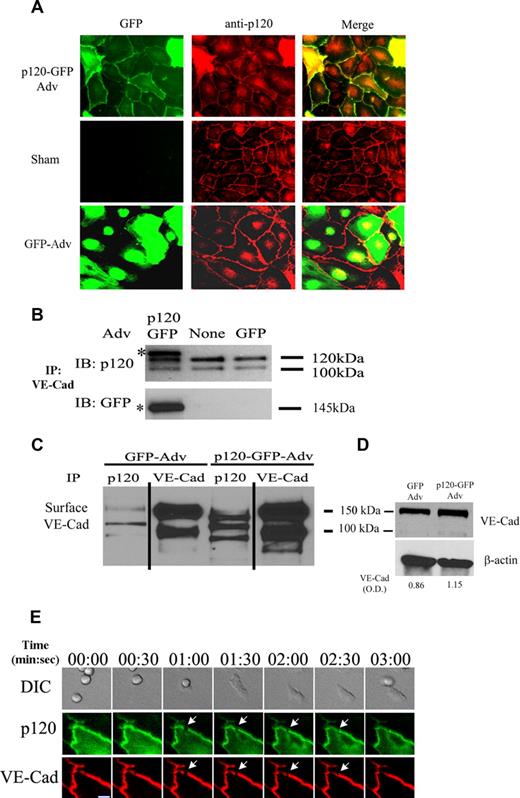
Uninfected HUVECs or those transduced with AdV-p120GFP (p120GFP) or AdvGFP (GFP) were examined by epifluorescence microscopy. p120GFP localized to cell-cell junctions, mimicking endogenous p120 (Figure 1A); the merge image for p120GFP shows colocalization of GFP signal and endogenous protein. Both endogenous isoforms of p120/p100 and the p120GFP protein (identified by asterisk when probed with anti-p120 mAb and as a specific single band when using anti-GFP mAb) are expressed in AdV-p120GFP–transduced HUVECs and coimmunoprecipitated with anti–VE-cad mAb (Figure 1B). To demonstrate association of p120GFP with surface VE-cad, HUVEC monolayers were surface biotinylated and lysed. mAb against p120 or VE-cad immunoprecipitated biotinylated VE-cad (bands of ∼ 110 kDa and 150 kDa, respectively) in HUVECs transduced with either AdV-p120GFP or AdV-GFP. Surface VE-cad and total VE-cad were increased in HUVECs transduced with AdV-p120GFP (Figure 1C,D) compared with AdV-GFP. These findings indicate that p120GFP mimics the behavior of endogenous p120 and associates with surface-expressed VE-cad at endothelial junctions.
Characterization of p120 expression in vascular endothelium and p120GFP/VE-cad gap formation during leukocyte TEM at the endothelial cell junctions. Confluent HUVECs were transduced with GFP or p120GFP adenovirues as described in “Adenovirus production and cell infection.” (A) p120GFP colocalizes with endogenous p120 at cell junctions. Monolayers were transduced with p120GFP, or sham treated, fixed with 10% buffered formalin, permeabilized, and stained with anti-p120 mAb. Representative fields were examined by epifluorescence microscopy and show junctional distribution of p120GFP and colocalization with endogenous p120 at cell junctions. (B) VE-cad was immunoprecipitated from the HUVEC lysates, and the material was immunoblotted for p120 to show endogenous isoforms of p120/p100, or with an anti-GFP mAb to detect expression of p120GFP. (C) HUVECs were surface biotinylated, lysed, and subjected to immunoprecipitation with anti-p120 mAb, or VE-cad mAb. The association of p120GFP with VE-cad was detected with streptavidin-peroxidase. Vertical lines have been inserted to indicate a repositioned gel lane. (D) Transduced HUVECs were lysed directly and blotted with Hec-1 to detect total VE-cad. Normalized VE-cad values versus β-actin are shown by OD numbers. Data are representative of 3 separate studies. (E) Three-channel live-time microscopy of PMNs in the process of transmigration. Paired 2-color fluorescence of VE-cad Alexa 568–stained HUVECs (red channel), p120GFP low-dose infected HUVECs (green channel), and simultaneous DIC images are presented. At t = 0, PMN approaches the brightly stained cell junction. At t = 1:00, PMN starts to transmigrate and a de novo gap is detected. At t = 3:00, the gap is sealed. This gap in p120-catenin GFP colocalized with the gap formed by VE-cadherin (red) during transmigration, as demonstrated in the merge panel. Bar represents 10 μm. The figure represents a typical sequence of events during PMN TEM. Five independent experiments using HUVECs and PMNs from multiple donors were analyzed.
Characterization of p120 expression in vascular endothelium and p120GFP/VE-cad gap formation during leukocyte TEM at the endothelial cell junctions. Confluent HUVECs were transduced with GFP or p120GFP adenovirues as described in “Adenovirus production and cell infection.” (A) p120GFP colocalizes with endogenous p120 at cell junctions. Monolayers were transduced with p120GFP, or sham treated, fixed with 10% buffered formalin, permeabilized, and stained with anti-p120 mAb. Representative fields were examined by epifluorescence microscopy and show junctional distribution of p120GFP and colocalization with endogenous p120 at cell junctions. (B) VE-cad was immunoprecipitated from the HUVEC lysates, and the material was immunoblotted for p120 to show endogenous isoforms of p120/p100, or with an anti-GFP mAb to detect expression of p120GFP. (C) HUVECs were surface biotinylated, lysed, and subjected to immunoprecipitation with anti-p120 mAb, or VE-cad mAb. The association of p120GFP with VE-cad was detected with streptavidin-peroxidase. Vertical lines have been inserted to indicate a repositioned gel lane. (D) Transduced HUVECs were lysed directly and blotted with Hec-1 to detect total VE-cad. Normalized VE-cad values versus β-actin are shown by OD numbers. Data are representative of 3 separate studies. (E) Three-channel live-time microscopy of PMNs in the process of transmigration. Paired 2-color fluorescence of VE-cad Alexa 568–stained HUVECs (red channel), p120GFP low-dose infected HUVECs (green channel), and simultaneous DIC images are presented. At t = 0, PMN approaches the brightly stained cell junction. At t = 1:00, PMN starts to transmigrate and a de novo gap is detected. At t = 3:00, the gap is sealed. This gap in p120-catenin GFP colocalized with the gap formed by VE-cadherin (red) during transmigration, as demonstrated in the merge panel. Bar represents 10 μm. The figure represents a typical sequence of events during PMN TEM. Five independent experiments using HUVECs and PMNs from multiple donors were analyzed.
Both p120 and VE-cad transiently form gaps at sites of leukocyte transmigration
Leukocyte TEM triggers the formation of transient gaps or enlargement of pre-existing gaps in VE-cad at cell junctions.18 To address whether leukocyte transmigration triggered gaps in junctional p120GFP, we monitored neutrophil transmigration in 4-hour TNF-α–activated HUVECs transduced with a low titer of Adv-p120GFP. Most PMNs (∼ 80%) adhered close to cell-cell junctions, and approached a continuous band of VE-cad and p120GFP staining. A gap formed in both VE-cad and p120GFP staining, and PMNs transmigrated through this gap (Figure 1E panels 00:30-02:30). The gap had its maximum opening as the bulk of the PMNs passed through the monolayer (Figure 1E panel 01:00) and resealed within minutes upon completion of transmigration (Figure 1E panels 02:30-03:00). This shows that VE-cad and p120 transiently disassociate at sites of PMN transmigration with essentially the same kinetics (Video S1, available on the Blood website; see the Supplemental Materials link at the top of the online article).
Overexpression of p120GFP augments VE-cad protein at cell junctions and extends VE-cad surface half-life
Recent reports that include overexpression of p120 indicate that p120 selectively controls VE-cad turnover at cell-cell borders.10,11 Based on this, we hypothesized that p120 overexpression stabilizes VE-cad at cell-cell junctions interfering with gap formation and hence preventing transmigration. We first assessed the effects of p120GFP on the surface level of VE-cad expression at cell-cell junctions (Figure 2A-D). The optimal dose of p120GFP (17.5 μL) augmented junctional VE-cad by 4-fold. A higher dose (25 μL) resulted in alteration of HUVEC size and shape accompanied by cytotoxicity (Figure 2A,B). GFP alone had no effect (Figure 2C) and AdV–VE-cad–GFP caused at best a 2-fold increase (Figure 2D). This indicates that the cytosolic pool of p120 is limiting and dictates the level of VE-cad surface expression, consistent with recent reports (reviewed in Reynolds25 ). The optimal concentration of each virus led to an infection rate of approximately 80% to 95% of cells. Monolayers retained normal morphology (Figure 2B), and a normal pattern of TNF-α–inducible adhesion molecules ICAM-1, VCAM-1, and E-selectin (data not shown), normal localization of JAM-A, ZO-1, catenins to cell-cell junctions, and the actin cytoskeleton organization was not altered (Figures S1,S2A). Because previous reports found that overexpression of p120 altered permeability, we performed the same dose-response analysis and measured transelectrical resistance (TER). Our data reveal a dose-response relationship: At low dose of p120GFP, TER was reduced, consistent with previous work24 ; the highest dose resulted in a slight enhancement in TER (Figure S2B,C). Interestingly, the half-life of surface VE-cad was extended when p120GFP was overexpressed (9.05 ± 2.0 hours), 1.7-fold longer compared with the AdVGFP-treated monolayers (5.2 ± 2.3 hours; Figure 2E). This confirms that a localized limiting pool of p120 is present in HUVECs, and when increased, can prevent VE-cad internalization, stabilizing VE-cad surface expression. Thus, we have devised a system that allows us to assess PMN transmigration in monolayers in which VE-cad is “locked” at junctions via overexpression of p120.
p120GFP overexpression increases VE-cad at the cell-cell junctions. HUVECs were infected with different doses of p120GFP (A,B), GFP (C), or VE-cadherin GFP (D). Hec-1–Alexa 568 Ab was used to detect VE-cad in each monolayer. The intensity of VE-cad at the junctions was quantified in live HUVECs by analyzing 20 cell-cell junctions including representative junctions of the heterogeneous population for each condition (in duplicate) for each experiment performed. (E) HUVECs were transduced with GFP or p120GFP (17.5 μL) surface biotinylated and lysed, or cultured for 2, 4, or 6 hours before being lysed and subjected to immunoprecipitation with Hec-1. A representative blot is shown. Values corresponding to the half-life were calculated according to the “exponential decay formula” and are the mean plus or minus SD of 3 different experiments.
p120GFP overexpression increases VE-cad at the cell-cell junctions. HUVECs were infected with different doses of p120GFP (A,B), GFP (C), or VE-cadherin GFP (D). Hec-1–Alexa 568 Ab was used to detect VE-cad in each monolayer. The intensity of VE-cad at the junctions was quantified in live HUVECs by analyzing 20 cell-cell junctions including representative junctions of the heterogeneous population for each condition (in duplicate) for each experiment performed. (E) HUVECs were transduced with GFP or p120GFP (17.5 μL) surface biotinylated and lysed, or cultured for 2, 4, or 6 hours before being lysed and subjected to immunoprecipitation with Hec-1. A representative blot is shown. Values corresponding to the half-life were calculated according to the “exponential decay formula” and are the mean plus or minus SD of 3 different experiments.
Overexpression of p120GFP in HUVECs inhibits PMN transmigration
The level of p120GFP expression was heterogeneous among the transduced cells. This heterogeneity may obscure or underestimate the extent to which PMN transmigration is inhibited. To address this issue, we performed 10-minute transmigration assays under defined shear flow capturing images of the GFP fluorescence and DIC of representative fields. We next mapped the intensity of p120GFP in each endothelial cell, separated these data into 3 levels of GFP expression (Figure 3A), and determined the location of leukocyte TEM relative to the level of GFP expression. Strikingly, little if any PMN transmigration occurred in regions of high p120GFP expression, whereas a lower but still significant reduction in TEM occurred at intermediate levels of p120GFP (Figure 3B). In regions with a high p120GFP signal, nearly all PMNs remained arrested at or adjacent to cell-to-cell borders, and failed to transmigrate and to trigger gap formation in VE-cad staining (Video S2). This effect was striking and is illustrated best in Figure 3C, in which PMNs are retained at junctions of p120GFP-transduced monolayers, but transmigrate and migrate away from junctions of GFP-transduced monolayers. In addition, the inhibitory effect was still observed even after 20 minutes of incubation, indicating that the block in TEM is not due to a delay in TEM (Figure 3D) or an increase in transcellular (nonjunctional) transmigration (data not shown); in fact, the majority of the cells that transmigrate do so at junctional locations where the levels of p120GFP and VE-cad are lowest (Figure 1E; Video S1), and the length of time for transmigration is prolonged (Figure S3). The inhibition of TEM is not due to an effect on neutrophil adhesion or ability of neutrophils to arrest at cell-cell junctions (Figure 3C).
Overexpression of p120 in HUVECs inhibits PMN transmigration. (A) The level of p120 GFP fluorescence and PMN transmigration was quantified as described in “Image acquisition and analysis” and “Quantitation of p120GFP and VE-cad fluorescence in endothelium,” using live cell fluorescence digital imaging. The p120GFP expression was grouped into 3 categories (low, intermediate, and high). (B) The amount of TEM was determined in each category. Values represent the mean plus or minus SD of 5 different experiments. P values are indicated, comparing each bar with GFP construct. ***P < .001; **P < .01. (C) PMNs remained bound at the cell-cell junctions on monolayers overexpressing p120GFP, but disappeared from the cell-cell junctions and efficiently transmigrated when perfused on HUVECs transduced with GFP. Values represent (number of cells bound to the junctions)/(number of total cells bound) × 100, and are the mean plus or minus SD of 5 different experiments. *P < .01. (D) The inhibitory effect of p120 overexpression was not due to a delay in PMN transmigration, as the block of TEM was sustained for more than 20 minutes. The percentage of TEM was normalized by dividing percentage of TEM at each time point by the number of cells that were bound initially at time 0. P values for p120GFP versus GFP at each time point are indicated. *P < .05; **P < .01. Data represent mean plus or minus SD of 3 different experiments. (E) Total accumulation of PMNs was similar on VE-cad–GFP– or GFP-transduced monolayers. (F) Overexpression of VE-cadherin GFP did not affect PMN TEM. Values in panels E and F represent the mean plus or minus SD from 3 different experiments using HUVECs and PMNs from different donors.
Overexpression of p120 in HUVECs inhibits PMN transmigration. (A) The level of p120 GFP fluorescence and PMN transmigration was quantified as described in “Image acquisition and analysis” and “Quantitation of p120GFP and VE-cad fluorescence in endothelium,” using live cell fluorescence digital imaging. The p120GFP expression was grouped into 3 categories (low, intermediate, and high). (B) The amount of TEM was determined in each category. Values represent the mean plus or minus SD of 5 different experiments. P values are indicated, comparing each bar with GFP construct. ***P < .001; **P < .01. (C) PMNs remained bound at the cell-cell junctions on monolayers overexpressing p120GFP, but disappeared from the cell-cell junctions and efficiently transmigrated when perfused on HUVECs transduced with GFP. Values represent (number of cells bound to the junctions)/(number of total cells bound) × 100, and are the mean plus or minus SD of 5 different experiments. *P < .01. (D) The inhibitory effect of p120 overexpression was not due to a delay in PMN transmigration, as the block of TEM was sustained for more than 20 minutes. The percentage of TEM was normalized by dividing percentage of TEM at each time point by the number of cells that were bound initially at time 0. P values for p120GFP versus GFP at each time point are indicated. *P < .05; **P < .01. Data represent mean plus or minus SD of 3 different experiments. (E) Total accumulation of PMNs was similar on VE-cad–GFP– or GFP-transduced monolayers. (F) Overexpression of VE-cadherin GFP did not affect PMN TEM. Values in panels E and F represent the mean plus or minus SD from 3 different experiments using HUVECs and PMNs from different donors.
To further prove that p120 regulates VE-cad gap formation and that increased junctional expression of VE-cad per se is not sufficient to block TEM, we performed the same set of studies using monolayers transduced with the optimal dose of AdV–VE-cad–GFP, which produces a 2-fold increase in VE-cad expression (Figure 2D). As we predicted, adhesion and TEM of PMNs (Figure 3E,F) or mononuclear cells (data not shown) were not affected. We conclude that p120 stabilization of VE-cad can regulate VE-cad gap formation and both PMN and mononuclear leukocyte TEM at cell-cell junctions.
Overexpression of p120GFP prevents phosphorylation of VE-cad and impairs TEM
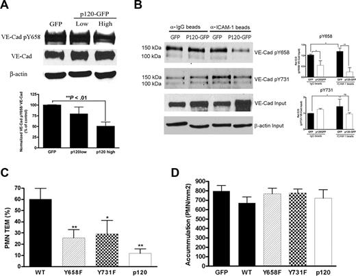
The association between p120 and cadherins is of low affinity.25 Phosphorylation of VE-cad at Tyr658 prevented binding of p120 to VE-cad in a system where CHO cells express different mutants of VE-cad.26 We and others have shown that rapid phosphorylation of proteins is triggered during the process of leukocyte adhesion and TEM.19,20,27,28 Recently, it has been shown that VE-cad becomes phosphorylated by Src and Pyk2 kinases at Tyr658 and Tyr731 during leukocyte adhesion and ICAM-1 engagement and that a nonphosphorylatable Tyr658 VE-cad mutant blocked PMN transmigration in a static transwell TEM assay. We, therefore, hypothesized that overexpression of p120 reduces constitutive phosphorylation of VE-cad Tyr658 and impairs ICAM-1–dependent phosphorylation of VE-cad and TEM.19,20 The phosphorylation status of VE-cad was examined in HUVECs transduced with increasing amounts of p120GFP using a phosphoTyr658–VE-cad–specific antibody. Figure 4A shows decreased VE-cad phosphorylation as p120 increases from a low (6 μL) to the optimal dose to obtain a 4-fold increase in surface VE-cad and to block TEM (17.5 μL). Further analysis showed the level of total VE-Cad increased 1.5-fold with a low dose and increased 2.2-fold with the high dose of p120-GFP. The densitometry data represent the ratio of phospho–VE-cad–Tyr658 to total VE-cad, and we interpret it to mean that in cells with the highest levels of p120, the phosphorylation of Tyr658 VE-cad is significantly attenuated and hence VE-cad–p120 association is retained at cell junctions. Consistent with our hypothesis, overexpression of p120 totally blocked ICAM-1–induced phosphorylation of Tyr658 in VE-cad (Figure 4B). Interestingly, these conditions did not alter constitutive or ICAM-1–triggered increase in phosphorylation of VE-cad–Tyr731. This indicates that p120-catenin is one mechanism regulating VE-cad stability at cell-cell borders and ICAM-1–dependent phosphorylation of VE-cad, and suggests additional as yet unidentified pathway(s) is involved. Furthermore, overexpression of nonphosphorylatable mutants of VE-cad at Tyr658 or Tyr731 decreased TEM to a similar extent seen with p120GFP overexpression (Figure 4C), whereas total accumulation of PMNs was not affected (Figure 4D). PMNs remain bound to the junctions and are unable to transmigrate (Video S3). These data highlight the role of p120 and phosphorylation of VE-cad in ICAM-1–dependent signaling and in PMN transmigration.
Overexpression of p120 results in hypophosphorylation of VE-cadherin, which in turn, regulates TEM. (A) HUVEC monolayers were infected with a low or a high dose of p120GFP (6 μL, 17.5 μL in Figure 2A) or GFP (2 μL in Figure 2C), lysed, and subjected to Western blot for phospho–VE-cad–Tyr658, total VE-cad, and β-actin. A representative blot is shown from 4 experiments performed. The graph represents normalized values obtained by densitometry analysis, indicating the relative absorbance of phospho–VE-cadherin–658 with respect to total VE-cadherin for each condition and normalized with the GFP values, and are the mean plus or minus SD of 4 different experiments. β-actin is shown as a loading control. (B) HUVECs were transduced with GFP or a high dose of p120GFP, treated with TNF-α, and incubated with control IgG beads or anti–ICAM-1 beads for 10 minutes and lysed in hot sample buffer. Samples were then diluted to allow immunoprecipitation with 4G10 mAb, and blotted for phospho–VE-cad–Tyr658 or phospho–VE-cad–Tyr731. A representative blot is shown from 3 independent experiments. The graph represents normalized values obtained by densitometry analysis corresponding to phospho–VE-cad divided by the total VE-cad input, and with respect to the GFP-IgG beads value, and is the mean plus or minus SD of 3 separate experiments. *P < .05; **P < .01. (C,D) HUVECs were infected with p120GFP, GFP, VE-cad, or the mutated version of VE-cad at Tyr658 or Tyr731. Overexpression of VE-cad Y658F and Y731F strongly inhibited TEM (C; **P < .01; *P < .05 with respect to WT), whereas total accumulation of PMNs was similar for every condition tested (D). Values represent the mean plus or minus SD from 3 different experiments.
Overexpression of p120 results in hypophosphorylation of VE-cadherin, which in turn, regulates TEM. (A) HUVEC monolayers were infected with a low or a high dose of p120GFP (6 μL, 17.5 μL in Figure 2A) or GFP (2 μL in Figure 2C), lysed, and subjected to Western blot for phospho–VE-cad–Tyr658, total VE-cad, and β-actin. A representative blot is shown from 4 experiments performed. The graph represents normalized values obtained by densitometry analysis, indicating the relative absorbance of phospho–VE-cadherin–658 with respect to total VE-cadherin for each condition and normalized with the GFP values, and are the mean plus or minus SD of 4 different experiments. β-actin is shown as a loading control. (B) HUVECs were transduced with GFP or a high dose of p120GFP, treated with TNF-α, and incubated with control IgG beads or anti–ICAM-1 beads for 10 minutes and lysed in hot sample buffer. Samples were then diluted to allow immunoprecipitation with 4G10 mAb, and blotted for phospho–VE-cad–Tyr658 or phospho–VE-cad–Tyr731. A representative blot is shown from 3 independent experiments. The graph represents normalized values obtained by densitometry analysis corresponding to phospho–VE-cad divided by the total VE-cad input, and with respect to the GFP-IgG beads value, and is the mean plus or minus SD of 3 separate experiments. *P < .05; **P < .01. (C,D) HUVECs were infected with p120GFP, GFP, VE-cad, or the mutated version of VE-cad at Tyr658 or Tyr731. Overexpression of VE-cad Y658F and Y731F strongly inhibited TEM (C; **P < .01; *P < .05 with respect to WT), whereas total accumulation of PMNs was similar for every condition tested (D). Values represent the mean plus or minus SD from 3 different experiments.
Internalization of VE-cad does not explain gap formation during TEM
In light of our data that p120 overexpression stabilizes junctional VE-cad and prevents VE-cad gap formation and TEM, we hypothesized that VE-cad is internalized at sites of leukocyte transmigration, forming gaps, and is recycled back to the junction once transmigration is completed. We tested this directly by live cell imaging studies as described earlier (Figure 1E; Video S1). Surprisingly, we did not detect any internalization or recycling of VE-cad during the time frame of PMN transmigration in HUVECs.
We used a second strategy to monitor internalization of VE-cad labeled with a modified biotinylation reagent that is susceptible to cleavage by MESNA, a non–cell-permeable reducing reagent. In pilot studies the biotin label is preserved on internalized VE-cad mAb and stripped on mAb at the surface29 (data not shown). We added mononuclear leukocytes, HL60 leukocytes that adhere but do not transmigrate, or buffer alone to HUVEC or MVEC monolayers immunostained with MESNA-cleavable biotin-tagged VE-cad mAb. Adhesion/transmigration was allowed to occur at 37°C for 10 minutes under static, no shear flow conditions. Nonadherent leukocytes were washed and monolayers lysed or fixed. Internalized biotinylated VE-cad mAb was measured by Western blot and immunofluorescence techniques, using streptavidin-HRP or streptavidin—Alexa 488 reagents, respectively. We found no significant differences in internalized VE-cad when comparing monolayers with many transmigration events with monolayers in which TEM did not take place (HL60; Figure S4A,B).
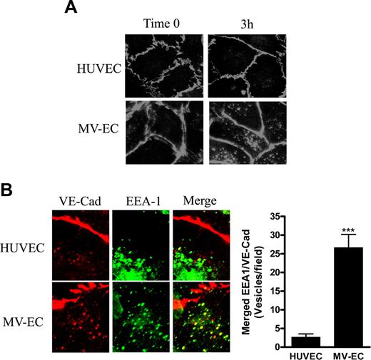
We nonetheless considered the possibility that VE-cad internalization occurs but the kinetics are too slow and/or the signal is below detectable levels by blotting approaches. The constitutive internalization and recycling of VE-cad is robust in MVECs.11 In a side-by-side comparison, more VE-cad containing intracellular vesicles was detected in MVECs compared with HUVECs after 3-hour incubation with labeled VE-cad mAb (Figure 5A). In MVECs, these were partially colocalized with the marker of early endosomes EEA-1 as reported previously.30 In HUVECs, internalized VE-cad was barely detectable and did not colocalize with EEA-1 (Figure 5B), suggesting that MVECs represent a good system through which to test our hypothesis by a live cell imaging approach.
Comparison of constitutive internalization of VE-cad in HUVECs and MVECs. (A) HUVEC or MVEC monolayers were stained with Alexa Fluor 568–conjugated mAb directed against the extracellular domain of VE-cad at 15°C (time 0). Cells were then transferred to 37°C for 3 hours. The location of VE-cad was examined by immunofluorescence microscopy. (B) Upon 3-hour incubation as in panel A, cells were fixed with 4% formaldehyde and stained for EEA-1. Colocalization of VE-cad and EEA-1 was determined by immunofluorescence microscopy, and quantified by counting merged (yellow) vesicles per field. Data represents the total number of merged vesicles per field using a 60× objective, and are the mean plus or minus SD of 2 different samples and 3 different experiments. ***P < .001.
Comparison of constitutive internalization of VE-cad in HUVECs and MVECs. (A) HUVEC or MVEC monolayers were stained with Alexa Fluor 568–conjugated mAb directed against the extracellular domain of VE-cad at 15°C (time 0). Cells were then transferred to 37°C for 3 hours. The location of VE-cad was examined by immunofluorescence microscopy. (B) Upon 3-hour incubation as in panel A, cells were fixed with 4% formaldehyde and stained for EEA-1. Colocalization of VE-cad and EEA-1 was determined by immunofluorescence microscopy, and quantified by counting merged (yellow) vesicles per field. Data represents the total number of merged vesicles per field using a 60× objective, and are the mean plus or minus SD of 2 different samples and 3 different experiments. ***P < .001.
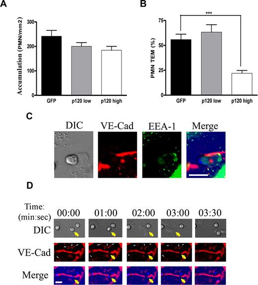
MVECs transduced with p120GFP resulted in up-regulated VE-cad expression due, in part, to decreased internalization as assessed by immunofluorescence techniques (data not shown). Further analysis showed significantly reduced PMN TEM and unaffected PMN adhesion in monolayers overexpressing high p120GFP (but not low levels; Figure 6A,B). MVECs were very homogeneous in terms of p120GFP expression, so it was not necessary to map the intensity of p120GFP as we did in HUVECs (Figure 3A). Transcellular and paracellular transmigration was similar in p120GFP- and GFP-transduced MVECs (data not shown).
Overexpression of p120 in MVECs inhibits PMN transmigration, and real-time imaging of VE-cad during PMN transmigration in MVECs. (A,B) MVEC monolayers were infected with different doses of AdV-p120GFP or AdV-GFP, and stimulated for 4 hours with TNF-α before the PMNs were perfused. Hec-1—Alexa 568 mAb was used to detect surface VE-cad by immunofluorescence staining. Total accumulation of PMNs was similar on p120GFP- or GFP-transduced monolayers (A). Overexpression of p120GFP (high dose) strongly inhibited TEM (B; P < .001). Values represent the mean plus or minus SD from 3 different experiments. (C,D) TNF-α–activated MVEC monolayers were immunolabeled with VE-cad—Alexa 568 mAb and inserted into the flow chamber. PMNs were perfused for 5 minutes and coverslips were fixed, permeabilized, and stained with EEA-1 Ab (C) or PMNs were allowed to transmigrate for 10 minutes and analyzed by 2-channel live-time microscopy (D). As the neutrophil begins to transmigrate, VE-cad forms a gap at the junction (panel 00:00) that widens (panel 02:00) and reseals (panel 03:00; yellow arrows), without apparent internalization of vesicles of VE-cad or recycling to the membrane of constitutively internalized VE-cad vesicles (thin white arrows). Merge panels show colocalization of the gap and the PMNs during the TEM process. Bar represents 10 μm. The figure represents a typical sequence of events during PMN transmigration from 3 independent experiments using HUVECs and PMNs from multiple donors.
Overexpression of p120 in MVECs inhibits PMN transmigration, and real-time imaging of VE-cad during PMN transmigration in MVECs. (A,B) MVEC monolayers were infected with different doses of AdV-p120GFP or AdV-GFP, and stimulated for 4 hours with TNF-α before the PMNs were perfused. Hec-1—Alexa 568 mAb was used to detect surface VE-cad by immunofluorescence staining. Total accumulation of PMNs was similar on p120GFP- or GFP-transduced monolayers (A). Overexpression of p120GFP (high dose) strongly inhibited TEM (B; P < .001). Values represent the mean plus or minus SD from 3 different experiments. (C,D) TNF-α–activated MVEC monolayers were immunolabeled with VE-cad—Alexa 568 mAb and inserted into the flow chamber. PMNs were perfused for 5 minutes and coverslips were fixed, permeabilized, and stained with EEA-1 Ab (C) or PMNs were allowed to transmigrate for 10 minutes and analyzed by 2-channel live-time microscopy (D). As the neutrophil begins to transmigrate, VE-cad forms a gap at the junction (panel 00:00) that widens (panel 02:00) and reseals (panel 03:00; yellow arrows), without apparent internalization of vesicles of VE-cad or recycling to the membrane of constitutively internalized VE-cad vesicles (thin white arrows). Merge panels show colocalization of the gap and the PMNs during the TEM process. Bar represents 10 μm. The figure represents a typical sequence of events during PMN transmigration from 3 independent experiments using HUVECs and PMNs from multiple donors.
In sham- or GFP-treated MVEC monolayers, we could clearly detect internalization (within minutes) of the immunolabeled VE-cad mAb when cells were incubated at 37°C (in the absence of leukocytes). We next performed live cell fluorescence imaging experiments of PMNs in 4-hour TNF-α–activated MVECs labeled with VE-cad—Alexa 568 mAb. In some experiments, we allowed the PMNs to transmigrate for 5 minutes, fixed the coverslips, permeabilized the cells, and stained them with EEA-1 Ab to study the presence of VE-cad in endocytic vesicles at the sites of transmigration (Figure 6C). In duplicate coverslips, we monitored PMN transmigration for 10 minutes, taking fluorescence and DIC images every 5 seconds using a 40× objective for a more detailed observation (Figure 6D). We observed a VE-cad gap during TEM, however, we did not observe VE-cad endocytic vesicles at the transmigration site, suggesting that VE-cad is not internalized during TEM (Figure 6C). We did not detect significant internalization of VE-cad–containing vesicles at sites of transmigration (Figure 6D panels 00:00-03:00, gap is identified by large yellow arrow; small white arrows identify vesicles). We did not observe recycling of the already internalized pools of VE-cad to close the gap (Figure 6D panels 01:00-03:30, indicated by white arrows). Review of many transmigration events showed no evidence of internalization (Videos S4,S5). We conclude VE-cad gaps formed during TEM are not likely to occur through the same internalization mechanism(s) that are responsible for constitutive turnover of surface VE-cad.
We noticed that VE-cad displacement was analogous to a “sliding curtain” within the junction as the gap widens during TEM (Figure 6D panels 01:00 and 02:00), which is consistent with our previous observations.18 This suggests that the VE-cad gap opening and subsequent resealing do not involve VE-cad internalization but, instead, involve a small opening that triggers a lateral displacement of VE-cad within the junction that diffuses back to reseal the junction after the leukocyte has transmigrated.
Discussion
We and others have shown disruption of the junctional VE-cad complex during leukocyte TEM in vitro.16,18,31 The mechanism underlying this process has not been defined, nor has it been proven that VE-cad gap formation is a necessary step in transmigration. In light of the recent demonstration that p120 regulates VE-cad surface expression at endothelial cell-cell junctions,11 we tested whether p120 was involved in disruption of junctional VE-cad, and by extension, in leukocyte TEM. We report for the first time that overexpression of a p120 fusion protein in HUVECs and MVECs stabilizes VE-cad expression at cell junctions and leads to a dose-dependent inhibition of both VE-cad gap formation and leukocyte TEM. Most PMNs fail to transmigrate and remain at cell-cell borders; nonjunctional TEM in HUVECs or MVECs does not increase. These data indicate that VE-cad disruption is essential for leukocyte paracellular transmigration, and that high cytosolic pools of p120-catenin stabilize VE-cad through reduced tyrosine phosphorylation. This, and studies with nonphosphorylatable VE-cad mutants, shows that TEM is strongly regulated by changes in VE-cad cytoplasmic tail tyrosine phosphorylation. We monitored VE-cad internalization to determine whether this mechanism is responsible for gap formation, using quantitative biochemical and high-resolution, live cell fluorescence imaging microscopy approaches. Both methods revealed no significant internalization of VE-cad over baseline at sites of PMN or monocyte transmigration. We conclude that VE-cad acts as a key gatekeeper for leukocyte paracellular transmigration and that p120 association with VE-cad is a critical interaction that regulates TEM through preventing tyrosine phosphorylation of VE-cad cytoplasmic tail.
The current data demonstrate that the cytosolic level of p120 is an important regulator of VE-cad disassembly and reassembly during leukocyte TEM in vitro, consistent with recent reports.32,33 Control experiments were performed to rule out trivial explanations for p120GFP inhibition of TEM. First, as p120GFP colocalizes at endothelial cell junctions with VE-cad, it mimics endogenous p120 (Figure 1). Second, as predicted, p120GFP causes a dose-dependent increase in the VE-cad level at cell junctions, which is explained by an extended half-life of surface-expressed VE-cad. Third, p120GFP has an intriguing dose-response effect on barrier function. Importantly, the dose of p120GFP required to inhibit TEM does not significantly affect HUVEC barrier function. Lower doses of p120GFP cause a significant decrease in TER, which is consistent with a previous study.24 Fourth, p120GFP does not disrupt the cortical actin cytoskeleton, the junctional localization of JAM-A or ZO-1, both of which are enriched at junctions and have been implicated in leukocyte TEM, or the localiztion of α-catenin and β-catenin. Fifth, approximately 2-fold overexpression of wild-type VE-cadGFP fusion protein does not inhibit TEM. Finally, the phosphorylation of Tyr658 in VE-cad was significantly reduced when p120GFP was overexpressed, even during ICAM-1 engagement. Because Potter et al26 found that phosphorylation of this tyrosine residue prevented p120 association with VE-cadherin, and more recently Allingham et al have shown that it becomes phosphorylated during leukocyte adhesion and ICAM-1 engagement,19 these data further corroborate our conclusion that p120GFP expression stabilizes VE-cad at cell-cell junctions, most likely by competing with nonreceptor tyrosine kinases Src and Pyk2 and possibly other kinases that phosphorylate cadherins (reviewed by Alemà and Salvatore9 ) as well as p120.6,7
These findings and prior studies provide insight into the mechanisms that regulate VE-cad gap formation and a link to early events in leukocyte transmigration. Prior studies demonstrated that p120 binding to cadherins is of low affinity6 and that phosphorylation of the VE-cad cytoplasmic tail at Tyr658 and Tyr731 prevents binding of p120 and β-catenin, respectively, suggesting that VE-cad and p120 interactions, as well as β-catenin, are amenable to dynamic regulation. Because expression of either Y731F or Y658F VE-cad mutants blocked most neutrophil transmigration but p120 overexpression prevented only ICAM-1–triggered phosphorylation of Y658-VE-cad, overexpression of p120-catenin must exert secondary effects that contribute to blocking TEM. For example, p120 overexpression can inhibit the small GTPase Rho A32 and endothelial cell Rho GTPases activation has been implicated in leukocyte transendothelial migration.34,35 Others have reported that neutrophil adhesion triggers disassociation of VE-cad from VE-PTP, a transmembrane phosphatase that stabilizes VE-cad–dependent cell permeability and reduces VE-cad tyrosine phosphorylation.36 It may also be possible that p120 overexpression stabilizes VE-cad association with VE-PTP, and this further contributes to TEM inhibition. Further investigation of these secondary affects should shed further light on the mechanisms by which p120 overexpression impairs leukocyte transmigration.
Initially, we hypothesized that tyrosine phosphorylation events lead to VE-cad internalization and result in gap formation. By direct live cell imaging, however, we could not detect any increase in VE-cad internalization or removal of VE-cad from the cell junctions during TEM events in MVECs or HUVECs. In fact, when imaging VE-cad redistribution during TEM under flow conditions in live time, we did not observe VE-cad–containing vesicles emanating from junctions at sites of gap formation or recycling back from the cytoplasm to the junctions when transmigration was completed and the gap was closed (Figure 6). Similarly negative results were obtained in live cell imaging samples that were fixed and reexamined under higher magnification fluorescence microscopy. VE-cad was not observed in early EEA-1–labeled endosomes near sites of TEM (Figure 6). The size of the VE-cad gap is approximately 4 to 5 μm, and its formation and resealing take place within minutes. Perhaps this is not enough time for this quantity of VE-cad to enter the endocytic pathway into early endosomes and recycle back to the junction. From studies of the related protein E-cadherin, this process of endocytosis and recycling to the membrane occurs over a longer time frame.12,37,38 Furthermore, biochemical approaches also failed to detect an increase in internalization of VE-cad during peak levels of TEM. If internalization is occurring, it is below detectable levels. Ultrastructural studies to measure this type of internalization are very difficult to interpret because current methods would not distinguish constitutive internalization of VE-cad from internalization occurring at TEM sites. We, therefore, suggest alternative mechanisms are responsible for gap formation during TEM. One possibility is based on the live cell imaging data and previous studies18 that supported a “shower curtain” model. This mechanism is likely to involve a lateral displacement of phosphorylated VE-cad to form the gap, and diffusion of junctional VE-cad to reseal the gap once transmigration is completed (Figure 7). Although proteolysis of VE-cad as a result of PMN adhesion has been reported,13,14 this does not appear to be a likely mechanisms for gap formation in our system because by live cell imaging, PMNs are retained at the cell-cell junctions in monolayers overexpressing p120 (Figure 3C) and no disappearance of VE-cad or gap formation is observed (Video S2). Understanding the role of phosphorylation in this complex will be essential for deciphering mechanisms underlying the regulation of junctional leukocyte TEM.
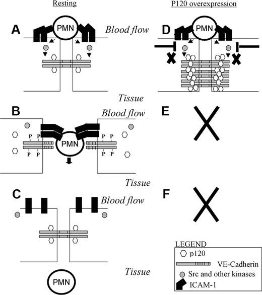
A model that envisions the p120/VE-cad complex as a key regulator of leukocyte TEM. (A) PMN interaction with adhesion molecules such as ICAM-1 on the endothelium surface triggers activation of src and other kinases. (B) Activation of kinases results in phosphorylation of VE-cad, dissociation of p120 from VE-cad, and opening of a junctional gap through which the PMN transmigrates. (C) Upon completion of TEM, p120 binds again to VE-cad as the gap reseals. (D) When p120 is overexpressed, receptor and nonreceptor tyrosine kinases fail to compete with the high levels of p120 present in the cytosol, VE-cad cannot be phosphorylated, and p120 is not released from the VE-cad complex. (E,F) This results in a lack of displacement of VE-cad and lack of gap formation, leading to diminished of PMN TEM.
A model that envisions the p120/VE-cad complex as a key regulator of leukocyte TEM. (A) PMN interaction with adhesion molecules such as ICAM-1 on the endothelium surface triggers activation of src and other kinases. (B) Activation of kinases results in phosphorylation of VE-cad, dissociation of p120 from VE-cad, and opening of a junctional gap through which the PMN transmigrates. (C) Upon completion of TEM, p120 binds again to VE-cad as the gap reseals. (D) When p120 is overexpressed, receptor and nonreceptor tyrosine kinases fail to compete with the high levels of p120 present in the cytosol, VE-cad cannot be phosphorylated, and p120 is not released from the VE-cad complex. (E,F) This results in a lack of displacement of VE-cad and lack of gap formation, leading to diminished of PMN TEM.
We propose a model incorporating these findings as follows. At sites of TEM, leukocyte binding to ICAM-119,20 and perhaps other adhesion molecules (PECAM-1, CD99, and VCAM-1) triggers localized tyrosine phosphorylation of VE-cad as recently reported. Ligation of ICAM-1 initiates src (and pyk-2) activation and phosphorylation of cortactin,23,28,39 and most likely, activated src mediates cross-phosphorylation of tyrosine residues in the cytoplasmic tail of VE-cad because both ICAM-1 and VE-cad molecules are in close proximity as leukocytes initially arrest at or near cell-cell junctions (reviewed in Alcaide et al40 ). Tyrosine phosphorylation of the cytoplasmic tail of VE-cad disrupts p120 association and destabilizes VE-cad interactions with the cytoplasmic catenins, resulting in loss of surface expression at the cell junctions via lateral displacement of phosphorylated VE-cad to form the gap. Once transmigration is completed, diffusion of junctional VE-cad reestablishes VE-cad at cell-cell borders (Figure 7). Under conditions of p120 overexpression, however, the tyrosine phosphorylation events in VE-cadherin normally induced during leukocyte binding to the endothelium and/or during TEM27,28 are not adequate to disrupt the enforced p120 association with VE-cad and, therefore, cannot induce the gap formation necessary for TEM (Video S2; Figure 7). Phosphorylation events mediated by src and other kinases take place at the endothelial cell junctions during TEM.28,41,42 These can also affect the stability and function of the VE-cad complex, including β-catenin,27 p120 (reviewed in Alemà and Salvatore8 ), and VE-cad itself.19,20,26 The literature supports our model, in which we propose that a low affinity p120–VE-cad interaction regulates leukocyte TEM by competing with tyrosine kinases for VE-cad juxtamembrane phosphorylation sites. The fact that p120 overexpression does not prevent phosphorylation of Tyr-731 and that the nonphosphorylatable VE-cadY731F mutant significantly blocked TEM suggest phosphorylation at Y731 is not controlled by a mechanism involving p120. One could speculate that sequential phosphorylation of these tyrosine residues occurs during ICAM-1 engagement/leukocyte adhesion/TEM and therefore preventing phosphorylation of either or both tyrosines blocks TEM. Alternatively, a more complicated sequence of events or broader targeting of regulatory proteins that associate with the VE-cad complex may also be involved. Future investigations are required to answer these questions.
Overexpression of p120 has different effects in different cell types: it enhances the motility of fibroblasts43 and epithelial cells43 and it inhibits the small Rho GTPase Rho A.32 In mice with conditional knockout of p120, the lack of p120 in skin epithelium resulted in infiltration of immune cells in the epidermis and constitutive activation of NFκB.44 Conditional knockout mice with loss of p120 in endothelium has not been reported, but we speculate that such mice would exhibit a similar or greater level of inflammation as well as disruption of vessel barrier function. Understanding the upstream signals induced by leukocyte adhesion to the activated endothelium, which can simultaneously trigger p120 dissociation from the VE-cad tail and VE-cad gap formation, will be crucial to understanding the role of p120 in controlling leukocyte transmigration.
The online version of this article contains a data supplement.
The publication costs of this article were defrayed in part by page charge payment. Therefore, and solely to indicate this fact, this article is hereby marked “advertisement” in accordance with 18 USC section 1734.
Acknowledgments
We thank Ms Kay Case, Deanna Lamont, and Vanessa Davis for providing well-characterized HUVECs; Richard Froio (Center for Excellence in Vascular Biology, Brigham and Women's Hospital) for technical assistance with adenovirus characterization; Dr Marianne Wessling-Resnick (Harvard School of Public Health) for technical advice on measurement of VE-cad internalization using NHS-S-S-biotin and MESNA treatments; Dr Keith Burridge for providing the adenovirus Y731F and Y658F VE-cad mutants; and Jeanne Zimmermann at the Brigham and Women's Hospital Editorial services for proofreading our paper.
This study was supported by NIH grants HL36028 (F.W.L.), HL36028 (T.N.M.), R01AR050501 (A.K.), HL077870 (P.V.), and HL32854 (D.E.G.).
National Institutes of Health
Authorship
Contribution: P.A. and F.W.L. designed, performed, and analyzed all the studies and wrote the paper; G.N. performed several immunofluorescence studies and contributed to experiments under flow conditions; S.A. performed several immunoprecipitation and Western blot experiments; P.V. provided vital reagents; A.K. provided MVECs and helped with the design and interpretation of the MVEC data; S.S. and T.N.M. designed and performed TER experiments; and D.E.G. and P.V. designed and performed FRAP experiments in support of the proposed alternative mechanisms for the VE-cad gap formation (data not shown).
Conflict-of-interest disclosure: The authors declare no competing financial interests.
Correspondence: Francis W. Luscinskas, Brigham and Women's Hospital, 77 Avenue Louis Pasteur, NRB752, Boston, MA 02115; e-mail: fluscinskas@rics.bwh.harvard.edu.

