Abstract
Nuclear factor-κB (NF-κB) transcription factors regulate B-cell development and survival. However, whether they also have a role during early steps of B-cell differentiation is largely unclear. Here, we show that constitutive activation of the alternative NF-κB pathway in p100−/− knockin mice resulted in a block of early B-cell development at the transition from the pre–pro-B to the pro–B-cell stage due to enhanced RelB activity. Expression of the essential B-cell transcription factors EBF and in particular Pax5 was reduced in p100−/− B-cell precursors in a RelB-dependent manner, resulting in reduced mRNA levels of B lineage-specific genes. Moreover, enhanced RelB function in p100−/− B-cell precursors was accompanied by increased expression of B lineage–inappropriate genes, such as C/EBPα, correlating with a markedly increased myeloid differentiation potential of p100−/− progenitor B cells. Ectopic expression of Pax5 in hematopoietic progenitors restored early B-cell development in p100−/− bone marrow, suggesting that impaired early B lymphopoiesis in mice lacking the p100 inhibitor may be due to down-regulation of Pax5 expression. Thus, tightly controlled p100 processing and RelB activation is essential for normal B lymphopoiesis and lymphoid/myeloid lineage decision in bone marrow.
Introduction
B-cell development in the bone marrow can be fractionated into sequential subsets. Pre–pro-B cells (Fr. A, according to Hardy's nomenclature) are the earliest B lineage precursors, characterized by the onset of surface expression of the relatively B lineage–restricted marker B220. The appearance of CD19 and rearrangement of the immunoglobulin (Ig) heavy chain gene locus coincides with the transition from pre–pro-B to pro-B cells (Fr. B/C).1-4 The pre–B-cell receptor (BCR) acts as an important checkpoint to control the transition from the pro-B to the pre–B-cell stage (Fr. C-prime/D). Successful Ig light chain gene rearrangement facilitates the emergence of immature IgM+ B cells (Fr. E) that emigrate to the periphery, where they undergo final maturation.
PU.1, E2A, EBF (early B-cell factor), and Pax5 (also called B cell–specific activator protein) are crucial transcription factors at the onset of B-cell development, regulating the expression of genes that maintain the B lineage differentiation program in a transcriptional hierarchy and a collaborative manner. Deficiency in any of these proteins blocks B-cell development at an early stage.5-7 Interestingly, Pax5−/− pro-B cells retain a lymphomyeloid potential and can be induced to differentiate into T, natural killer (NK), dendritic cells, macrophages, granulocytes, and osteoclasts upon IL-7 withdrawal and appropriate cytokine stimulation.8 Ectopic expression of C/EBPα, a member of CCAAT/enhancer-binding protein family, strongly promotes myeloid differentiation of Pax5−/− pro-B cells.9 The enforced expression of C/EBPα reprograms splenic B cells rapidly and efficiently into macrophages by inhibiting Pax5.10 These findings indicate that Pax5 plays an essential role in B lineage commitment by suppressing alternative lineage choices.
Another transcription factor that plays a pivotal role in B cells is nuclear factor (NF)–κB.11,12 The NF-κB family of transcription factors in vertebrates consists of 2 classes: whereas RelA, RelB, and c-Rel are synthesized as mature forms, the NF-κB1 (p105/p50) and NF-κB2 (p100/p52) proteins are synthesized as precursor molecules. In most cells, NF-κB proteins are inactive in the cytoplasm through their association with inhibitory IκB polypeptides. Conserved ankyrin repeats in the C-terminal halves of NF-κB1/p105 and NF-κB2/p100 render these precursors functionally similar to IκB. Potent inducers, such as tumor necrosis factor (TNF), interleukin-1 (IL-1), and lipopolysaccharide, activate classical p50-RelA heterodimers in an IκB kinase (IKK) complex-dependent manner. The recently described alternative NF-κB pathway targets the p100 inhibitor via the upstream kinases NF-κB–inducing kinase and IKKα. Signal-induced phosphorylation and proteolytic processing of p100 causes the degradation of its inhibitory C-terminal half. Consequently, the N-terminal portion of p100, that is NF-κB2/p52, is released and translocates to the nucleus as p52-RelB heterodimer.13,14 Therefore, p100 functions as a specific and potent inhibitor of RelB. In addition, it has recently been shown that p100 can also block RelA complexes.15 Processing of p100 to p52 is triggered by a specific set of stimulators, including BAFF (B cell-activating factor), CD40 ligand, lymphotoxin (LT)α1β2, and RANK (receptor activator of NF-κB) ligand, and is involved in the generation of peripheral B lymphocytes as well as in secondary lymphoid organogenesis.16-20
NF-κB is essential for peripheral B-cell maturation and activation. For example, in mice reconstituted with c-rel−/−relA−/− fetal liver cells, splenic B cells are reduced approximately 10-fold.21,22 The combined loss of NF-κB1 and NF-κB2 arrests B-cell development at the T2 transitional stage as a result of defects in BAFF signaling.23 While the alternative NF-κB2–dependent pathway enhances long-term B-cell survival, the classic NF-κB1–dependent pathway promotes Ig class switching and the generation of antibodies triggered by BAFF signaling.24 Finally, B lineage-specific ablation of IKKγ, and thereby classical NF-κB activity, reduces splenic B-cell numbers similar to BAFF-R deficiency without disturbing early B-cell development.25 In bone marrow, NF-κB is likely to increase survival downstream of signaling through the pre-BCR.11,12 However, it is unclear whether proper regulation of NF-κB has a cell-autonomous role in early B-cell development: that is before the expression of the pre-BCR.
To explore the consequences of constitutive alternative NF-κB signaling for B-cell development, we investigated knockin mice specifically lacking the p100 inhibitor (p100−/−) compared with nfkb2−/− mice, which are deficient for both p100 and p52. We found that in p100−/− bone marrow B-cell development was blocked at the pre–pro-B to pro–B-cell transition as a result of enhanced RelB activity. Expression of EBF and in particular Pax5 was reduced and B lymphopoiesis in p100−/− bone marrow was rescued by ectopic expression of Pax5. Strikingly, p100−/− B lineage precursors maintained a myeloid-lineage differentiation potential associated with induced C/EBPα expression. These findings indicate that signal-independent, constitutive activation of p52-RelB heterodimers via the alternative NF-κB pathway interferes with early B lymphopoiesis and promotes myeloid differentiation.
Methods
Mice
Generation of p100−/−,26 relB−/−,27 nfkb2−/−,28 nfkb1−/−,29 IkPax5/+,30 and vav-cretg31 mice has been described previously. All animals were housed and bred under standardized conditions with water and food ad libitum in a specific pathogen-free mouse facility at the Leibniz-Institute for Age Research—Fritz-Lipmann-Institute (Jena, Germany). The procedures for performing animal experiments were in accordance with the principles of the ATBW and were approved by the TLLV Thüringen (Erfurt, Germany).
Flow cytometric analyses and cell sorting
Flow cytometric analysis was performed using a FACSCalibur or FACSAria flow cytometer (BD Biosciences, Heidelberg, Germany). Bone marrow single cells and splenocytes were isolated, and red blood cells (RBCs) were lysed according to standard procedures. Single-cell suspensions were washed twice with staining buffer (phosphate-buffered saline [PBS] and 2% fetal calf serum [FCS]) and incubated with Fc Block (clone 2.4 G2). The following fluorochrome-conjugated monoclonal antibodies (mAbs) were used: anti–B220-Biotin/phycoerythrin (PE)/fluorescein isothiocyanate (FITC; RA3-6B2), anti–CD19-allophycocyanin (APC)/PE (1D3), anti–Sca-1-PE (D7), anti–CD43-Biotin/FITC (S7), anti-CD117 (c-Kit)–Biotin/APC (2B8), anti-CD127 (IL-7Rα)-Biotin (B12-1), anti–CD25-Biotin (7D4), anti–IgM-Biotin/FITC/APC (R6-60.2), anti–IgD-FITC/PE (11-26), anti–Gr-1-PE (RB6-8C5), anti-CD11b (Mac-1)–FITC (M1/70), anti–CD45.1-Biotin (A20), and anti-CD45.2-Biotin (104). Biotinylated mAbs were detected with Streptavidin-PerCP or PE-Cy7 (1:300; BD Biosciences). All mAbs were purchased from BD Biosciences or eBioscience (San Diego, CA). Apoptotic cells were detected by annexin-V–FITC staining (BD Biosciences) according to the manufacturer's recommendations. All incubations were for 30 minutes on ice, followed by two washes with FACS buffer. Data were analyzed using CellQuest Pro software. Cell sorting was performed with a FACSAria cell sorter.
qRT-PCR analysis
Total RNA isolation using the NucleoSpin RNA II kit (Macherey & Nagel, Düren, Germany) was performed according to the manufacturer's instructions. Contaminating DNA was removed by DNase I treatment. Equivalent amounts of RNA were reverse-transcribed with Superscript Moloney murine leukemia virus (Promega, Mannheim, Germany). Primers for quantitative revers transcription-polymerase chain reaction (qRT-PCR) were designed using Primer Express Software v2.0 (Applied Biosystems, Darmstadt, Germany) with a melting temperature of 58°C to 60°C. Triplicates were performed for all qRT-PCR reactions using reagents supplied by Quantace (Watford, United Kingdom) with an iCycler real-time PCR machine (Bio-Rad Laboratories, Munich, Germany). Primer sequences are shown in Document S1 (available on the Blood website; see the Supplemental Materials link at the top of the online article).
Bone marrow chimeras
Donor bone marrow cells were isolated from femora of 3-week-old wild-type or p100−/− mice and injected (4-6 × 10⁶ cells intravenously per mouse) into 2- to 3-month-old C57BL/6 recipients. Before injection, recipient mice were irradiated twice (3-hour interval, 5.5 Gy; Nordion International, Vancouver, BC) and rested for 3 to 4 hours after the second irradiation. Chimeric mice were analyzed 6 weeks after bone marrow reconstitution. For competitive chimeras, lethally irradiated Ly5.1+ C57BL/6 mice were transplanted with 4 × 10⁶ cells comprising a 1:1 mixture of Ly5.2+p100−/− and Ly5.1+ C57BL/6 bone marrow. Origin and composition of lymphoid cells was determined by Ly5.1 and Ly5.2 markers.
Cell culture
Purified murine pre–pro-B cells were grown on a semiconfluent layer of γ-irradiated (30 Gy) ST2 cells in Iscove modified Dulbecco medium supplemented with 10% heat-inactivated fetal calf serum, 2 mM glutamine, 50 μM β-mercaptoethanol, 0.03% (vol/vol) primatone, 100 U/mL penicillin, and 100 μg/mL streptomycin. IL-7, IL-3, Flt-3L, stem cell factor (SCF), and macrophage-colony-stimulating factor (M-CSF) (10 ng/mL each; PeproTech, Hamburg, Germany) were added to the culture for differentiation experiments. To assess the phagocytic function of macrophages, 5 μg/mL FITC-labeled Escherichia coli bioparticles (Invitrogen, Karlsruhe, Germany) were added directly to the cell culture medium. Four hours later, cells were washed 3 times with PBS and analyzed by flow cytometry for the presence of ingested bioparticles.
Statistics
Data are expressed as means (± SD). Differences were analyzed by Student t test. Sample sizes were chosen to produce statistically unambiguous results. A P value of .05 or less was considered significant (*) and a P value of .005 or less was considered highly significant (**).
Results
Disturbed B lymphopoiesis in mice lacking NF-κB2/p100
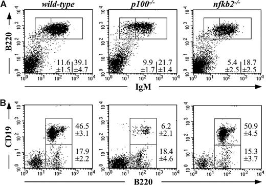
Spleens of 3-week-old p100−/− mice were anemic, approximately 10-fold smaller than in wild-type mice, and dramatically diminished in cellularity26 (data not shown). The relative frequency of B220+ B cells in p100−/− spleens was reduced approximately 2-fold as detected by flow cytometry. The reduction of B cells occurred mainly in the B220+IgM+ compartment. B220+IgM− B cells were less affected (Figure 1A). Similar results were obtained with lymph nodes and peripheral blood (data not shown). Thus, lack of the NF-κB2/p100 inhibitor resulted in severely decreased peripheral B-cell numbers.
Disturbed B lymphopoiesis in mice lacking NF-κB2/p100. (A) Splenocytes from 3-week-old wild-type, p100−/−, and nfkb2−/− mice were analyzed for B220 and IgM expression by flow cytometry. (B) B220 and CD19 expression on bone marrow cells from 3-week-old wild-type, p100−/−, and nfkb2−/− mice was analyzed. Numbers indicate mean values plus or minus SD (%) from 5 mice.
Disturbed B lymphopoiesis in mice lacking NF-κB2/p100. (A) Splenocytes from 3-week-old wild-type, p100−/−, and nfkb2−/− mice were analyzed for B220 and IgM expression by flow cytometry. (B) B220 and CD19 expression on bone marrow cells from 3-week-old wild-type, p100−/−, and nfkb2−/− mice was analyzed. Numbers indicate mean values plus or minus SD (%) from 5 mice.
An even more pronounced decrease of B220+ cells was evident in bone marrow from p100−/− mice compared with littermate controls (Figure 1B). To determine more precisely the stage at which B lymphopoiesis was arrested in p100−/− bone marrow, we used the marker CD19, which is expressed on B lineage precursors after the pre-pro-B stage. Figure 1B shows that B220+CD19− cells could be detected at normal frequency in p100−/− bone marrow, whereas B220+CD19+ B cells were strongly reduced (6.2% compared with 46.5%). These abnormalities did not occur in bone marrow from age-matched nfkb2−/− mice (lacking both p100 and p52), although splenic B cells were also diminished (Figure 1A,B). These results suggest that the absence of the p100 inhibitor caused a p52-dependent block in bone marrow B lymphopoiesis.
Constitutive NF-κB2 activation blocks B lymphopoiesis at the pre–pro-B-cell stage
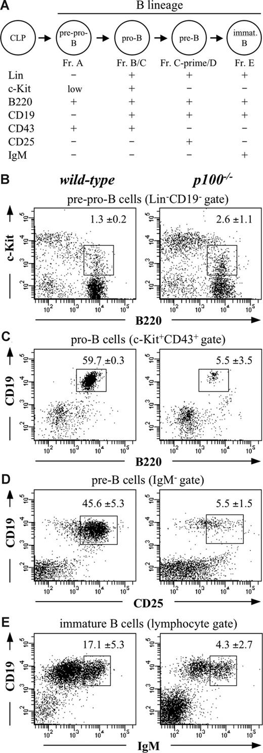
Because the B220+CD19+ B-cell pool consists of pro-B, pre-B, and immature B cells (Figure 2A), we performed detailed flow cytometric analyses to more precisely define the point in B-cell development affected by the lack of NF-κB2/p100. Whereas the frequency of common lymphoid progenitors (CLPs; Lin−Sca-1+IL-7R+c-Kitlow) was normal (data not shown), pre–pro-B-cell percentages were moderately increased (2-fold) in p100−/− bone marrow (Figure 2B). In contrast, the frequency of pro-B cells was more than 10-fold reduced compared with wild-type controls (Figure 2C). Thus, NF-κB2/p100 deficiency blocked B-cell development at the pre–pro-B to pro–B-cell transition.
B-cell development is arrested at the pre–pro-B to pro–B-cell transition in the absence of NF-κB2/p100. (A) Schematic illustration of B-cell development in bone marrow. The expression of characteristic cell surface proteins for successive progenitor stages in mouse B lymphopoiesis are indicated. Lin−, negative for the expression of lineage-specific markers (CD3ε−CD8α−CD19−DX5−Gr-1−Ly6C−Mac-1−Ter119−). (B) Flow cytometric analysis of c-Kit and B220 expression on Lin−CD19− wild-type and p100−/− bone marrow to define pre–pro-B cells (Fr. A). (C) Analysis of CD19 and B220 expression on c-Kit+CD43+ wild-type and p100−/− bone marrow to define pro-B cells (Fr. B/C). (D) Analysis of CD19 and CD25 expression on IgM− wild-type and p100−/− bone marrow to define pre-B cells (Fr. C-prime/D). (E) Analysis of CD19+IgMhigh immature B cells (Fr. E) in wild-type and p100−/− bone marrow. Percentages of relevant populations are indicated. Numbers represent mean values (± SD) from 5 mice.
B-cell development is arrested at the pre–pro-B to pro–B-cell transition in the absence of NF-κB2/p100. (A) Schematic illustration of B-cell development in bone marrow. The expression of characteristic cell surface proteins for successive progenitor stages in mouse B lymphopoiesis are indicated. Lin−, negative for the expression of lineage-specific markers (CD3ε−CD8α−CD19−DX5−Gr-1−Ly6C−Mac-1−Ter119−). (B) Flow cytometric analysis of c-Kit and B220 expression on Lin−CD19− wild-type and p100−/− bone marrow to define pre–pro-B cells (Fr. A). (C) Analysis of CD19 and B220 expression on c-Kit+CD43+ wild-type and p100−/− bone marrow to define pro-B cells (Fr. B/C). (D) Analysis of CD19 and CD25 expression on IgM− wild-type and p100−/− bone marrow to define pre-B cells (Fr. C-prime/D). (E) Analysis of CD19+IgMhigh immature B cells (Fr. E) in wild-type and p100−/− bone marrow. Percentages of relevant populations are indicated. Numbers represent mean values (± SD) from 5 mice.
Within the IgM− gate, an 8-fold reduction in CD19+CD25+ pre-B-cell frequency was detected in p100−/− bone marrow (Figure 2D). The frequency of p100−/− IgM+ immature B cells was reduced approximately 4-fold compared with wild-type controls (Figure 2E). These observations were confirmed when absolute B-cell numbers were calculated (Figure S1A). Thus, consistent with the severely reduced pro–B-cell pool in p100−/− bone marrow, there was also a significant decrease in the number of pre-B cells. However, the reduction of p100−/− immature B cells was less compared with other B-cell subsets, indicating that the severe block at the pre–pro-B to pro–B-cell transition was partially compensated at later stages of B-cell development.
It has been shown that mice lacking the antiapoptotic protein MCL-1 arrest B-cell development at the pro–B-cell stage as a result of increased apoptosis.32 In addition, Bcl-xL overexpression rescues impaired pre–B-cell generation caused by NF-κB inactivation.33 When apoptosis was examined in p100−/− bone marrow B-cell subpopulations, we observed a 2- to 3-fold reduction in annexin V staining (Figure S1B), correlating with up-regulated Bcl-2 expression (data not shown). Therefore, the block in B lymphopoiesis at the pre–pro-B to pro–B-cell transition in mice lacking the NF-κB2/p100 inhibitor is unlikely to be due to decreased cell survival. Moreover, attenuated apoptosis in p100−/− B-cell precursors may contribute to overcome the developmental block at later maturation stages.
Mice lacking NF-κB2/p100 have increased numbers of myeloid cells in bone marrow and peripheral lymphoid organs.26 To exclude the possibility that the defective B-cell development in p100−/− bone marrow was caused by myeloid hyperplasia, we depleted myeloid cells by treating mutant animals with the anti-granulocyte monoclonal antibody RB6-8C5. Although myeloid cells were strongly reduced, numbers of CD19+ B cells did not increase (data not shown), indicating that the defective B lymphopoiesis in p100−/− mice was not caused by myeloid hyperplasia.
NF-κB2/p100 acts cell autonomously to maintain B lymphopoiesis
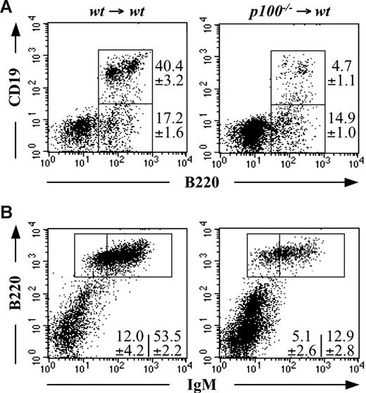
To define whether the impaired B-cell development resulting from the lack of NF-κB2/p100 is a hematopoietic defect or a defect in the stromal compartment that supports B-cell differentiation, bone marrow reconstitution experiments were performed. After lethal irradiation, wild-type recipients were reconstituted with wild-type (wt → wt) or p100−/− bone marrow cells (p100−/− → wt) and chimeras were analyzed 6 weeks after transplantation. The developmental block at the pre–pro-B to pro–B-cell transition was also observed in p100−/− → wt chimeras as indicated by the pronounced loss of B220+CD19+ cells compared with wt → wt controls. The percentage of B220+CD19− cells was not significantly affected (Figure 3A). The reduction of B cells was also observed in the periphery of p100−/− → wt chimeras (Figure 3B). Therefore, the intact radiation-resistant stromal microenvironment in wild-type recipients could not support proper B-cell development when reconstituted with p100−/− bone marrow cells. These observations suggest that the B-cell development abnormality in mice lacking NF-κB2/p100 was derived from the hematopoietic rather than nonhematopoietic (stromal) compartment. A similar result was obtained in competitive mixed chimeras using CD45.1+ wild-type and CD45.2+p100−/− donors (data not shown), indicating that p100−/− B-cell precursors could not undergo normal maturation even with the support of wild-type bone marrow cells. Thus, inhibition of the alternative NF-κB pathway by p100 within B cells is required for normal B lymphopoiesis in the bone marrow.
Analysis of B-cell development in bone marrow chimeras. Mice were lethally irradiated and reconstituted with wild-type or p100−/− bone marrow cells as indicated. Lymphocytes in either bone marrow (A) or spleen (B) were analyzed for CD19 and B220 or B220 and IgM expression, respectively. Numbers represent mean values (± SD) from 3 mice.
Analysis of B-cell development in bone marrow chimeras. Mice were lethally irradiated and reconstituted with wild-type or p100−/− bone marrow cells as indicated. Lymphocytes in either bone marrow (A) or spleen (B) were analyzed for CD19 and B220 or B220 and IgM expression, respectively. Numbers represent mean values (± SD) from 3 mice.
Altered NF-κB DNA-binding activity in p100−/− B cells
Constitutively increased binding to κB sites was found in nuclear extracts from p100−/− lymphoid or nonlymphoid tissues.26 To further investigate the contribution of individual family members to the elevated NF-κB activity in the B-cell lineage, we quantified NF-κB DNA binding in sorted B220+ cells from wild-type and p100−/− mice. In line with a previous report,34 p50 and c-Rel binding was most abundant. RelB and also p52 activity was markedly elevated in p100−/− B cells, whereas p50 activity was only moderately increased. Binding of RelA and c-Rel was similar in wild-type and p100−/− B cells (Figure S2). Interestingly, when in addition to p100 only one allele of the relB gene was deleted, as in B cells from p100−/−relB−/+ mice, the elevated RelB activity was restored to wild-type levels. As expected, when both alleles of the relB gene were inactivated (p100−/−relB−/−), RelB activity was abolished to background levels (Figure S2). We also observed a modest increase (2-fold) in RelA activity in p100−/−relB−/+ and p100−/−relB−/− B cells. Together, these results suggest that increased RelB activity may negatively regulate B-cell development.
Enhanced RelB activity limits early B-cell development
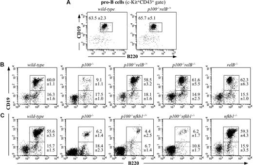
Flow cytometric analysis of 3-week-old p100−/−relB−/+ mice revealed that within the c-Kit+CD43+ compartment, the frequency of B220+CD19+ pro-B cells was equivalent between p100−/−relB−/+ and wild-type bone marrow (Figure 4A). The pre-B and immature B-cell compartments were also restored to wild-type levels (data not shown). When the relB gene was completely inactivated in p100−/− mice (p100−/−relB−/−), the frequency of B220+CD19+ cells was also similar to that in wild-type controls (Figure 4B). However, splenic B220+ B-cell numbers were decreased in p100−/−relB−/− mice (data not shown), probably as a result of abnormal B-cell proliferation and increased apoptosis.35 Moreover, p100−/−relB−/− mice showed phenotypic abnormalities, such as splenomegaly, lack of Peyer patches, and poorly developed lymph nodes, similar to relB−/− mice.20 Thus, complete ablation of the relB gene in p100−/− mice rescued the defective early B-cell development in bone marrow but resulted in a phenotype resembling that of relB−/− mice.
B-cell development in p100−/− bone marrow is rescued by partial inactivation of RelB but not of p50/NF-κB1. (A) Examination of bone marrow CD19+B220+ pro-B cells (c-Kit+CD43+ gate) by flow cytometry in wild-type and p100−/−relB−/+ mice. (B) B-cell development in bone marrow was analyzed in 3-week-old wild-type, p100−/−, p100−/−relB−/+, p100−/−relB−/−, and relB−/− mice for expression of CD19 and B220 by flow cytometry. (C) CD19 and B220 expression on bone marrow cells was examined in wild-type, p100−/−, p100−/−nfkb1−/+, p100−/−nfkb1−/−, and nfkb1−/− mice. Lymphocytes were gated for flow cytometric analysis. Percentages indicate mean values (± SD) from 3 to 5 mice.
B-cell development in p100−/− bone marrow is rescued by partial inactivation of RelB but not of p50/NF-κB1. (A) Examination of bone marrow CD19+B220+ pro-B cells (c-Kit+CD43+ gate) by flow cytometry in wild-type and p100−/−relB−/+ mice. (B) B-cell development in bone marrow was analyzed in 3-week-old wild-type, p100−/−, p100−/−relB−/+, p100−/−relB−/−, and relB−/− mice for expression of CD19 and B220 by flow cytometry. (C) CD19 and B220 expression on bone marrow cells was examined in wild-type, p100−/−, p100−/−nfkb1−/+, p100−/−nfkb1−/−, and nfkb1−/− mice. Lymphocytes were gated for flow cytometric analysis. Percentages indicate mean values (± SD) from 3 to 5 mice.
To examine whether p50 complexes, such as classical p50-RelA heterodimers, also contribute to the impaired B-cell development in p100−/− bone marrow, we generated p100−/− mice that carried a targeted deletion of the nfkb1 gene. Both p100−/−nfkb1−/+ and p100−/−nfkb1−/− mice were clinically indistinguishable from age-matched p100−/− mice. The B220+CD19− and B220+CD19+ B-cell compartments were comparable between 3-week-old wild-type and nfkb1−/− mice. However, a marked reduction of B220+CD19+ pro-B cells was determined in p100−/−nfkb1−/+ and p100−/−nfkb1−/− bone marrow, which was similar to that of p100−/− mice (Figure 4C). These data show that deleting either 1 or 2 alleles of the nfkb1 gene did not rescue the B-cell developmental block in p100−/− mice. Together, these results indicate that enhanced activity of alternative p52-RelB heterodimers due to the lack of the p100 inhibitor, rather than classic p50-RelA complexes, were responsible for the arrest of B lymphopoiesis at the pre–pro-B to pro–B-cell transition in p100−/− mice.
Efficient differentiation of p100−/− pre–pro-B cells into myeloid cells
To examine the plasticity of p100−/− B-cell precursors to differentiate into other lineages, we cultured pre–pro-B cells from wild-type and p100−/− mice on ST2 stromal cells in the presence of cytokines. After 14 days, almost all wild-type B-cell precursors differentiated along the B lineage. However, only few CD19+ B cells were detected in p100−/− cultures, and the majority of p100−/− precursors lost their B lineage potential and differentiated into CD19−Mac-1+ cells. Biphenotypic cells expressing both CD19 and Mac-1 appeared only in p100−/− cultures (Figure 5A). CD19−Mac-1+ cells were negative for CD11c, indicating that they represent myelomonocytic and not dendritic cells. A similar result was obtained when cells were cultured for only 8 days (data not shown). Moreover, the in vitro–generated Mac-1+ myeloid cells showed uptake of fluorescently labeled E coli, suggesting that p100−/− pre–pro-B cells could efficiently differentiate into functional macrophage-like cells (Figure 5B).
Efficient differentiation of p100−/− pre–pro-B cells into myeloid cells. (A) Lin−B220+CD19−c-Kitlow pre–pro-B cells were sorted from bone marrow of 18- to 20-day-old wild-type and p100−/− mice and cultured as described in “Methods.” After 14 days, cells were collected, stained for CD19 and Mac-1 expression, and analyzed by flow cytometry. Numbers represent mean values (± SD) from 3 mice. (B) Efficient uptake of E coli-FITC bioparticles by in vitro–differentiated Mac-1+p100−/− myeloid cells. Cultured p100−/− B220+CD19− pre-pro-B cells were incubated after 14 d with FITC-labeled E coli and uptake by CD19+ (----) or Mac-1+ cells (—) was measured by flow cytometry.
Efficient differentiation of p100−/− pre–pro-B cells into myeloid cells. (A) Lin−B220+CD19−c-Kitlow pre–pro-B cells were sorted from bone marrow of 18- to 20-day-old wild-type and p100−/− mice and cultured as described in “Methods.” After 14 days, cells were collected, stained for CD19 and Mac-1 expression, and analyzed by flow cytometry. Numbers represent mean values (± SD) from 3 mice. (B) Efficient uptake of E coli-FITC bioparticles by in vitro–differentiated Mac-1+p100−/− myeloid cells. Cultured p100−/− B220+CD19− pre-pro-B cells were incubated after 14 d with FITC-labeled E coli and uptake by CD19+ (----) or Mac-1+ cells (—) was measured by flow cytometry.
Disturbed expression of B cell–specific genes in p100−/− bone marrow
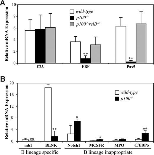
The transcription factors E2A and EBF are required for proper development of early B progenitors and for the cooperative activation of a B lymphoid gene expression program at the onset of B-cell development.5 In E2A−/− or EBF−/− bone marrow, B-cell development is blocked at the pre–pro-B-cell stage.36-38 EBF mRNA levels were 4- to 5-fold reduced in sorted B220+CD19−Ly6C−DX5− pre-pro-B cells from p100−/− compared with control mice, whereas E2A expression was unchanged (Figure 6A). The transcription factor Pax5 is crucial for maintaining B lymphocyte commitment. In p100−/− mice, B lymphopoiesis was arrested at a similar stage as in Pax5−/− mice.39 Therefore, one possibility is that the lack of NF-κB2/p100 affects the expression of Pax5. To address this point, Pax5 mRNA levels were determined. Strikingly, very little Pax5 expression was detected in p100−/− compared with wild-type B220+CD19−Ly6C−DX5− pre–pro-B cells (Figure 6A). In p100−/−relB−/+mice, the elevated RelB DNA-binding activity was reduced to wild-type levels, correlating with restored early B-cell development. Interestingly, expression levels of Pax5 and EBF were also restored to wild-type levels in these animals, indicating that expression of Pax5 and EBF was affected by the strength of RelB activity (Figure 6A).
Disturbed expression of B lineage–specific and B lineage–inappropriate genes in p100−/− bone marrow. (A) Quantitative real-time PCR (qRT-PCR) analysis of B-lineage transcription factors in sorted B220+CD19−DX5−Ly6C− B-cell precursors from wild-type, p100−/−, and p100−/−relB−/+ bone marrow. (B) qRT-PCR analysis for expression of B lineage–specific and B lineage–inappropriate genes in sorted wild-type and p100−/− B220+CD19−DX5−Ly6C− B-cell precursors.
Disturbed expression of B lineage–specific and B lineage–inappropriate genes in p100−/− bone marrow. (A) Quantitative real-time PCR (qRT-PCR) analysis of B-lineage transcription factors in sorted B220+CD19−DX5−Ly6C− B-cell precursors from wild-type, p100−/−, and p100−/−relB−/+ bone marrow. (B) qRT-PCR analysis for expression of B lineage–specific and B lineage–inappropriate genes in sorted wild-type and p100−/− B220+CD19−DX5−Ly6C− B-cell precursors.
Expression of Pax5 target genes that code for essential components of the pre-BCR signaling pathway, such as Igα (mb-1) and BLNK (B-cell linker), was also attenuated in p100−/− B220+CD19−Ly6C−DX5− bone marrow cells. On the other side, mRNA levels of B lineage–inappropriate genes, such as Notch1, M-CSFR, and MPO, were elevated compared with wild-type controls (Figure 6B). It has been shown that Pax5 represses Notch1, M-CSFR, and MPO, thereby limiting developmental options of early B-cell precursors.8 Moreover, expression of C/EBPα, which has been reported to repress Pax540 and to cause myeloid reprogramming of B-cell precursors,41 was up-regulated 5-fold in p100−/− compared with wild-type cells (Figure 6B). Expression of IL-7Rα, Flt-3, and PU.1 mRNA was not affected by the lack of p100 (data not shown). Thus, the disturbed expression pattern of B lineage–specific and –inappropriate genes in p100−/− early B-cell precursors correlated with strongly reduced Pax5 expression.
Restoration of Pax5 expression rescues early B-cell development in p100−/− mice
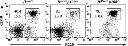
The phenotypic similarities between p100−/− and Pax5−/− mice prompted us to determine whether the disturbed B-cell development in p100−/− bone marrow was caused by the down-regulation of Pax5. Therefore, we expressed a conditional Pax5 minigene in p100−/− mice under the control of the Ikaros (Ik) locus.30 Upon Cre-mediated recombination in hematopoietic stem cell–derived progenitors, the inactive Ikneo allele can be converted to the active IkPax5 allele, expressing Pax5 in all hematopoietic cells. Pan-hematopoietic Pax5 expression increases B lymphopoiesis at the expense of T cell development and, to a lesser extent, of erythropoiesis.30 In contrast to Ikneo/+p100−/− mice, ectopic Pax5 expression in IkPax5/+p100−/− littermates rescued the developmental block from the pre–pro-B to the pro–B-cell stage in the bone marrow as indicated by the significant presence of B220+CD19+ cells (Figure 7). Therefore, the impaired early B lymphopoiesis in mice lacking the p100 inhibitor may be due to the down-regulation of Pax5 expression, which is triggered by enhanced RelB activity.
Reconstitution of Pax5 expression rescues early B-cell development in p100−/− bone marrow. Bone marrow from 3-week-old mice was analyzed by flow cytometry for CD19 and B220 (c-Kit+CD43+ gate). Ikneo/+, transgenic mice expressing an inactive Pax5 transgene under the control of the Ikaros locus; Ikneo/+p100−/−, Ikneo/+ mice on a p100−/− background; IkPax5/+p100−/−, p100−/− mice in which the inactive Ikneo allele has been converted into the active IkPax5 transgene upon Cre-mediated recombination using a Vav-Cre mouse line. Numbers represent mean values (± SD) from 3 mice per genotype.
Reconstitution of Pax5 expression rescues early B-cell development in p100−/− bone marrow. Bone marrow from 3-week-old mice was analyzed by flow cytometry for CD19 and B220 (c-Kit+CD43+ gate). Ikneo/+, transgenic mice expressing an inactive Pax5 transgene under the control of the Ikaros locus; Ikneo/+p100−/−, Ikneo/+ mice on a p100−/− background; IkPax5/+p100−/−, p100−/− mice in which the inactive Ikneo allele has been converted into the active IkPax5 transgene upon Cre-mediated recombination using a Vav-Cre mouse line. Numbers represent mean values (± SD) from 3 mice per genotype.
Discussion
In this study, we investigated the consequences of signal-independent, constitutive activation of the alternative NF-κB pathway on early B-cell development. We show that proper control of p100 processing was critical for the regulation of B lineage–specific transcription factor networks, B lymphopoiesis, and lymphoid-myeloid lineage decision. B lymphopoiesis was blocked at the pre–pro-B to pro–B-cell transition in p100−/− mice that lack the inhibitory p100 precursor but still express the p52 subunit of NF-κB. In contrast, we did not observe defects in early B-cell development in young nfkb2−/− animals lacking both p100 and p52, although reduced B-cell numbers in bone marrow and spleen have been reported for adult nfkb2−/− mice.28,42 B-cell development in p100−/− bone marrow could be rescued by deleting one or both relB alleles, whereas the nfkb1 gene was not involved. Biochemical analysis of B-cell nuclear extracts revealed that the loss of one relB allele was sufficient to reduce the RelB activity to wild-type levels. Together, these results suggest that increased activity of p52-RelB heterodimers as a result of the lack of the p100 inhibitor was responsible for the impaired B lymphopoiesis. The specific role of p100 is further supported by the absence of an early B-cell phenotype in p105−/− mice, which lack the inhibitory p105 precursor but still express the p50 subunit of NF-κB43 (data not shown).
Mice lacking p100 show myeloid hyperplasia and enhanced cytokine production.26 Elevated TNF levels produced by myeloid cells have been suggested to interfere with normal lymphocyte development in NF-κB–deficient mice.44 Our results from competitive repopulation and Pax5 reconstitution experiments as well as from the culture of purified pre–pro-B cells strongly argue against indirect effects mediated by altered cytokine levels as the cause for the impaired B lymphopoiesis in p100−/− bone marrow.
The block in B-cell development was due to cell-intrinsic mechanisms as shown by bone marrow transfer experiments. Although pro–B-cell numbers were 10-fold reduced in p100−/− bone marrow, B-cell development was not completely arrested, as indicated by the few CD19+ pro-B cells that differentiated into pre-B and immature B cells. Annexin V staining suggested that reduced apoptosis at later stages partially compensated for the severe loss of p100−/− pro–B cells. These data indicate that the constitutive activation of the alternative NF-κB pathway resulted in impaired entry into the pro–B-cell stage rather than in generally impaired progression through early B-cell developmental stages. Reduced apoptosis frequency correlated with increased Bcl-2 expression in p100−/− B-cell subpopulations. Putative NF-κB binding sites are located in the Bcl-2 promoter, and overexpression of p52 increases endogenous Bcl-2 expression.45 In addition, BAFF signaling induces Bcl-2 expression in transitional B cells via p100 processing.16 Thus, our findings are in line with the current model that p52 functions as a survival factor by stimulating the expression of Bcl-2 and related antiapoptotic genes.11
Expression of EBF and in particular Pax5 was down-regulated in p100−/− pre–pro-B cells in a RelB-dependent manner, unraveling a novel connection between NF-κB proteins and B lineage–specific transcription factors. Genetic evidence places Pax5 in the transcriptional hierarchy downstream of the genes encoding the transcription factors PU.1, Ikaros, E2A, and EBF,5 but a linear hierarchical model may not fully explain the transcriptional control of B lineage commitment.7 On one side, the Pax5 gene is a direct target of EBF,46 and retroviral EBF expression in E2A-deficient progenitors induces differentiation to committed pro-B cells by activating the Pax5 gene.47 The EBF promoter region harbors sequences with similarities to NF-κB binding sites (data not shown). The reduced EBF expression in p100−/− B-cell precursors could therefore be a consequence of direct repression of EBF by the enhanced RelB activity and may contribute to the markedly reduced Pax5 levels. On the other side, loss of Pax5 in DT40 B cells results in diminished EBF transcripts, suggesting that Pax5 is needed for the sustained expression of EBF.48 In addition, ectopic Pax5 expression in pro-T cells activates expression of the EBF gene,49 and Pax5 has been shown to regulate the stronger proximal EBF promoter.50 Thus, increased RelB activity as a result of the loss of the p100 inhibitor may also repress Pax5 (and as a consequence EBF) expression, although a direct regulation of the Pax5 gene by RelB/NF-κB remains to be shown.
An alternative explanation of the impaired B-cell development is offered by the strongly up-regulated C/EBPα expression in p100−/− pre–pro-B cells. C/EBPα has been shown to convert committed B cells into myeloid cells.41 Ectopic expression of C/EBPα and GATA transcription factors efficiently redirect Pax5−/− pro-B cells to the myeloid lineage.9 C/EBPα extinguishes CD19 expression by inhibiting Pax5 activity and induces myeloid gene expression by cooperating with endogenous PU.1.10 Furthermore, the loss of Pax5 triggered by the up-regulation of C/EBPα is critical for the diversion of CLPs from the lymphoid to myeloid lineages.40 In addition, the loss of Pax5 in cultured Myc5 cells correlates with the acquisition of myeloid markers, such as Mac-1 and F4/80.51 We observed that enhanced RelB function in p100−/− B-cell precursors correlated with the up-regulation of B lineage–inappropriate genes, such as Notch1, MCSFR, and C/EBPα. Moreover, sorted pre–pro-B cells from p100−/−, but not wild-type, bone marrow could efficiently differentiate into Mac-1+ myeloid cells that engulfed fluorescently labeled E coli bacteria. In this context, it is important to note that p100−/− mice develop myeloid hyperplasia with increased numbers of Mac-1+ cells in bone marrow26 (data not shown). It is therefore conceivable that the induction of C/EBPα expression in p100−/− pre–pro-B cells blocked the B lymphoid potential by repressing Pax5 and EBF, whereas it promoted myeloid development, resulting in reduced B-cell numbers and myeloid hyperplasia in the bone marrow of p100−/− mice.
So far, the alternative NF-κB pathway, in terms of p52-RelB complexes, has largely been involved in terminal differentiation, survival, and activation of peripheral B cells.16,24,28,35,42,52-54 In this study, we showed that constitutively elevated RelB activity due to p100 deficiency disturbed B lymphopoiesis at the pre–pro-B to pro–B-cell transition: that is before expression of the pre-BCR. RelB-mediated repression of Pax5 may be responsible for this phenotype because ectopic expression of Pax5 in hematopoietic cells fully rescued the impaired B lymphopoiesis in p100−/− bone marrow. Alternatively, expression of the Pax5 transgene may prevent C/EBPα-driven myeloid differentiation, thereby promoting B-cell development.9 Together, our data suggest that reduced Pax5 levels in p100−/− B-cell precursors in combination with induced C/EBPα facilitated the conversion of pre–pro-B cells to myeloid cells. Thus, proper control of p100 processing and RelB activation is obligatory for the maintenance of an intact network of B lineage transcription factors and B lineage commitment. How the NF-κB family member RelB manipulates this complex network of B lymphopoiesis and lineage-commitment requires further analysis.
The online version of this article contains a data supplement.
The publication costs of this article were defrayed in part by page charge payment. Therefore, and solely to indicate this fact, this article is hereby marked “advertisement” in accordance with 18 USC section 1734.
Acknowledgments
We gratefully acknowledge Debra Weih, Elke Meier, and Agnes Lovas for excellent technical assistance and suggestions. We thank Jan Tuckermann for critically reading the manuscript. We are indebted to Sabine Matz and all the staff in the animal facility of the Leibniz-Institute for Age Research—Fritz-Lipmann-Institute.
This work was supported by the Deutsche Forschungsgemeinschaft (Grant We2224/4 to F.G. and F.W.).
Authorship
Contribution: F.G. designed and performed research, collected, analyzed, and interpreted data, and drafted the manuscript. S.T. performed research (flow cytometry) and collected data. M.B. provided essential mouse lines, interpreted data, and improved the manuscript. F.W. designed research, analyzed and interpreted data, and wrote the manuscript.
Conflict-of-interest disclosure: The authors declare no competing financial interests.
Correspondence: Falk Weih, Leibniz-Institute for Age Research—Fritz-Lipmann-Institute (FLI), Research Group Immunology, 07745 Jena, Germany; e-mail: fweih@fli-leibniz.de.


This feature is available to Subscribers Only
Sign In or Create an Account Close Modal