Abstract
Protein tyrosine phosphatase 1B (PTP1B) is a ubiquitously expressed enzyme shown to negatively regulate multiple tyrosine phosphorylation-dependent signaling pathways. PTP1B can modulate cytokine signaling pathways by dephosphorylating JAK2, TYK2, and STAT5a/b. Herein, we report that phosphorylated STAT6 may serve as a cytoplasmic substrate for PTP1B. Overexpression of PTP1B led to STAT6 dephosphorylation and the suppression of STAT6 transcriptional activity, whereas PTP1B knockdown or deficiency augmented IL-4–induced STAT6 signaling. Pretreatment of these cells with the PTK inhibitor staurosporine led to sustained STAT6 phosphorylation consistent with STAT6 serving as a direct substrate of PTP1B. Furthermore, PTP1B-D181A “substrate-trapping” mutants formed stable complexes with phosphorylated STAT6 in a cellular context and endogenous PTP1B and STAT6 interacted in an interleukin 4 (IL-4)–inducible manner. We delineate a new negative regulatory loop of IL-4–JAK-STAT6 signaling. We demonstrate that IL-4 induces PTP1B mRNA expression in a phosphatidylinositol 3-kinase–dependent manner and enhances PTP1B protein stability to suppress IL-4–induced STAT6 signaling. Finally, we show that PTP1B expression may be preferentially elevated in activated B cell–like diffuse large B-cell lymphomas. These observations identify a novel regulatory loop for the regulation of IL-4–induced STAT6 signaling that may have important implications in both neoplastic and inflammatory processes.
Introduction
Interleukin 4 (IL-4) is a type I cytokine that has an important role in the regulation of Th2 cells and B cells during an immune response. IL-4 regulates cellular differentiation, proliferation, and apoptosis and plays an important role in the pathogenesis of allergic and autoimmune diseases.1,2 Furthermore, recent studies have suggested that IL-4 signaling may have an important role in neoplastic diseases. IL-4 may affect malignant cells and can elicit potent antitumor activity against carcinoma and lymphoma cell lines in vitro and in animal models.3-6 We have recently demonstrated qualitatively different IL-4 effects on gene expression, cell proliferation, and intracellular signaling in germinal center B-cell (GCB)–like versus activated B-cell (ABC)–like diffuse large B-cell lymphomas (DLBCLs).7 Further, our preliminary data have suggested that IL-4 may enhance chemotherapy and complement-dependent rituximab-mediated cytotoxicities in GCB-like but not in ABC-like DLBCLs.8
The pleiotropic but specific effects of IL-4 on different cell types result from the activation of distinct signaling pathways that are tightly regulated.1 IL-4 signaling is initiated when the cytokine binds its cell surface receptor activating receptor-associated Janus-activated protein kinases (JAKs) that phosphorylate specific tyrosine residues in the IL-4Rα chain. This is followed by recruitment and phosphorylation of signal transducer and activator of transcription 6 (STAT6) and insulin receptor substance-2 (IRS-2), and activation of phosphatidylinositol 3-kinase (PI3K) and p38 mitogen-activated protein kinase (MAPK) signaling.1,9 Once phosphorylated, the STAT6 molecule disengages from the receptor and forms homodimers that translocate to the nucleus where they bind specific DNA motifs in the promoter of IL-4–responsive genes. The JAK-STAT signaling pathway is regulated at multiple intracellular levels.10 JAKs can be suppressed or inactivated by suppressor of cytokine signaling (SOCS) proteins, protein tyrosine phosphatases (PTPs) and ubiquitin-mediated protein degradation. STAT proteins can be regulated by protein inhibitor of activated STAT (PIAS) proteins (PIAS1, PIAS3, PIASX, and PIASY) that inhibit transcriptional activity of STATs, but to date no PIAS proteins have been shown to affect the transcriptional activity of STAT6. Both cytoplasmic (SHP1 and PTP-BL) and nuclear (PTP-BL and TCPTP) PTPs11-13 have been shown to dephosphorylate STAT6 to attenuate signaling. The necessity for multiple PTPs regulating STAT6 dephosphorylation may stem from differences in tissue and subcellular distribution of the PTPs and/or biologic requirement for redundancy in the regulation of this important and multifunctional signaling pathway.
PTP1B (encoded by Ptpn1) is a widely expressed tyrosine-specific phosphatase that contains a small C-terminal hydrophobic stretch that localizes the enzyme to the endoplasmic reticulum14 and has previously been implicated in the regulation of JAK/STAT signaling.15,16 Herein we report that cytoplasmic STAT6 is a physiologic substrate for PTP1B. We demonstrate that PTP1B and STAT6 interact in cells and delineate the protein domains responsible for this interaction. We establish that IL-4 increases PTP1B levels by stimulating its RNA transcription and increasing protein stability, identifying a new negative feedback loop for the inactivation of STAT6 signaling. We show that the endoplasmic reticulum-targeted PTP1B acts in concert with the nuclear 45-kDa variant of TCPTP to dephosphorylate cytoplasmic and nuclear STAT6, respectively. Finally, we demonstrate preferential expression of PTP1B in ABC-like DLBCLs, thus potentially contributing to the enhanced STAT6 dephosphorylation that is observed in these tumors on IL-4 stimulation7 and leading to decreased expression of IL-4 target genes, that play an important role in lymphomagenesis and the biology of distinct DLBCL subtypes.7,17,18
Methods
Reagents
Recombinant human and mouse IL-4 were purchased from R&D Systems (Minneapolis, MN) and PeproTech (Rocky Hill, NJ), respectively. SMART pool small-interfering RNAs (siRNAs) for PTP1B, TCPTP, and Stat6 and nontargeting siRNAs were from Dharmacon RNA Technologies (Lafayette, CO); PD98059 was from EMD Biosciences (San Diego, CA); Ly294002, sodium orthovanadate, and staurosporine were from Sigma-Aldrich (St Louis, MO). Phospho-STAT6 (pSTAT6-Tyr641), phospho-Akt (pAkt-Ser473), and Akt were detected with rabbit antibodies from Cell Signaling Technology (Danvers, MA). Polyclonal STAT6 (M200 or S20), JAK1 and JAK2, and monoclonal PTP-PEST antibodies were from Santa Cruz Biotechnology (Santa Cruz, CA). Monoclonal PTP1B antibody was from EMD Biosciences, antiactin from Sigma-Aldrich, antiphosphotyrosine from Millipore (Billerica, MA), anti-MYC from Roche Applied Science (Indianapolis, IN), aminomethyl-cyclohexane-1-carboxylic acid–conjugated rabbit antimouse from Chemicon (Temecula, CA), Alexa Fluor 488 F(ab′)2 antirabbit, Alexa Fluor 555 antimouse, and 7-aminoactinomycin D (7AAD) from Invitrogen (Carlsbad, CA), and 4′,6′-diamidino-2-phenylindole (DAPI) from Invitrogen. The IL-4Rα antibody for flow cytometric analysis was purchased from BD Biosciences (San Jose, CA).
Cell culture
HEK (human embryonic kidney) 293 and HeLa cervical adenocarcinoma cells (ATCC, Manassas, VA) and immortalized PTP1B-deficient (Ptpn1−/−) mouse embryo fibroblasts and those reconstituted with wild-type PTP1B19 were cultured at 37°C and 5% CO2 in Dulbecco modified Eagle medium (Mediatech, Herdon, VA) supplemented with 10% fetal bovine serum (FBS) (HyClone Laboratories, Logan, UT), 100 units/mL penicillin and 100 μg/mL streptomycin (Invitrogen). DLBCL cell-lines SUDHL6, VAL, and RCK8 were grown in RPMI 1640 medium (Mediatech), supplemented with 10% fetal bovine serum, 2 mM glutamine (Invitrogen), and penicillin/streptomycin. OCILY10 cells were grown in Iscove's modified Dulbecco's medium essential medium (Mediatech), supplemented with 20% fresh human plasma and 50 μM 2-β mercaptoethanol (Invitrogen).
Plasmid constructs, cell transfections, Western blot analysis, and immunoprecipitations
pMT2, pMT2-PTP1B, pMT2-PTP1B-D181A pMT2-PTP-PEST, and pMT2-PTP-PEST-D199A plasmid constructs have been described previously.20,21 pcDNA3 (Invitrogen)-Stat6 was cloned using standard techniques from the pVL1393-STAT6.myc plasmid generously provided by Dr Paul Rothman (Department of Medicine, Columbia University, New York, NY). pMT2-PTP1B plasmid was used as a template for the polymerase chain reaction (PCR) to generate the pIRES-hrGFPII (Stratagene, La Jolla, CA) PTP1B construct. All constructs were sequence-verified. The C-terminal truncated STAT6 (STAT6ΔC) pcDNA3 construct, designated TPU547, was generously provided by Amgen (Thousand Oaks, CA).22 The STAT6-driven luciferase reporter construct designated C/EBP-N4 (TPU474) was generously provided by Amgen (Thousand Oaks, CA).22
The transfection of cell lines was reported previously.11 For knocking down the expression of TCPTP or PTP1B, HeLa cells, grown in 10% FBS in RPMI 1640 medium, were transfected with 200 pmol SMART pool TCPTP siRNAs, PTP1B siRNAs, or control scrambled siRNA (Dharmacon RNA Technologies) using the Lipofectamine 2000 (Invitrogen), according to the manufacturer's protocol. At 72 hours after transfection, PTP1B- and IL-4–induced pSTAT6 protein expression was examined by immunoblotting.
VAL cells were transiently transfected with Amaxa Nucleofector methodology (Amaxa Biosystems, Gaithersburg, MD) as was reported previously.11 The cells were incubated in a humidified 37°C/5% CO2 incubator for 24 hours and sorted for green fluorescent protein (GFP) expression by flow cytometry before proceeding with further experiments.
Western blot analysis and immunoprecipitation were performed as reported previously.11
Luciferase reporter transactivation assays
HEK 293 cells were cotransfected with the STAT6-driven luciferase reporter construct designated C/EBP-N4 (TPU474, 4 μg), the constitutively active Renilla reniformis luciferase-producing vector pRL (0.2 μg; Promega, Madison, WI), and the expression plasmids pMT2-PTP1B or pMT2 (8 μg each) with the Polyfect transfection reagent (QIAGEN, Valencia, CA) according to the manufacturer's instructions. Forty-eight hours after transfection, the cells were either left untreated or stimulated for 6 hours with 100 U/mL IL-4. Firefly (Photinus pyralis) and R reniformis luciferase activities were detected with the Dual Luciferase assay kit according to manufacturer's instructions (Promega).
RNA isolation, reverse transcription reaction, and real-time PCR
Isolation of RNA, its quantification, and the reverse transcription (RT) reactions were performed as reported previously.23 CD23 and PTP1B mRNA expression in transfected and untransfected VAL cell lines cultured with and without 100 U/mL IL-4, 10 μM Ly294002, or 30 μM PD098059 were measured by real-time PCR as we reported previously.11
Microscopy
Vector control-transfected HeLa cells or cells transiently expressing PTP1B D181A mutant were used for PTP1B and pSTAT6 colocalization studies. Cells were stimulated with IL-4 (100 U/mL) for 15 minutes, fixed in 4% formaldehyde, and stained with DAPI and antibodies to pSTAT6 and PTP1B. Cells were visualized by Zeiss LSM510 confocal microscope with plan-apochromat 40×/1.3 NA oil-immersion lens, 364, 488, and 555 nm laser lines, for DAPI, Alexa Fluor 488 detection of anti-STAT6, and Alexa Fluor 555 detection of anti-PTP1B, respectively. Images were acquired using 1024 × 1024 pixel scan size, 12-bit digitization, and 4-frame averaging. The LSM510 images were exported as 16-bit RGB TIFF image files into MetaMorph 5.07 (Universal Imaging/MDS Analytical Technologies, Downingtown, PA) after resaving the images in Adobe Photoshop 6.0. In MetaMorph 5.07, cell and nuclear outlines were drawn by hand, using the DAPI and reflected light images to determine edges. Binary masks of entire cells and nuclei were used in MetaMorph to select pixels in background-subtracted images to measure expression of pSTAT6 and PTP1B. The Measure objects command was used to measure and export to Microsoft Excel the area and total gray level (integrated intensity) of the background-subtracted green and red channels. Cytoplasmic pixels originating from the pSTAT6 were calculated in 175 cells from each experimental condition, and mean plus or minus SE for each group was calculated.
Flow cytometry of IL-4Rα surface expression
Immunofluorescence analysis of surface expression of IL-4Rα was performed as reported previously.7 Briefly, 1.0 × 10⁶ Rck-8 cells were incubated with either isotype control (BD Biosciences) or antihuman IL-4R mouse IgG1 antibodies labeled with phycoerythrin in a total volume of 100 μL on ice for 30 minutes. After staining, the cells were washed 3 times with cold PBS containing 1% BSA. Cells were analyzed by flow cytometry on an LSR analyzer (BD Biosciences).
Pulse-chase immnunoprecipitation assays
For assessing PTP1B protein stability, pulse-chase experiments were performed; 5 × 10⁶ RCK8 cells were rinsed with PBS and starved in methionine-free RPMI 1640 medium (Sigma-Aldrich) for 2 hours. Cells were pulsed with 10 μCi [35S]methionine (SJ204; GE Healthcare, Little Chalfont, United Kingdom) for an additional 2 hours. The cells were rinsed with PBS, incubated in complete RPMI 1640 medium containing 10% FBS, and either left unstimulated or stimulated with IL-4 (100 U/mL) for up to 24 hours. Cells were lysed in immunoprecipitation lysis buffer (1× phosphate-buffered saline, 1% Nonidet P-40 [NP-40], 0.5% sodium deoxycholate, 0.1% sodium dodecyl sulfate [SDS], 10 mM phenylmethylsulfonyl fluoride, 1 μg/mL aprotinin, 100 mM sodium orthovanadate) and PTP1B was immunoprecipitated with anti-PTP1B antibody and resolved by sodium dodecyl sulfate–polyacrylamide gel electrophoresis (SDS-PAGE) (12%).
Immunohistochemistry
Serial 4-μm–thick sections from paraffin-embedded lymphoid tissue or tissue microarray blocks were deparaffinized in xylene and hydrated in a series of graded alcohols. Heat-induced antigen retrieval by microwave pretreatment was performed in citric acid buffer (10 mM, pH 6.0, for 10 minutes). Endogenous peroxidase was blocked by preincubation with 1% hydrogen peroxide in phosphate-buffered saline. A mouse monoclonal antibody directed against PTP1B (clone AE4-2J; Calbiochem, San Diego, CA) was used at a dilution of 1:150. Detection was carried out using the DakoCytomation EnVision + System-HRP labeled polymer (Dako North America, Carpinteria, CA). Materials and methods for HGAL, BCL6, CD10, BCL2, LMO2, and MUM1 immunostaining have been described previously.24,25 The “Deconvoluter” algorithm was used for hierarchical clustering, as previously reported.24
Statistical analysis
A 2-tailed Student t test was used and P less than .05 was considered statistically significant.
Results
STAT6 is a PTP1B substrate
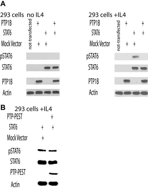
In our previous study, we demonstrated that STAT6 is dephosphorylated by TCPTP in the nucleus.11 Given the high degree of sequence identity and the previously reported capability of these 2 phosphatases to dephosphorylate common substrates,26 we examined whether PTP1B may also regulate IL-4–induced STAT6 signaling. Plasmid constructs for the expression of STAT6 and either wild-type PTP1B, or PTP-PEST used as a control, were cotransfected in HEK 293 cells, which express low levels of endogenous STAT6 and PTP1B (Figure 1). At 48 hours after transfection, STAT6 phosphorylation was assessed by immunoblot analysis using antibodies specific for the tyrosyl phosphorylated STAT6 (pSTAT6) in IL-4–stimulated (30 minutes) and unstimulated cells. In unstimulated cells, no STAT6 phosphorylation was detected. In cells cotransfected with vector control, IL-4 induced the phosphorylation of the overexpressed STAT6. In contrast, STAT6 phosphorylation was not observed in cells overexpressing PTP1B (Figure 1A). No effect on STAT6 phosphorylation was observed in cells transfected with PTP-PEST (Figure 1B). Comparable expression of STAT6 was confirmed by immunoblotting with anti-STAT6 antibody, and equal loading was confirmed by immunoblotting with an actin antibody (Figure 1A,B).
PTP1B dephosphorylates IL-4–induced STAT6. (A,B) HEK293 cells were transfected with PTP1B (A) or PTP-PEST (B) plasmids and either STAT6 plasmid or empty vector. Forty-eight hours after transfection, cells were stimulated with IL-4 (100 U/mL) for 30 minutes. As a control, nontransfected HEK293 cells were similarly stimulated (A). Cellular proteins from unstimulated and IL-4–stimulated cells were resolved by SDS-PAGE and immunoblotted for tyrosine-phosphorylated STAT6 (pSTAT6), STAT6, PTP1B, or PTP-PEST and actin. The result is representative of 3 independent experiments.
PTP1B dephosphorylates IL-4–induced STAT6. (A,B) HEK293 cells were transfected with PTP1B (A) or PTP-PEST (B) plasmids and either STAT6 plasmid or empty vector. Forty-eight hours after transfection, cells were stimulated with IL-4 (100 U/mL) for 30 minutes. As a control, nontransfected HEK293 cells were similarly stimulated (A). Cellular proteins from unstimulated and IL-4–stimulated cells were resolved by SDS-PAGE and immunoblotted for tyrosine-phosphorylated STAT6 (pSTAT6), STAT6, PTP1B, or PTP-PEST and actin. The result is representative of 3 independent experiments.
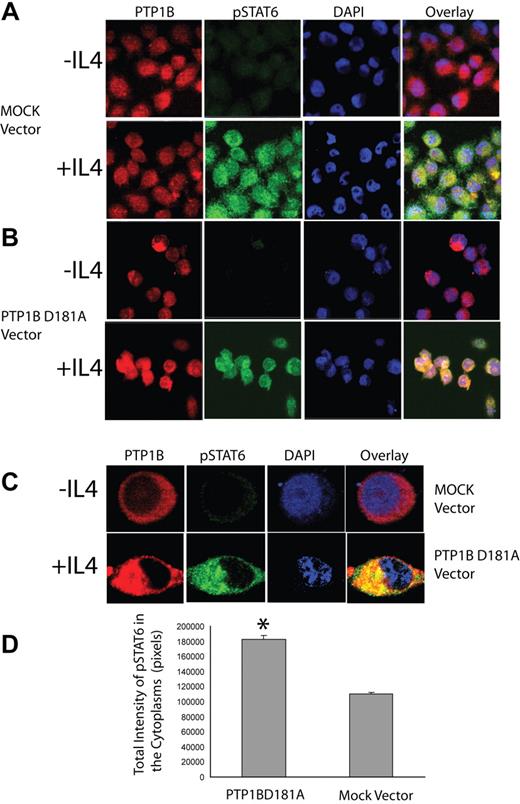
To determine whether PTP1B might act directly on pSTAT6, we used PTP1B-D181A substrate-trapping mutants that can form stable complexes with tyrosine-phosphorylated substrates in a cellular context.27 We determined by immunofluorescence microscopy whether the endoplasmic reticulum–targeted PTP1B-D182A may prevent the nuclear translocation of endogenous phosphorylated STAT6 in response to IL-4 stimulation. In unstimulated vector control-transfected HeLa cells, STAT6 and PTP1B-D182A did not colocalize (data not shown), and there was no evidence for pSTAT6 (Figure 2A). On IL-4 stimulation, endogenous pSTAT6 was readily detected and accumulated in the cytoplasm colocalizing with the PTP1B-D181A mutant (Figure 2B,C). Furthermore, there was a marked and statistically significant (P < .001) increase in the cytoplasmic pSTAT6 in cells expressing PTP1B-D181A compared with control vector-transfected cells (Figure 2D) consistent with PTP1B-D181A trapping and preventing the nuclear translocation of pSTAT6.
PTP1B-D181A colocalizes and traps pSTAT6 in the cytoplasm. Control vector-transfected HeLa cells (A) or cells transiently expressing PTP1B D181A mutant (B) were stimulated with IL-4 (100 U/mL) for 15 minutes, fixed in 4% formaldehyde, and stained with DAPI and antibodies to pSTAT6 and PTP1B as described in “Methods” in “Microscopy.” Cells were visualized by Zeiss LSM510 confocal microscope with a plan-apochromat 40×/1.3 NA oil-immersion lens, 364, 488, and 555 nm laser lines, for DAPI, Alexa Fluor 488 detection of anti-STAT6, and Alexa Fluor 555 detection of anti-PTP1B, respectively. Images were acquired using 1024 × 1024 pixel scan size, 12-bit digitization, and 4-frame averaging. The measurements were performed as described in “Methods” in “Microscopy.” (C) Four-fold magnification of individual cells from experimental conditions shown in panels A and B. (A-C) Slides were viewed with a Zeiss LSM 510/UV confocal Axiovert 200M microscope (Zeiss, Thornwood, NY) using a Zeiss plan-apochrome oil-immersion lens at 40×/1.3 NA and ProLong Gold antifade reagent (Invitrogen, Eugene, OR) as mounting medium. Images were acquired using a Zeiss LSM510/UV confocal scanner, and were processed with Zeiss LSM510 AIM 3.2 SP2 confocal microscope software and Adobe Photoshop version 7.0 (Adobe, San Jose, CA). (D) Pixel intensity corresponding to pSTAT6 was quantified as described in “Methods” in “Microscopy” in 175 control HeLa cells and 175 HeLa cells expressing the PTP1B D181A mutant. The results shown are mean plus or minus SE; P < .001. The results in panels A, B, and D are representative of 3 independent experiments.
PTP1B-D181A colocalizes and traps pSTAT6 in the cytoplasm. Control vector-transfected HeLa cells (A) or cells transiently expressing PTP1B D181A mutant (B) were stimulated with IL-4 (100 U/mL) for 15 minutes, fixed in 4% formaldehyde, and stained with DAPI and antibodies to pSTAT6 and PTP1B as described in “Methods” in “Microscopy.” Cells were visualized by Zeiss LSM510 confocal microscope with a plan-apochromat 40×/1.3 NA oil-immersion lens, 364, 488, and 555 nm laser lines, for DAPI, Alexa Fluor 488 detection of anti-STAT6, and Alexa Fluor 555 detection of anti-PTP1B, respectively. Images were acquired using 1024 × 1024 pixel scan size, 12-bit digitization, and 4-frame averaging. The measurements were performed as described in “Methods” in “Microscopy.” (C) Four-fold magnification of individual cells from experimental conditions shown in panels A and B. (A-C) Slides were viewed with a Zeiss LSM 510/UV confocal Axiovert 200M microscope (Zeiss, Thornwood, NY) using a Zeiss plan-apochrome oil-immersion lens at 40×/1.3 NA and ProLong Gold antifade reagent (Invitrogen, Eugene, OR) as mounting medium. Images were acquired using a Zeiss LSM510/UV confocal scanner, and were processed with Zeiss LSM510 AIM 3.2 SP2 confocal microscope software and Adobe Photoshop version 7.0 (Adobe, San Jose, CA). (D) Pixel intensity corresponding to pSTAT6 was quantified as described in “Methods” in “Microscopy” in 175 control HeLa cells and 175 HeLa cells expressing the PTP1B D181A mutant. The results shown are mean plus or minus SE; P < .001. The results in panels A, B, and D are representative of 3 independent experiments.
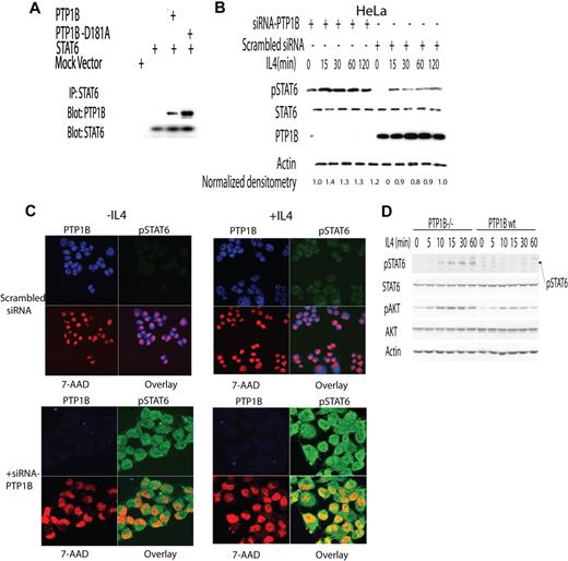
We further assessed whether PTP1B-D181A and pSTAT6 may form a complex in PTP1B immunoprecipitates from IL-4-stimulated cells coexpressing wild-type PTP1B or PTP1B-D181A and Myc-tagged STAT6 (Figure 3A). STAT6 was detected in wild-type PTP1B immunoprecipitates, but this was significantly enhanced in immunoprecipitates of the PTP1B-D181A substrate trapping mutant. Taken together, these results indicate that PTP1B has the capacity to recognize STAT6 as a cellular substrate.
STAT6 is the physiologic substrate of PTP1B. (A) HEK293 cells were transfected with Myc-tagged STAT6 and PTP1B or PTP1B D181A plasmids. At 48 hours after transfection, cells were stimulated with IL-4 (100 U/mL) for 30 minutes. Cellular lysates were extracted and subjected to immunoprecipitation with anti-MYC antibody. (B) HeLa cells were transfected with either SMART pool siRNA for PTP1B or scrambled control (200 pmol). At 72 hours after transfection, the cells were stimulated with IL-4 (100 U/mL) for 15 to 120 minutes. Cellular lysates were extracted at indicated time points and blotted for pSTAT6, STAT6, PTP1B, and actin. Mean relative densitometry of pSTAT6/STAT6 ratio from 3 independent experiments is depicted. The value in specimen siRNA-PTP1B at time point 0 was arbitrarily defined as 1. (C) HeLa cells were transfected with either SMART pool siRNA for PTP1B or scrambled control (200 pmol). At 48 hours after transfection, the cells were stimulated with IL-4 (100 U/mL) for 15 minutes and fixed in 4% formaldehyde and stained with 7AAD (nuclear staining) and antibodies to pSTAT6 and PTP1B. Slides were viewed with a Zeiss LSM510/UV confocal Axiovert 200M microscope (Zeiss, Thornwood, NJ) using a Zeiss plan-apochrome oil-immersion lens at 40×/1.3 NA and ProLong Gold antifade reagent (Invitrogen, Eugene, OR) as mounting medium. Images were acquired using a Zeiss LSM510/UV confocal scanner, and were processed with Zeiss LSM510 AIM 3.2 SP2 confocal microscope software and Adobe Photoshop version 7.0 (Adobe, San Jose, CA). (D) IL-4 induces enhanced STAT6 phosphorylation in PTP1B-deficient cells. Mouse embryo fibroblasts from PTP1B-deficient (−/−) and wild-type (+/+) mice were serum starved for 4 hours and then stimulated with 50 ng/mL IL-4 for the indicated times. Activation of STAT6 and AKT was assessed with phosphotyrosine-specific STAT6 and phosphoserine-specific AKT antibodies. Equal loading was confirmed by immunoblotting with an actin antibody. Representative results of 3 independent experiments are shown.
STAT6 is the physiologic substrate of PTP1B. (A) HEK293 cells were transfected with Myc-tagged STAT6 and PTP1B or PTP1B D181A plasmids. At 48 hours after transfection, cells were stimulated with IL-4 (100 U/mL) for 30 minutes. Cellular lysates were extracted and subjected to immunoprecipitation with anti-MYC antibody. (B) HeLa cells were transfected with either SMART pool siRNA for PTP1B or scrambled control (200 pmol). At 72 hours after transfection, the cells were stimulated with IL-4 (100 U/mL) for 15 to 120 minutes. Cellular lysates were extracted at indicated time points and blotted for pSTAT6, STAT6, PTP1B, and actin. Mean relative densitometry of pSTAT6/STAT6 ratio from 3 independent experiments is depicted. The value in specimen siRNA-PTP1B at time point 0 was arbitrarily defined as 1. (C) HeLa cells were transfected with either SMART pool siRNA for PTP1B or scrambled control (200 pmol). At 48 hours after transfection, the cells were stimulated with IL-4 (100 U/mL) for 15 minutes and fixed in 4% formaldehyde and stained with 7AAD (nuclear staining) and antibodies to pSTAT6 and PTP1B. Slides were viewed with a Zeiss LSM510/UV confocal Axiovert 200M microscope (Zeiss, Thornwood, NJ) using a Zeiss plan-apochrome oil-immersion lens at 40×/1.3 NA and ProLong Gold antifade reagent (Invitrogen, Eugene, OR) as mounting medium. Images were acquired using a Zeiss LSM510/UV confocal scanner, and were processed with Zeiss LSM510 AIM 3.2 SP2 confocal microscope software and Adobe Photoshop version 7.0 (Adobe, San Jose, CA). (D) IL-4 induces enhanced STAT6 phosphorylation in PTP1B-deficient cells. Mouse embryo fibroblasts from PTP1B-deficient (−/−) and wild-type (+/+) mice were serum starved for 4 hours and then stimulated with 50 ng/mL IL-4 for the indicated times. Activation of STAT6 and AKT was assessed with phosphotyrosine-specific STAT6 and phosphoserine-specific AKT antibodies. Equal loading was confirmed by immunoblotting with an actin antibody. Representative results of 3 independent experiments are shown.
To determine whether PTP1B might regulate STAT6 signaling in a physiologic context, we transiently knocked down the expression of PTP1B in HeLa cells using siRNAs and assessed the phosphorylation of STAT6 before and after IL-4 stimulation. The siRNA-induced suppression in PTP1B protein expression was associated with the appearance of pSTAT6 in unstimulated cells as shown by immunoblot analysis (Figure 3B) and by immunofluorescence microscopy (Figure 3C). The increase in basal pSTAT6 was similar to the maximal pSTAT6 occurring in scrambled siRNA-transfected control cells after IL-4 stimulation (Figure 3B). Furthermore, PTP1B knockdown resulted in enhanced STAT6 phosphorylation in response to IL-4 (Figure 3B) beyond that occurring in IL-4–stimulated control cells. The increase in IL-4–stimulated pSTAT6 in PTP1B knockdown cells was associated with a marked increase in cytoplasmic pSTAT6 and a mild increase in nuclear pSTAT6 as assessed by immunofluorescence microscopy (Figure 3C). These results indicate that PTP1B may regulate IL-4–induced STAT6 signaling in vivo. To explore this possibility further, we examined IL-4–induced STAT6 tyrosine phosphorylation in immortalized PTP1B-deficient (Ptpn1−/−) fibroblasts versus those reconstituted wild-type PTP1B.19 IL-4 induced the phosphorylation of STAT6 in PTP1B-deficient fibroblasts but not in the PTP1B-reconstituted cells (Figure 3D). Simultaneous examination of IL-4–induced AKT phosphorylation, which occurs downstream of JAK-mediated PI3K signaling in parallel to STAT6 phosphorylation, demonstrated elevated Akt phosphorylation in PTP1B-deficient fibroblasts (Figure 3D). Notably, the increase in PI3K/Akt signaling was not as pronounced as that for STAT6 phosphorylation consistent with the notion that PTP1B might act directly on STAT6, but raising the possibility that PTP1B may also affect the JAK PTKs that are activated by IL-4.
Previous studies have demonstrated that PTP1B can dephosphorylate JAK2 and TYK2, but not JAK1 or JAK3,16 which are the principal JAK PTKs responsible for propagating the IL-4 signal. Although JAK3 is restricted to the hematopoietic compartment, JAK1 is expressed ubiquitously. To test whether overexpressed PTP1B might dephosphorylate and inactivate JAK1 in response to IL-4 and thus contribute to the suppression of IL-4 signaling, JAK1 or JAK2 tyrosine phopshorylation was assessed in HEK293 cells overexpressing STAT6 plus or minus PTP1B. At 48 hours after transfection, cells were stimulated with IL-4 for 30 minutes and the phosphorylation of JAK1 and JAK2 was assessed by immunoprecipitation with specific antibodies and immunoblot analysis using antibodies specific for the phosphotyrosine (pTyr). In cells cotransfected with STAT6 and control vector, IL-4 induced the phosphorylation of STAT6 and JAK1 but not JAK2, as expected. In contrast, phosphorylation of JAK1 and STAT6 was not observed in cells overexpressing PTP1B (Figure 4A). Furthermore, in specific immunoprecipitates, we noted that PTP1B coimmunoprecipitated with JAK1 in an IL-4–inducible manner (Figure 4B). Thus, these results are consistent with the potential for PTP1B to act directly on JAK PTKs to regulate IL-4 signaling.
Dephosphorylation of pSTAT6 by PTP1B is independent of PTP1B effects on JAK1. (A) HEK293 cells were transfected with STAT6 plasmid and either PTP1B plasmid or empty vector. Forty-eight hours after transfection, cells were stimulated with IL-4 (100 U/mL) for 30 minutes. Cellular lysates were extracted and subjected to immunoprecipitation with anti-JAK1 or JAK2 antibodies followed by anti- pTyr or anti-JAK1 or JAK2 Western immunoblotting. Corresponding cellular lysates were also immunoblotted with STAT6 and pSTAT6-specific antibodies. (B) HeLa cells were either left unstimulated or were stimulated with IL-4 (100 U/mL) for 30 minutes. Cellular lysates were extracted and subjected to immunoprecipitation with anti- JAK1 and PTP1B followed by anti-PTP1B and anti-JAK1 immunoblotting. (C) HeLa cells were transfected with either SMART pool of siRNA for PTP1B or scrambled control (200 pmol). At 72 hours after transfection, the cells were stimulated with IL-4 (100 U/mL) for 15 to 45 minutes with or without the addition of staurosporine starting 15 minutes after IL-4 stimulation. Cellular proteins were immunoblotted for pSTAT6, STAT6, and actin at the indicated times. Representative results of 3 independent experiments are shown.
Dephosphorylation of pSTAT6 by PTP1B is independent of PTP1B effects on JAK1. (A) HEK293 cells were transfected with STAT6 plasmid and either PTP1B plasmid or empty vector. Forty-eight hours after transfection, cells were stimulated with IL-4 (100 U/mL) for 30 minutes. Cellular lysates were extracted and subjected to immunoprecipitation with anti-JAK1 or JAK2 antibodies followed by anti- pTyr or anti-JAK1 or JAK2 Western immunoblotting. Corresponding cellular lysates were also immunoblotted with STAT6 and pSTAT6-specific antibodies. (B) HeLa cells were either left unstimulated or were stimulated with IL-4 (100 U/mL) for 30 minutes. Cellular lysates were extracted and subjected to immunoprecipitation with anti- JAK1 and PTP1B followed by anti-PTP1B and anti-JAK1 immunoblotting. (C) HeLa cells were transfected with either SMART pool of siRNA for PTP1B or scrambled control (200 pmol). At 72 hours after transfection, the cells were stimulated with IL-4 (100 U/mL) for 15 to 45 minutes with or without the addition of staurosporine starting 15 minutes after IL-4 stimulation. Cellular proteins were immunoblotted for pSTAT6, STAT6, and actin at the indicated times. Representative results of 3 independent experiments are shown.
To determine whether the increased pSTAT6 after PTP1B knockdown might also be attributable to PTP1B acting directly on STAT6, as opposed to acting exclusively on the upstream JAK PTKs, we determined the intensity and extent of STAT6 phosphorylation after the JAK protein tyrosine kinases were inhibited with staurosporine (Figure 4C). Staurosporine suppressed IL-4–induced STAT6 phosphorylation in cells tranfected with control scrambled siRNAs and PTP1B-specific siRNAs. However, STAT6 phosphorylation in staurosporine-treated cells was sustained in cells in which PTP1B was knocked down. Therefore, these results are consistent with PTP1B being a direct negative regulator of STAT6 phosphorylation in vivo.
PTP1B interacts with STAT6
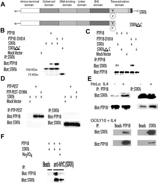
Previously, we reported that TCPTP interacts directly with STAT6,11 whereas others have shown the same to be true for TCPTP and STAT1.28 Accordingly, we determined whether PTP1B may also interact with STAT6. To ascertain whether STAT6 and PTP1B interact directly, STAT6 and PTP1B coimmunoprecipitation experiments were performed. Constructs for the expression of either Myc-fused STAT6 or a STAT6 mutant missing the C terminal transactivation domain but containing Tyr641 that is phosphorylated on IL-4 stimulation (STAT6ΔC, Figure 5A) and either PTP1B or its substrate-trapping mutant PTP1B D181A or PTP-PEST and its trapping mutant PTP-PEST D199A used as controls, were transiently transfected into HEK293 cells (Figure 5B-D). After IL-4 stimulation, cellular lysates were prepared, STAT6 (Figure 5B), PTP1B (Figure 5C), or PTP-PEST (Figure 5D) immunoprecipitated, resolved by SDS-PAGE, and immunoblotted with anti-PTP1B (Figure 5B), anti-STAT6 (Figure 5C), or anti-PTP-PEST (Figure 5D) antibodies. Interactions were noted between STAT6 and wild-type PTP1B (Figure 4B,C) but not PTP-PEST (Figure 5D). Higher amounts of STAT6 were present in PTP1B D181A substrate-trapping mutant immunoprecipitates and vice versa (Figure 5B,C), consistent with the formation of enzyme-substrate complexes and STAT6 serving as a PTP1B substrate. The C terminal–deleted STAT6ΔC mutant was not able to interact with PTP1B (Figure 5B,C), indicating that the C-terminal domain of STAT6 may be required for the interaction with PTP1B, as was also previously shown for the interaction between TCPTP and STAT6.11 Furthermore, because the STAT6ΔC mutant contains tyrosine 641, these observations suggest that the latter is not sufficient for the interaction between phosphorylated STAT6 and PTP1B. Importantly, endogenous STAT6 interacted with endogenous PTP1B in HeLa cells and lymphoma OCILY10 cells (Figure 5E). Notably, in contrast to our previous findings on the interaction between TCPTP and STAT6,11 an interaction between PTP1B and STAT6 was detected in unstimulated cells, and it increased after IL-4 stimulation (Figure 5D; and data not shown). Therefore, these results are consistent with PTP1B regulating STAT6 phosphorylation both under basal conditions and after IL-4 stimulation.
PTP1B interacts with the transactivation domain of STAT6. (A) Schematic representation of STAT6 and its mutant missing the C-terminal transactivation domain (STAT6ΔC). (B,C) HEK293 cells were transfected with MYC-fused STAT6 or STAT6 ΔC plasmids and vector control, PTP1B, or PTP1B D181A plasmids. At 48 hours after transfection, cells were stimulated with IL-4 (100 U/mL) for 30 minutes. Cellular lysates were extracted and subjected to immunoprecipitation with (B) anti-MYC or (C) anti-PTP1B antibodies followed by anti-PTP1B and anti-STAT6 Western blotting. (D) HEK293 cells were transfected with PTP-PEST, PTP-PEST-D199A, STAT6 plasmids, and vector control. At 48 hours after transfection, cells were stimulated with IL-4 (100 U/mL) for 30 minutes. Cellular lysates were extracted and subjected to immunoprecipitation with anti–PTP-PEST or anti-STAT6 followed by blotting with anti-STAT6 and anti–PTP-PEST antibodies, respectively. (E) HeLa and OCILY10 cells were stimulated with IL-4 (100 U/mL) for 30 minutes. Cellular lysates were extracted and subjected to immunoprecipitation with anti-STAT6 followed by anti-PTP1B or anti-STAT6 blotting. (F). STAT6 and PTP1B were transiently expressed in the HEK293 cells, as described in “Methods” in “Cell transfections.” At 48 hours after transfection, the cells were left untreated or incubated for 30 minutes with sodium orthovanadate (2 mM). The cells were then stimulated with IL-4 (100 U/mL) for 30 minutes, and cellular lysates were extracted and subjected to immunoprecipitation with anti-MYC followed by blotting with either anti-PTP1B or anti-STAT6 antibodies. Representative results of 3 independent experiments are shown in panels B-F.
PTP1B interacts with the transactivation domain of STAT6. (A) Schematic representation of STAT6 and its mutant missing the C-terminal transactivation domain (STAT6ΔC). (B,C) HEK293 cells were transfected with MYC-fused STAT6 or STAT6 ΔC plasmids and vector control, PTP1B, or PTP1B D181A plasmids. At 48 hours after transfection, cells were stimulated with IL-4 (100 U/mL) for 30 minutes. Cellular lysates were extracted and subjected to immunoprecipitation with (B) anti-MYC or (C) anti-PTP1B antibodies followed by anti-PTP1B and anti-STAT6 Western blotting. (D) HEK293 cells were transfected with PTP-PEST, PTP-PEST-D199A, STAT6 plasmids, and vector control. At 48 hours after transfection, cells were stimulated with IL-4 (100 U/mL) for 30 minutes. Cellular lysates were extracted and subjected to immunoprecipitation with anti–PTP-PEST or anti-STAT6 followed by blotting with anti-STAT6 and anti–PTP-PEST antibodies, respectively. (E) HeLa and OCILY10 cells were stimulated with IL-4 (100 U/mL) for 30 minutes. Cellular lysates were extracted and subjected to immunoprecipitation with anti-STAT6 followed by anti-PTP1B or anti-STAT6 blotting. (F). STAT6 and PTP1B were transiently expressed in the HEK293 cells, as described in “Methods” in “Cell transfections.” At 48 hours after transfection, the cells were left untreated or incubated for 30 minutes with sodium orthovanadate (2 mM). The cells were then stimulated with IL-4 (100 U/mL) for 30 minutes, and cellular lysates were extracted and subjected to immunoprecipitation with anti-MYC followed by blotting with either anti-PTP1B or anti-STAT6 antibodies. Representative results of 3 independent experiments are shown in panels B-F.
To examine whether the PTP1B catalytic domain contributes to the interaction with STAT6, we assessed whether the PTP active site inhibitor sodium orthovanadate could abrogate the interaction. PTP1B and STAT6 were coimmunoprecipitated in the presence or absence of sodium orthovanadate and its effects on association monitored by immunoblot analysis. Sodium orthovanadate markedly decreased the binding of STAT6 to PTP1B (Figure 5F), thus suggesting that the catalytic domain of PTP1B is required for the interaction with STAT6.
PTP1B inhibits the expression of IL-4–STAT6-induced genes
To examine whether PTP1B has the capacity to modulate IL-4–STAT6-dependent signaling, we tested the effects of overexpression of PTP1B on STAT6 transcriptional activity as assessed in a luciferase reporter assay and by assessing the expression of the endogenous STAT6 target, CD23. PTP1B overexpression markedly reduced IL-4–induced STAT6 reporter luciferase activity (Figure 6A). Furthermore, overexpression of PTP1B in the GCB-like VAL DLBCL cells, which express low levels of PTP1B, markedly reduced the IL-4–induced mRNA expression of CD23, a classic IL-4 target gene (Figure 6B). These data support a potential regulatory role for PTP1B in IL-4–induced STAT6 signaling.
PTP1B inhibits IL-4–induced gene transcription. (A) Mock or PTP1B encoding plasmids were cotransfected into HEK293 cells with the STAT6-driven luciferase reporter-construct C/EBP-N4 and STAT6 and pRL plasmids. Luciferase activity was determined 48 hours after transfection in either unstimulated cells or cells that had been stimulated with IL-4 (100 U/mL) for 6 hours. Numbers refer to relative luciferase activities, the average value obtained for the reporter without activator being taken as 1. Numbers are means and SD of 3 independent experiments, each performed in triplicate. (B) VAL cells were transiently transfected with either control pIRES-hrGFPII vector or pIRES-hrGFPII PTP1B vectors as described in “Methods” in “Cell transfections.” At 48 hours after transfection, cells were sorted for GFP expression and incubated with or without IL-4 (100 U/mL) for 6 hours. RNA was extracted, and CD23 and PTP1B RNA expression was measured by real-time RT-PCR in triplicate. The result is representative of 3 independent experiments.
PTP1B inhibits IL-4–induced gene transcription. (A) Mock or PTP1B encoding plasmids were cotransfected into HEK293 cells with the STAT6-driven luciferase reporter-construct C/EBP-N4 and STAT6 and pRL plasmids. Luciferase activity was determined 48 hours after transfection in either unstimulated cells or cells that had been stimulated with IL-4 (100 U/mL) for 6 hours. Numbers refer to relative luciferase activities, the average value obtained for the reporter without activator being taken as 1. Numbers are means and SD of 3 independent experiments, each performed in triplicate. (B) VAL cells were transiently transfected with either control pIRES-hrGFPII vector or pIRES-hrGFPII PTP1B vectors as described in “Methods” in “Cell transfections.” At 48 hours after transfection, cells were sorted for GFP expression and incubated with or without IL-4 (100 U/mL) for 6 hours. RNA was extracted, and CD23 and PTP1B RNA expression was measured by real-time RT-PCR in triplicate. The result is representative of 3 independent experiments.
PTP1B expression is induced by IL-4 stimulation
Cytokine-induced JAK/STAT signaling pathways are exquisitely sensitive to negative feedback loops. Previously, we demonstrated that IL-4 can induce an increase in TCPTP protein levels, thus generating a negative feedback loop for the suppression of nuclear STAT6 phosphorylation.11 Therefore, we examined whether IL-4 may regulate PTP1B mRNA and protein levels. DLBCL cell lines (RCK8 and VAL) were stimulated with IL-4 for 3 to 12 hours and PTP1B mRNA and protein expression were measured by real-time RT-PCR and immunoblotting, respectively. IL-4 induced PTP1B mRNA as early as 3 hours after stimulation, and this was accompanied by an increase in PTP1B protein levels (Figure 7A). To determine whether the increase in PTP1B protein levels may also be attributed to changes in protein stability, we undertook pulse-chase studies monitoring for the loss of [35S]methionine-labeled protein. We found that IL-4 stimulation significantly decreased the degradation of PTP1B protein (Figure 7B). Overall, these results suggest that IL-4 may affect PTP1B protein levels by stimulating mRNA and protein expression and decreasing PTP1B degradation. To delineate the intracellular signaling pathway mediating the effects of IL-4 on PTP1B mRNA expression, we measured PTP1B mRNA by real-time PCR after siRNA mediated STAT6 knockdown in lymphoma cells (Figure 7C). STAT6 knockdown did not abrogate IL-4–induced increase in PTP1B mRNA, suggesting that the IL-4–induced increase in PTP1B mRNA levels was STAT6 independent. IL-4 stimulation is also known to activate PI3K- and MAPK-signaling pathways.1,9 Therefore, the effects of specific PI3K and MAPK inhibitors on IL-4–induced PTP1B mRNA levels were assessed. Two PI3K inhibitors, Ly294002 (Figure 7D) and wortmannin (data not shown), ameliorated the IL-4–induced increase in PTP1B mRNA, whereas the MAPK inhibitor PD98059 only slightly decreased PTP1B mRNA levels. These findings suggest that IL-4 regulates PTP1B mRNA expression predominantly through the activation of the PI3K pathway.
PTP1B mRNA and protein levels are increased by IL-4 stimulation. (A) DLBCL cell lines RCK8 or VAL (not shown) were stimulated with IL-4 (100 U/mL) for 3, 6, and 12 hours. RNA was extracted and PTP1B mRNA was measured by real-time RT-PCR in triplicate. In addition, cells lysates were prepared and PTP1B and actin were immunoblotted. (B) To monitor for the effect of IL-4 on protein stability, pulse-chase experiments were performed as described in “Methods” in “Pulse-chase immunoprecipitation assays.” Briefly, RCK8 cells were starved of methionine and then labeled with [;35S]methionine for 2 hours, after which they were either left unstimulated or stimulated with IL-4 (100 U/mL) for up to 24 hours and incorporated [35S]methionine monitored in PTP1B immunoprecipitates by autoradiography. MD indicates mean densitometry of 3 independent experiments. The value at time point 0 was arbitrarily defined as 1. (C) VAL DLBCL cells were transfected with control or STAT6 siRNA. At 48 hours after siRNA transfection, the cells were stimulated with IL-4 for 6 hours. RNA was extracted, and PTP1B and CD23 RNA expression was measured by real-time RT-PCR in triplicate. Cells lysates were immunoblotted for STAT6 and actin. (D) VAL DLBCL cells were grown in complete media supplemented with Ly294002 (10 μM) or PD98059 (30 μM) for 30 minutes. The cells were stimulated with IL-4 for 6 hours, RNA was extracted, and PTP1B RNA expression was measured by real-time RT-PCR in triplicate. (E) RCK8 DLBCL cells were grown in complete media either without or with IL-4 (100 U/mL) for 15 hours. The cells were then stimulated with IL-4 (100 U/mL) for 30 minutes, and cellular lysates were extracted and blotted for pSTAT6, STAT6, PTP1B, and actin. (F) RCK8 DLBCL cells were grown in complete media either without (green) or with IL-4 (100 U/mL; red) for 15 hours. The cells were stained with phycoerythrin-conjugated antihuman IL-4R antibody or appropriate anti isotype control antibody (black) and analyzed by flow cytometry. Representative results from 3 independent experiments are shown.
PTP1B mRNA and protein levels are increased by IL-4 stimulation. (A) DLBCL cell lines RCK8 or VAL (not shown) were stimulated with IL-4 (100 U/mL) for 3, 6, and 12 hours. RNA was extracted and PTP1B mRNA was measured by real-time RT-PCR in triplicate. In addition, cells lysates were prepared and PTP1B and actin were immunoblotted. (B) To monitor for the effect of IL-4 on protein stability, pulse-chase experiments were performed as described in “Methods” in “Pulse-chase immunoprecipitation assays.” Briefly, RCK8 cells were starved of methionine and then labeled with [;35S]methionine for 2 hours, after which they were either left unstimulated or stimulated with IL-4 (100 U/mL) for up to 24 hours and incorporated [35S]methionine monitored in PTP1B immunoprecipitates by autoradiography. MD indicates mean densitometry of 3 independent experiments. The value at time point 0 was arbitrarily defined as 1. (C) VAL DLBCL cells were transfected with control or STAT6 siRNA. At 48 hours after siRNA transfection, the cells were stimulated with IL-4 for 6 hours. RNA was extracted, and PTP1B and CD23 RNA expression was measured by real-time RT-PCR in triplicate. Cells lysates were immunoblotted for STAT6 and actin. (D) VAL DLBCL cells were grown in complete media supplemented with Ly294002 (10 μM) or PD98059 (30 μM) for 30 minutes. The cells were stimulated with IL-4 for 6 hours, RNA was extracted, and PTP1B RNA expression was measured by real-time RT-PCR in triplicate. (E) RCK8 DLBCL cells were grown in complete media either without or with IL-4 (100 U/mL) for 15 hours. The cells were then stimulated with IL-4 (100 U/mL) for 30 minutes, and cellular lysates were extracted and blotted for pSTAT6, STAT6, PTP1B, and actin. (F) RCK8 DLBCL cells were grown in complete media either without (green) or with IL-4 (100 U/mL; red) for 15 hours. The cells were stained with phycoerythrin-conjugated antihuman IL-4R antibody or appropriate anti isotype control antibody (black) and analyzed by flow cytometry. Representative results from 3 independent experiments are shown.
To examine the potential physiologic consequences of the IL-4–PTP1B negative feedback loop, we have examined STAT6 phosphorylation on restimulation with IL-4 in RCK8 cells (Figure 7E). Prior exposure to IL-4 resulted in increased PTP1B protein levels that were associated with reduced pSTAT6 levels on IL-4 restimulation at 15 hours. The reduced pSTAT6 levels on IL-4 restimulation were not the result of IL-4R internalization and down-regulation because flow cytometric analyses demonstrated that cell surface IL-4R expression increased in response to IL-4 (Figure 7F), consistent with previous reports demonstrating that the IL-4R is an IL-4 target gene.7,29 Overall, these observations suggest that the IL-4–PTP1B negative feedback loop might control the magnitude and duration of the IL-4–induced intracellular signaling.
Coordinated regulation of STAT6 by PTP1B and TCPTP
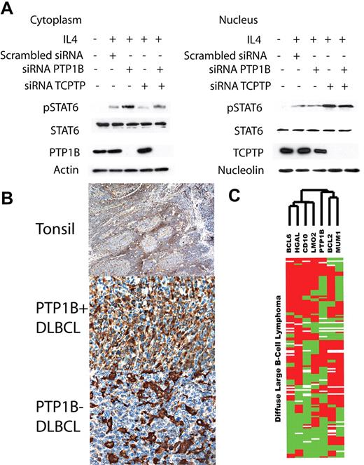
Our current and previous studies11 suggest that both cytoplasmic PTP1B and nuclear TCPTP participate in STAT6 dephosphorylation. To examine whether the 2 PTPs act in collaborative manner to regulate STAT6 signaling, we transiently knocked down the expression of PTP1B, TCPTP, or both in HeLa cells using siRNAs and assessed the phosphorylation of STAT6 before and after IL-4 stimulation (Figure 8A). Knockdown of PTP1B led to a marked increase in cytoplasmic pSTAT6, but only a mild increase in the nuclear pSTAT6. In contrast, knockdown of TCPTP resulted in a marked increase in the nuclear pSTAT6 and a decrease in the cytoplasmic pSTAT6, which may be the result of nuclear accumulation of STAT6. Simultaneous knockdown of both PTP1B and TCPTP resulted in increased pSTAT6 in both cytoplasm and nucleus. Overall, these results suggest that PTP1B and TCPTP coordinately regulate STAT6 phosphorylation by acting in different subcellular compartments.
Coordinated regulation of STAT6 by PTP1B and TCPTP and PTP1B expression in DLBCL. (A) HeLa cells were transfected with either SMART pool siRNA for PTP1B, TCPTP, or scrambled control (200 pmol). At 72 hours after transfection, the cells were stimulated with IL-4 (100 U/mL) for 15 minutes. Cytoplasmic and nuclear lysates were extracted and blotted for tyrosine-phosphorylated STAT6 (pSTAT6), STAT6, PTP1B, TCPTP, actin, and nucleolin. (B) Immunohistochemical staining for PTP1B in tonsils and DLBCL. Low magnification image of normal tonsil sections shows PTP1B-specific staining mainly outside germinal centers (original magnification ×40); PTP1B staining is localized to the cytoplasm. Also demonstrated are representative examples of PTP1B immunostaining in DLBCL (original magnification ×400). Images of immunohistologic staining were acquired using a Nikon Eclipse E400 microscope (Nikon, Tokyo, Japan) and a Nikon DS-L1 digital camera. (C) Hierarchical cluster analysis of immunohistologic data. The expression patterns of 7 proteins (LMO2, HGAL, CD10, BCL6, MUM1/IRF4 (MUM1), BCL2, and PTP1B) in 80 cases of DLBCL are shown. Positive staining is indicated in red, lack of staining in green, and uninformative data in white. PTP1B protein expression is clustered on the same branch of the dendrogram as nongerminal-center proteins MUM1 and BCL2 and away from germinal-center proteins HGAL, BCL6, LMO2, and CD10.
Coordinated regulation of STAT6 by PTP1B and TCPTP and PTP1B expression in DLBCL. (A) HeLa cells were transfected with either SMART pool siRNA for PTP1B, TCPTP, or scrambled control (200 pmol). At 72 hours after transfection, the cells were stimulated with IL-4 (100 U/mL) for 15 minutes. Cytoplasmic and nuclear lysates were extracted and blotted for tyrosine-phosphorylated STAT6 (pSTAT6), STAT6, PTP1B, TCPTP, actin, and nucleolin. (B) Immunohistochemical staining for PTP1B in tonsils and DLBCL. Low magnification image of normal tonsil sections shows PTP1B-specific staining mainly outside germinal centers (original magnification ×40); PTP1B staining is localized to the cytoplasm. Also demonstrated are representative examples of PTP1B immunostaining in DLBCL (original magnification ×400). Images of immunohistologic staining were acquired using a Nikon Eclipse E400 microscope (Nikon, Tokyo, Japan) and a Nikon DS-L1 digital camera. (C) Hierarchical cluster analysis of immunohistologic data. The expression patterns of 7 proteins (LMO2, HGAL, CD10, BCL6, MUM1/IRF4 (MUM1), BCL2, and PTP1B) in 80 cases of DLBCL are shown. Positive staining is indicated in red, lack of staining in green, and uninformative data in white. PTP1B protein expression is clustered on the same branch of the dendrogram as nongerminal-center proteins MUM1 and BCL2 and away from germinal-center proteins HGAL, BCL6, LMO2, and CD10.
PTP1B expression in non-Hodgkin lymphoma
We have previously reported rapid nuclear and cytoplasmic dephosphorylation of STAT6 in ABC-like DLBCL.7 Furthermore, we have demonstrated that, compared with GCB-like DLBCL, ABC-like DLBCL express higher levels of TCPTP that may account for nuclear STAT6 dephosphorylation. To examine PTP1B protein expression in non-Hodgkin lymphoma, we performed immunohistochemical analysis of PTP1B protein in 371 cases of hematolymphoid malignancies (Table 1) and normal tonsils. In normal tonsils, PTP1B immunostaining was seen in a subset of B cells and T cells mainly outside of germinal centers (Figure 8B). Staining was also seen in histiocytes/macrophage lineage cells (not shown). Staining was localized to the cytoplasm of cells in tonsils and hematopoietic neoplasms (Figure 8B). Interestingly, whereas grade 1 follicular lymphomas (FLs) did not express PTP1B, grade 2 and 3 FLs expressed PTP1B in 14% and 29% of cases, respectively. PTP1B expression was observed in 58% of DLBCL, 62% of peripheral T-cell lymphomas, and 33% of marginal zone lymphomas but was less frequent in other lymphoid tumors. To further elucidate PTP1B expression in DLBCL subtypes, we compared its expression with that of 6 additional markers (HGAL, BCL6, CD10, LMO2, BCL2, and MUM1) documented in our previous work.25 Hierarchical cluster analysis demonstrated that PTP1B protein correlated with the expression patterns of ABC-specific markers BCL2 and MUM1, but not with germinal center markers. Thus, these results suggest that PTP1B is more commonly expressed the ABC-like DLBCL.
Immunohistologic analysis of PTP1B protein expression in hematolymphoid neoplasia
| Lymphoma subtype . | Total positive* . | % positive . |
|---|---|---|
| B-cell lymphoma (N = 339) | ||
| Follicular lymphoma | ||
| Grade 1 | 0/48 | 0 |
| Grade 2 | 8/59 | 14 |
| Grade 3 | 20/68 | 29 |
| Diffuse large B-cell lymphoma | 46/80 | 58 |
| Burkitt lymphoma | 0/4 | 0 |
| Marginal zone lymphoma | 7/21 | 33 |
| Mantle cell lymphoma | 3/18 | 17 |
| Small lymphocytic lymphoma/CLL | 2/32 | 6 |
| Precursor B-lymphoblastic lymphoma | 1/7 | 14 |
| Plasma cell neoplasms | 0/2 | 0 |
| T-cell lymphoma (N = 35) | ||
| Precursor T-lymphoblastic lymphoma | 0/11 | 0 |
| Peripheral T-cell lymphoma | 10/16 | 62 |
| Anaplastic large cell lymphoma | 1/5 | 20 |
| NK lymphoma | 0/3 | 0 |
| Histiocytic neoplasms (N = 7) | ||
| Langerhan cell histiocytosis | 3/4 | 75 |
| Rosai-Dorfman disease | 3/3 | 100 |
Cases were scored positive if more than 30% of lymphoma cells stained for PTP1B.
Discussion
PTP1B is the prototype for the PTP superfamily of enzymes and has been implicated in multiple signaling pathways. PTP1B is a critical physiologic regulator of receptor PTKs for insulin,30,31 insulin-like growth factor 1,30,32 colony-stimulating factor 1,33 epidermal growth factor,19,27,34 and platelet growth factor.19,34 Furthermore, PTP1B has been implicated in the regulation of cytokine signaling, including that mediated by leptin and growth hormone, by attenuating the phosphorylation of JAK2 and TYK2.35,36 However, PTP1B was reported not to recognize JAK1 and JAK3 as substrates that are thought to be responsible for IL-4 signaling16,37 ; because there is a 72% amino acid identity in the PTP domains of PTP1B and TCPTP, these enzymes share some of their targets, such as epidermal growth factor and insulin receptors as well as STAT5a/b.15,26,38 In our previous work, we have demonstrated that TCPTP can dephosphorylate STAT6 in the nucleus in response to IL-411 in addition to its ability to dephosphorylate JAK1 and JAK3.37 In the present study, we extend these observations and demonstrate that PTP1B dephosphorylates STAT6 and has the capacity to regulate the JAK PTKs in response to IL-4. Similarly to TCPTP, PTP1B's regulation of STAT6 phosphorylation, was direct and independent of its ability to inactivate JAK PTKs because STAT6 phosphorylation was prolonged in PTP1B knockdown cells relative to control cells in the presence of staurosporine, a potent inhibitor of tyrosine kinases. Furthermore, we have shown that a direct interaction occurs between residues in the STAT6 C-terminal transactivation domain distal to tyrosine 641 and the PTP1B catalytic domain. We have demonstrated that complex formation between PTP1B and STAT6 is enhanced in the context of the PTP1B D181A substrate trapping mutation and that the interaction between PTP1B and STAT6 can be suppressed by the PTP active site inhibitor orthovanadate. In addition, we have shown that PTP1B overexpression or deficiency as a result of RNAi-mediated knockdown or genetic manipulation can lead to decreased and increased IL-4-induced STAT6 phosphorylation, respectively. Finally, we have demonstrated previously that recombinant PTP1B can dephosphorylate STAT6 in vitro.7 Taken together, these findings establish STAT6 as a bona fide PTP1B substrate conforming to all of the recently established criteria.26
Our results suggest that PTP1B and TCPTP may coordinately regulate STAT6 signaling in the cellular context by dephosphorylating STAT6 in the cytoplasm and nucleus, respectively. The individual or combined knockdown of PTP1B and TCPTP resulted in distinct but additive effects on the phosphorylation of STAT6 in the cytoplasm and nucleus. Similar to our findings in this study, PTP1B and TCPTP have been reported to act in concert to regulate the intensity and duration of insulin receptor activation by differentially modulating the phosphorylation of specific insulin receptor sites.39 Although STAT6 harbors a single tyrosine phosphorylation site, thus excluding the possibility that PTP1B and TCPTP act on different sites, some differences in the interaction between STAT6 with PTP1B and TCPTP were observed. In contrast to the TCPTP-STAT6 interactions, which were detected only on IL-4 stimulation, interactions of PTP1B with STAT6 were also observed in resting cells and increased on IL-4 stimulation. This may suggest that the PTP1B and STAT6 proteins interact in 2 different modes: phosphorylation-dependent and -independent. Consistent with this notion, previous studies have shown that PTP1B may interact with unphosphorylated proteins through its carboxy-terminal proline-rich motif,40,41 whereas other have reported that PTP1B may interact with the insulin receptor via the PTP1B catalytic domain but independent of its active site.42 Alternatively, but not mutually exclusive, it is also possible that binding in resting cells may reflect the ongoing process of phosphorylation by JAK PTKs and dephosphorylation by PTP1B. A similar interaction was not seen previously for TCPTP and STAT6 in unstimulated cells,11 consistent with the notion that TCPTP acts on STAT6 in the nucleus.11
Previous studies have demonstrated that YB-1 may induce transcription from the PTP1B promoter in response to insulin and IL-6 family cytokines.43 In addition, the Bcr-Abl oncogene has been shown to promote binding of Sp1 and Sp3 to the PTP1B promoter, thereby increasing PTP1B expression.44 Moreover, recent studies indicate that the NAD-dependent protein deacetylase SIRT1 can repress PTP1B transcription,45 whereas the proinflammatory cytokine TNF can induce the expression of PTP1B in insulin responsive tissue to contribute to insulin and leptin resistance.46 Herein we have demonstrated that IL-4 can induce the expression of PTP1B, thus establishing the existence of a new negative feedback loop, which may restrict the time-course of IL-4–induced STAT6 phosphorylation and signaling. The IL-4–induced increase in PTP1B mRNA levels was mediated by PI3K signaling and was independent of STAT6, whereas the mechanism for the IL-4–induced increase in PTP1B protein stability remains unclear. Elucidating this new negative regulatory feedback loop that may regulate the extent of STAT6 phosphorylation, coupled with previously reported IL-4–induced TCPTP negative regulatory loop mediating STAT6 and JAK dephosphorylation11 as well as cytokine-induced SOCS pathways,10 further highlights the complexity and fine-tuning potential of the intracellular regulation of cytokine signaling.
The identification of STAT6 as a PTP1B substrate might have important clinical implications. For example the augmentation of PTP1B expression and/or activity may serve as new therapeutic strategy to control allergic and autoimmune diseases by the suppression of IL-4 signaling. Furthermore, PTP1B may also play an important role in the responses of lymphoma cells to their microenvironment and possibly play a role in lymphomagenesis. Consistent with the latter, recent studies have demonstrated that PTP1B may also play an important role in lymphomagenesis in mice.47 In our studies, we found that, whereas PTP1B was expressed in only a small fraction of cells in germinal center and not expressed in grade 1 FL, it was expressed in higher grades of FL and in 58% of DLBCL. Previously, we reported distinct responsiveness of GCB-like and ABC-like DLBCL cell lines to IL-4 stimulation7 with enhanced STAT6 dephosphorylation in the ABC-like cell lines resulting in noninducibility of IL-4 target genes in these tumors. Furthermore, we have demonstrated that IL-4 may increase doxorubicin and complement-mediated rituxmab cellular cytotoxicity in GCB-like cell-lines but protects the ABC-like DLBCL.8 At least some of the observed differences were attributed to the differential expression of TCPTP. However, PTP1B mRNA was also reported to be differentially expressed in DLBCL subtypes, being highly expressed in the ABC-like tumors.48-50 Herein we extend these observations and demonstrate that PTP1B protein is preferentially expressed in ABC-like DLBCL and may also contribute to the differential response to IL-4 in these lymphomas through the direct regulation of cytoplasmic STAT6 phosphorylation.
The publication costs of this article were defrayed in part by page charge payment. Therefore, and solely to indicate this fact, this article is hereby marked “advertisement” in accordance with 18 USC section 1734.
Acknowledgments
We thank Dr Benjamin Neel (Ontario Cancer Institute, Toronto) for the PTP1B−/− and reconstituted cells and Dr Nicholas Tonks (Cold Spring Harbor Laboratory, NY) for the PTP-PEST constructs.
This work was supported by the United States Public Health Service, National Institutes of Health (RO1 CA109335 and RO1 CA122105) (I.S.L.), the Dwoskin Family Foundation (I.S.L.), and the National Health and Medical Research Council of Australia (T.T.).
National Institutes of Health
Authorship
Contribution: X.L. and Y.N. performed experiments and analyzed the data; R.M., B.S., X.J., and K.A.S. performed experiments; T.T. designed the study, analyzed the data, and wrote the paper; I.S.L. planned and designed the study, analyzed the data, and wrote the paper; all the authors approved the final version of the manuscript.
Conflict-of-interest disclosure: The authors declare no competing financial interests.
Correspondence: Tony Tiganis, Department of Biochemistry and Molecular Biology, Monash University, Victoria, Australia; e-mail: Tony.Tiganis@med.monash.edu.au; and Izidore S. Lossos, Sylvester Comprehensive Cancer Center, Department of Hematology and Oncology, University of Miami, 1475 NW 12th Avenue (D8-4), Miami, FL 33136; e-mail: ilossos@med.miami.edu.



![Figure 7. PTP1B mRNA and protein levels are increased by IL-4 stimulation. (A) DLBCL cell lines RCK8 or VAL (not shown) were stimulated with IL-4 (100 U/mL) for 3, 6, and 12 hours. RNA was extracted and PTP1B mRNA was measured by real-time RT-PCR in triplicate. In addition, cells lysates were prepared and PTP1B and actin were immunoblotted. (B) To monitor for the effect of IL-4 on protein stability, pulse-chase experiments were performed as described in “Methods” in “Pulse-chase immunoprecipitation assays.” Briefly, RCK8 cells were starved of methionine and then labeled with [;35S]methionine for 2 hours, after which they were either left unstimulated or stimulated with IL-4 (100 U/mL) for up to 24 hours and incorporated [35S]methionine monitored in PTP1B immunoprecipitates by autoradiography. MD indicates mean densitometry of 3 independent experiments. The value at time point 0 was arbitrarily defined as 1. (C) VAL DLBCL cells were transfected with control or STAT6 siRNA. At 48 hours after siRNA transfection, the cells were stimulated with IL-4 for 6 hours. RNA was extracted, and PTP1B and CD23 RNA expression was measured by real-time RT-PCR in triplicate. Cells lysates were immunoblotted for STAT6 and actin. (D) VAL DLBCL cells were grown in complete media supplemented with Ly294002 (10 μM) or PD98059 (30 μM) for 30 minutes. The cells were stimulated with IL-4 for 6 hours, RNA was extracted, and PTP1B RNA expression was measured by real-time RT-PCR in triplicate. (E) RCK8 DLBCL cells were grown in complete media either without or with IL-4 (100 U/mL) for 15 hours. The cells were then stimulated with IL-4 (100 U/mL) for 30 minutes, and cellular lysates were extracted and blotted for pSTAT6, STAT6, PTP1B, and actin. (F) RCK8 DLBCL cells were grown in complete media either without (green) or with IL-4 (100 U/mL; red) for 15 hours. The cells were stained with phycoerythrin-conjugated antihuman IL-4R antibody or appropriate anti isotype control antibody (black) and analyzed by flow cytometry. Representative results from 3 independent experiments are shown.](https://ash.silverchair-cdn.com/ash/content_public/journal/blood/112/10/10.1182_blood-2008-03-148726/7/m_zh80210826420007.jpeg?Expires=1769085220&Signature=1CbSeMwpQmREa-82t2aR8HMCdH8VIbSXUDBh0lAZM28nXKATrBiBlQcECo055FYSvKcrH8kSBrf~pJ0xBd6pyFC14S06RIeioECtRxSBPafcG9a1WrQQKOT16aDzfEX~-Juv~CtjZ7PbHM6fUs-KuY7SkTlsQkUGGrKvNIZcgFKxMHSFX7nWxWiZADkpzgrij4E0mxEj-fTPOEoFSeIr1rNeNN1kKDX6bva7~w1IGKUovaP51Nx69HYWL5wZKrKXigeIQC-q6pAplZUZfC0qTjLQOLN0J4Ug0Og-KrLfoVbCxoybYL2ro7jfo5zNprtZdgUNndKdRKtutSx7HfJvVg__&Key-Pair-Id=APKAIE5G5CRDK6RD3PGA)
This feature is available to Subscribers Only
Sign In or Create an Account Close Modal