Abstract
The emergence of resistance to imatinib (IM) mediated by mutations in the BCR-ABL domain has become a major challenge in the treatment of chronic myeloid leukemia (CML). Here, we report on studies performed with a novel small molecule inhibitor, PHA-739358, which selectively targets Bcr-Abl and Aurora kinases A to C. PHA-739358 exhibits strong antiproliferative and proapoptotic activity against a broad panel of human BCR-ABL–positive and –negative cell lines and against murine BaF3 cells ectopically expressing wild-type (wt) or IM-resistant BCR-ABL mutants, including T315I. Pharmacologic synergism of IM and PHA-739358 was observed in leukemia cell lines with subtotal resistance to IM. Treatment with PHA-739358 significantly decreased phosphorylation of histone H3, a marker of Aurora B activity and of CrkL, a downstream target of Bcr-Abl, suggesting that PHA-739358 acts via combined inhibition of Bcr-Abl and Aurora kinases. Moreover, strong antiproliferative effects of PHA-739358 were observed in CD34+ cells derived from untreated CML patients and from IM-resistant individuals in chronic phase or blast crisis, including those harboring the T315I mutation. Thus, PHA-739358 represents a promising new strategy for treatment of IM-resistant BCR-ABL-positive leukemias, including those harboring the T315I mutation. Clinical trials investigating this compound in IM-resistant CML have recently been initiated.
Introduction
Chronic myeloid leukemia (CML) is a hematopoietic disorder characterized by the presence of a reciprocal translocation involving the long arms of chromosome 9 and 22. The resultant BCR-ABL fusion gene encodes a constitutively activated tyrosine kinase, which phosphorylates a broad range of substrates, many of which are key enzymes in cellular signal transduction.1
The tyrosine kinase inhibitor imatinib (IM, formerly STI571, Gleevec) selectively targets the ATP binding site of Bcr-Abl.2 Based on numerous clinical studies,3-6 IM became the new “gold standard” in conventional treatment of CML. However, the emergence of resistance to IM remains a major problem. In particular, patients with advanced stages of CML either do not respond or often develop resistance to IM, resulting in remissions lasting for only 6 to 12 months. Clones expressing mutant forms of BCR-ABL are frequently detected in these patients.2 Thus, mutations in the kinase domain of Abl preventing the adoption of the inactive conformation required for IM binding or directly interfering with the inhibitor interaction have been identified as the most prevalent reason for resistance in IM-treated patients. More than 70 point mutations have been described so far and can be assigned to 4 major groups based on their location in the kinase domain: the ATP binding loop (∼40% of the mutations), the gatekeeper residue Thr315 (25% of the mutations), the catalytic domain (25% of the mutations), or the activation loop (5% of the mutations).2,7,8 Second-generation Bcr-Abl inhibitors, such as dasatinib,9 nilotinib,10 and bosutinib,11 are capable of overcoming the majority of these mutations. However, so far none of these novel tyrosine kinase inhibitors significantly suppresses the proliferation of leukemia cells harboring the T315I mutation.12 Because this mutation is predicted to play an even more important clinical role in the future resulting from an increased selection pressure on resistant leukemic subclones, the need for alternative therapeutic strategies is obvious.
Aurora kinases play a critical role in the regulation of mitotic processes during cell division.13,14 However, inappropriate expression of these enzymes may result in aneuploidy and carcinogenesis.15 Consequently, the potential therapeutic value of targeting Aurora kinases has become a focus of anticancer therapy.16,17 Thus, growth inhibition of various malignancies has been demonstrated both in in vitro and in vivo experiments.18 Most recently, the Aurora kinase inhibitor VX-680/MK-0457 was found to be active ex vivo against cells from patients harboring the BCR-ABL-T315I mutation19 ; in parallel, clinical responses were achieved in patients bearing T315I mutated BCR-ABL.20
Here we report on in vitro studies performed with a novel kinase inhibitor, PHA-739358, which exhibits potent inhibitory activity against all 3 of the known Aurora kinases (A, B, and C) as well as Bcr-Abl tyrosine kinase.21 To study the therapeutic potential of this new compound, we examined the antiproliferative and proapoptotic effects in IM-sensitive and -resistant leukemic cell lines. Furthermore, the efficacy of PHA-739358 was evaluated in primary CD34+ cells derived from patients with newly diagnosed CML, in IM-resistant blast crisis, and from a patient harboring an IM- and dasatinib-resistant T315I mutation. By studying the signal transduction pathways involved in defined resistance-conferring BCR-ABL mutants, we could demonstrate that the antiproliferative effects of PHA-739358 in CML cells are mediated by functional inhibition of both Bcr-Abl and Aurora kinases.
Methods
Reagents
IM was kindly provided by Novartis Pharma (Basel, Switzerland). For combination studies, IM was obtained commercially from Toronto Research Chemicals (Toronto, ON). PHA-739358 was kindly provided by Nerviano Medical Sciences (Milan, Italy). Stock solutions of IM (10 mg/mL; in dimethyl sulfoxide [DMSO]/H2O [1:1]) and PHA-739358 (10 mg/mL; in DMSO) were stored at −20°C. The highest concentration of DMSO in cell culture medium was less than or equal to 0.1% and did not have any effect on cell growth.
Cell culture techniques
Human cell lines K562, HL60, and murine BaF3 cells were obtained from DSMZ (Bielefeld, Germany). BaF3-p210, -M351T, -E255K, and -T315I cells were kindly provided by N. P. Shah and C. L. Sawyers (University of California–Los Angeles). Cells were cultured under standard conditions (37°C/humidified atmosphere/5%CO2) in RPMI 1640 medium (Invitrogen, Paisley, United Kingdom) containing 10% fetal bovine serum (Biochrom, Berlin, Germany). Medium for IL3-dependent BaF3 cells was supplemented with 1 ng/mL of recombinant murine interleukin-3.
Purification of stem and progenitor cells
Peripheral blood or bone marrow samples from CML patients and healthy donors were collected with informed consent according to institutional guidelines of the University of Hamburg Medical Center. CD34+ cells were purified using a Midi-MACS CD34 Isolation Kit (Miltenyi Biotec, Bergisch Gladbach, Germany) as previously described.22 Purity of CD34+ cells quantified by flow cytometry ranged between 93% and 99% in all samples.
Short-term expansion of CD34+ cells
For short-term expansion assays, 103 CD34+ cells were plated in triplicates in 96-well plates (Nunc A/S, Roskilde, Denmark) containing 100 μL of serum-free medium per well supplemented with human stem-cell factor (100 ng/mL), human Flt-3 Ligand (100 ng/mL), human thrombopoietin (50 ng/mL), human interleukin-3 and -6 (IL-3 and IL-6, respectively, both 20 ng/mL), and granulocyte colony-stimulating factor (20 ng/mL) plus PHA-739358 or IM at the indicated concentrations. After 5 days, an additional 100 μL of cytokine and PHA-739358 or IM containing medium were added. Cell numbers within each individual well were evaluated at days 3, 6, and 9 or at days 3, 6, and 12 for healthy donor samples.
MTT assay
Cells were seeded in triplicates in 96-well flat-bottomed microtiter plates (BD Biosciences, Heidelberg, Germany) at a density of 1.5 × 10⁴ cells/well in 150 μL of their respective media. After 24 hours increasing concentrations of PHA-739358 (0-5.0 μM) or/and IM (0-20 μM) were added. After 48 hours of treatment, the viable cells were assayed for their ability to transform 3-(4,5-dimethylthiazol-2-yl)-2,5-diphenyltetrazolium bromide (MTT) into purple formazan, as described previously.23 The compound concentration that inhibits response by 50% (IC50) is defined as the concentration resulting in 50% growth inhibition that corresponds to the fraction affected (Fa value) of 0.5. All tested cells were exposed to 10 different concentrations of each compound. Fraction affected (Fa) and dose-effect relationship at the point of IC50 were analyzed by CalcuSyn Software (Biosoft, Cambridge, United Kingdom).
Analysis of DNA content and apoptosis by flow cytometry
Cells were plated in triplicates in 6-well plates and cultured in 2 mL of their respective media. After 24 hours, cells were exposed to increasing concentrations of PHA-739358 for 48 hours, then washed with phosphate-buffered saline (PBS), and fixed in cold 70% ethanol overnight at −20°C. For flow cytometric analysis, cells were washed twice with PBS, resuspended in PBS containing RNAse A (100 μg/mL) and propidium iodide (10 μg/mL), and incubated for 30 minutes on ice; 10 000 cells were analyzed from each sample.
Assessment of phosphorylation status by intracellular flow cytometry
Cells exposed to 5 μM PHA-739358 or 5 μM IM for 2 hours or 24 hours were collected, fixed in 2% formaldehyde for 10 minutes at 37°C, chilled on ice for 1 minute, and then permeabilized with ice-cold 90% methanol for 30 minutes on ice. From each sample, 5 × 10⁵ cells were washed with 2 mL incubation buffer (PBS/0.5% bovine serum albumin), centrifuged at 50g for 5 minutes, and resuspended in 100 μL of incubation buffer with 2.0 μL of Phospho-CrkL, Phospho-Stat5, or Phospho-Histone H3-(Ser10) specific antibody (all antibodies from Cell Signaling Technology, Danvers, MA). After 45 minutes of incubation at RT, cells were washed twice, resuspended in 100 μL incubation buffer with 0.5 μL of the secondary antibody (antirabbit IgG FITC-conjugate; Jackson ImmunoResearch Europe, Newmarket, United Kingdom), and incubated at RT for 30 minutes in the dark. Again, cells were washed twice with washing buffer. Samples stained with specific Phospho-Histone H3-(Ser10) antibody were also stained with propidium iodide.
All samples were analyzed by flow cytometry, and isotype controls were included for cytometer setup. The amount of phosphorylated proteins (P-CrkL and P-Stat5) was determined as the geometric mean fluorescence intensity, and the changes of the phosphorylation status were expressed as a percentage of the no-drug control.
CML CD34+ cells were incubated with either 5 μM PHA-739358 for 72 hours and 96 hours or with 5 μM IM for 96 hours. After collection, cells were prepared as described above.
Western blotting
K562 cells cultured under standard conditions were exposed for 1 hour to 2 μM and 5 μM of PHA-739358 or IM. Collected cells were lysed in sodium dodecyl sulfate (SDS) buffer (125 mM Tris-HCl, pH 6.8, 2% SDS) and after sonication and boiling, 20 μg total extract of the indicated samples was loaded on SDS-polyacrylamide gel electrophoresis precast gels (Invitrogen) and immunoblotted. The following antibodies were used: c-Abl (Sigma-Aldrich, Chemie, Munich, Germany), Phospho-H3-(Ser10; Upstate), Histone-H3 (Abcam, Cambridge, United Kingdom), Stat5 (Cell Signaling Technology). Anti-PathScan Bcr-Abl activity assay (Cell Signaling Technology) was used for the detection of Phospho-Bcr-Abl, Phospho-Stat5, and Phospho-CrkL. Immunoreactive signals were detected with SuperSignal West Pico Chemiluminescent Substrate (Pierce Chemical, Rockford, IL).
Evaluation of PHA-739358 and IM in vivo
To evaluate the efficacy and toxicity of PHA-739358 in vivo, a subcutaneous animal model for CML was used; 5 × 10⁷ K562 cells were injected into the flanks of female SCID mice and tumor growth was monitored daily by palpation. On day 7, when tumors reached an estimated weight of 100 to 150 mg, animals were assigned to 3 experimental groups by random selection and received the following treatment for a period of 10 days: group 1, control, vehicle solution (7 mice); group 2, PHA-739358 twice a day intraperitoneally at a dose of 15 mg/kg (7 mice); and group 3, IM twice a day per os at 100 mg/kg. Tumor growth was assessed by caliper, and tumor weight was calculated according to the following formula: Tumor weight = [length (mm) × width2 (mm)]/2. Toxicity was monitored by changes in body weight and vitality of the animals. Data were expressed as mean plus or minus SE, and results were compared by Dunnett test. P less than .05 was considered significant. All experiments were done in accordance with the institutional guidelines for the welfare of experimental animals.
Results
PHA-739358 inhibits proliferation of leukemic cell lines independent of BCR-ABL mutational status
The potential effects of PHA-739358 treatment on cellular proliferation were evaluated using MTT assays with a panel of human and murine leukemic and nonleukemic cell lines. PHA-739358 strongly inhibited proliferation of all cell lines tested, with IC50 values ranging from 0.05 μM to 3.06 μM (Table 1).
Aurora kinase inhibitor PHA-739358 has potent effects on proliferation of leukemic cell lines
| Cell line . | PHA-739358, IC50 (μM) . | IM, IC50 (μM) . |
|---|---|---|
| K562* | 0.16 | 0.19 |
| BV173* | 0.11 | 0.23 |
| LAMA-84* | 0.12 | 0.03 |
| KYO-1* | 0.21 | 0.38 |
| KU-812* | 0.05 | 0.03 |
| KCL-22* | 0.60 | 0.50 |
| JURL-MK-1* | 0.07 | 0.03 |
| EM-2* | 0.17 | 0.12 |
| MEG-01* | 0.65 | 0.20 |
| HL60† | 3.06 | 25.00 |
| BaF3† | 0.33 | 29.60 |
| BaF3-p210 wt* | 0.36 | 0.90 |
| BaF3-p210 M351T* | 0.51 | 1.57 |
| BaF3-p210 E255K* | 0.47 | 11.20 |
| BaF3-p210 T315I* | 0.12 | 18.39 |
BCR-ABL-positive and BCR-ABL-negative cell lines were treated with PHA-739358 or IM for 48 hours. Cell viability was determined by MTT assay.
BCR-ABL-positive cells.
BCR-ABL-negative cells.
We also performed trypan blue exclusion assays in human K562, BV173, and HL60 cells. Increasing concentrations of PHA-739358 produced a dose-dependent reduction of cell growth after 48 hours in BCR-ABL–positive (K562, BV173) and BCR-ABL–negative (HL60) cells (data not shown).
PHA-739358 induces antiproliferative effects in BaF3-p210 cells, including IM-resistant M351T, E255K, and T315I mutants
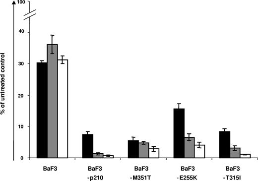
To determine the impact of the BCR-ABL mutational status on the efficacy of PHA-739358, we treated murine BaF3 and BaF3-p210 cells, including their IM-resistant mutants M351T, E255K, and T315I with increasing concentrations of PHA-739358. As already observed in human leukemic cell lines, PHA-739358 strongly inhibited cell proliferation of wt BaF3 cells and BaF3 cells expressing BCR-ABL, the latter independently of its mutational status, in a dose-dependent manner. In these experiments, sensitivity of BCR-ABL mutants to PHA-739358 did not seem to correlate with the degree of resistance to IM as the inhibition of proliferation in highly IM-resistant T315I mutant was within the ranges of wt BCR-ABL and lower-grade IM-resistant mutants when exposed to PHA-739358 (Figure 1).
PHA-739358 impairs proliferation of BaF3 and BaF3-p210, including -M351T, -E255K, and -T315I mutants. BaF3 and BaF3-p210, cells, including −M351T, −E255K, and −T315I mutants were exposed to 0.2 μM (■), 0.8 μM ( ), and 3.2 μM (□) of PHA-739358 for 48 hours and the number of viable cells was assessed by trypan blue exclusion assay. PHA-739358 inhibited proliferation of all cells tested in a dose-dependent manner, independent of their BCR-ABL mutational status. The percentage of cell growth was normalized to the growth of the no-drug control. The data shown are means of 3 independent experiments (± SD).
PHA-739358 impairs proliferation of BaF3 and BaF3-p210, including -M351T, -E255K, and -T315I mutants. BaF3 and BaF3-p210, cells, including −M351T, −E255K, and −T315I mutants were exposed to 0.2 μM (■), 0.8 μM ( ), and 3.2 μM (□) of PHA-739358 for 48 hours and the number of viable cells was assessed by trypan blue exclusion assay. PHA-739358 inhibited proliferation of all cells tested in a dose-dependent manner, independent of their BCR-ABL mutational status. The percentage of cell growth was normalized to the growth of the no-drug control. The data shown are means of 3 independent experiments (± SD).
To exclude the possibility that retroviral transduction impacts significantly on proliferation of BaF3 cells under PHA-739358 treatment, wt BaF3 were transduced with MSCV-IRES-puromycin empty vector (generous gift from C. Stocking, Heinrich-Pette-Institute (HPI), Hamburg, Gemany), as a non-BCR-ABL control. Compared with parental BaF3 cells, no significant differences in proliferation of transduced cells were detected under PHA-739358 treatment (data not shown).
Pharmacologic synergism of PHA-739358 and IM in BCR-ABL–positive IM-sensitive cell lines
To analyze any potential pharmacologic synergism of PHA-739358 with IM, we performed MTT assays in human K562 and BV173 cells and in murine IM-sensitive BaF3-p210 cells or BaF3 cells expressing BCR-ABL mutants with different degree of IM resistance. In both human and murine cell lines exhibiting no or subtotal resistance to IM, synergistic interactions of PHA-739358 and IM were observed for 50% inhibition (ED50), 75% inhibition (ED75), and 90% inhibition (ED90; Figure 2A-D; Table 2: Combination Index values < 0.9). Instead, no synergistic effects were found in BCR-ABL–negative HL60 or BaF3 cells as well as in BaF3-T315I cells with high-grade IM resistance (data not shown and Figure 2F).
PHA-739358 and IM synergize to induce cytotoxicity in BCR-ABL–positive IM-sensitive cell lines. Dose-effect curves for monotherapy with PHA-739358 and IM and combination of both substances in K562 (A), BV173 (B), BaF3-p210 (C), BaF3-M351T (D), BaF3-E255K (E), and BaF3-T315I (F) cells after 48 hours. Proliferation was assessed by MTT assay and results were analyzed with Calcusyn software.
PHA-739358 and IM synergize to induce cytotoxicity in BCR-ABL–positive IM-sensitive cell lines. Dose-effect curves for monotherapy with PHA-739358 and IM and combination of both substances in K562 (A), BV173 (B), BaF3-p210 (C), BaF3-M351T (D), BaF3-E255K (E), and BaF3-T315I (F) cells after 48 hours. Proliferation was assessed by MTT assay and results were analyzed with Calcusyn software.
Combination Index values for the effects of PHA-739358 and imatinib
| PHA-738359 plus IM . | ED50 . | ED75 . | ED90 . |
|---|---|---|---|
| K562 | 0.25 | 0.44 | 0.79 |
| BV173 | 0.37 | 0.51 | 0.70 |
| BaF3-p210 | 0.40 | 0.54 | 0.73 |
| BaF3-M351T | 0.56 | 0.59 | 0.63 |
| BaF3-E255K | 0.89 | 0.86 | 0.83 |
| BaF3-T315I | 0.90 | 1.16 | 1.51 |
| PHA-738359 plus IM . | ED50 . | ED75 . | ED90 . |
|---|---|---|---|
| K562 | 0.25 | 0.44 | 0.79 |
| BV173 | 0.37 | 0.51 | 0.70 |
| BaF3-p210 | 0.40 | 0.54 | 0.73 |
| BaF3-M351T | 0.56 | 0.59 | 0.63 |
| BaF3-E255K | 0.89 | 0.86 | 0.83 |
| BaF3-T315I | 0.90 | 1.16 | 1.51 |
PHA-739358 induces apoptosis, accumulation of cells with more than or equal to 4N DNA content, and G2/M arrest in BCR-ABL–positive BaF3 cells
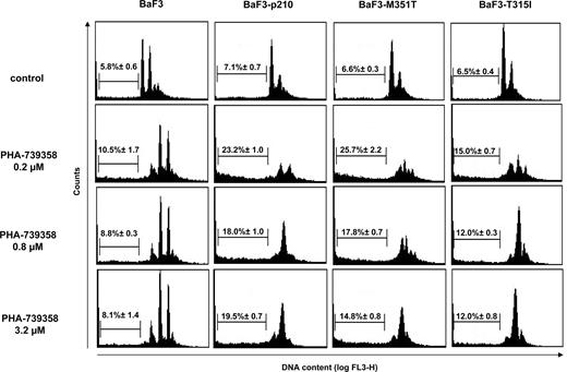
Inhibition of Aurora kinases induces endoreduplication and generates polyploid cells. We therefore analyzed the effects of PHA-739358 on cell-cycle progression. PHA-739358 treatment strongly inhibited proliferation of all cell lines tested. As expected, however, accumulation of cells containing more than 4N DNA was mainly found in BaF3 cells. Instead, in BaF3-p210 cells, including their IM-resistant mutants, treatment with 0.2 μM PHA-739358 generated polyploid cells and produced a marked loss of viability resulting from apoptosis, measured as the percentage of cells with sub-G1 DNA content. Treatment with higher concentrations of PHA-739358 primarily resulted in G2/M growth arrest (Figure 3).
PHA-739358 induces apoptosis, accumulation of cells with more than or equal to 4N DNA content, and G2/M arrest in BCR-ABL–positive BaF3 cells. BaF3, BaF3-p210, BaF3-M351T, and BaF3-T315I cells were exposed for 48 hours to indicated concentrations of PHA-739358. Cell-cycle analysis and apoptotic fraction of propidium iodide-stained cells were assessed by flow cytometry. Numbers on the plots are means (± SD) of the apoptotic fraction.
PHA-739358 induces apoptosis, accumulation of cells with more than or equal to 4N DNA content, and G2/M arrest in BCR-ABL–positive BaF3 cells. BaF3, BaF3-p210, BaF3-M351T, and BaF3-T315I cells were exposed for 48 hours to indicated concentrations of PHA-739358. Cell-cycle analysis and apoptotic fraction of propidium iodide-stained cells were assessed by flow cytometry. Numbers on the plots are means (± SD) of the apoptotic fraction.
PHA-739358 shares activity of both Aurora and Abl kinase inhibitors
The influence of PHA-739358 on Aurora kinase activity was assessed by the phosphorylation status of histone H3 at Ser10 (Figures 4,5C). K562 cells exposed to PHA-739358 showed strong reduction of phosphorylation with only 0.1% phospho-H3 (Figure 4C) compared with 3.5% in untreated (Figure 4A) and 3.7% in IM-treated cells (Figure 4B).
PHA-739358 reduces phosphorylation of histone H3 at Ser10. K562 cells were exposed to 5 μM PHA-739358 or 5 μM IM for 2 hours. Flow cytometric analysis of cells double stained with specific phospho-H3-(Ser10) antibody and propidium iodide (DNA content) was performed. The gates indicate phospho-histone H3-(Ser10)-positive cells in untreated control cells (A), IM-treated cells (B), or cells exposed to PHA-739358 (C).
PHA-739358 reduces phosphorylation of histone H3 at Ser10. K562 cells were exposed to 5 μM PHA-739358 or 5 μM IM for 2 hours. Flow cytometric analysis of cells double stained with specific phospho-H3-(Ser10) antibody and propidium iodide (DNA content) was performed. The gates indicate phospho-histone H3-(Ser10)-positive cells in untreated control cells (A), IM-treated cells (B), or cells exposed to PHA-739358 (C).
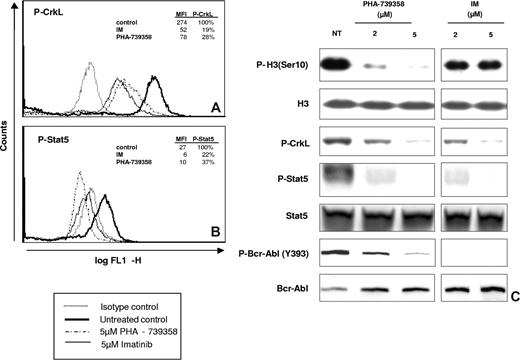
Inhibitory activity of PHA-739358 on Bcr-Abl kinase was evaluated by determining phosphorylation of CrkL and Stat5, 2 well-known downstream targets of Bcr-Abl (Figure 5). PHA-739358 treatment produced distinct inhibition of c-Abl autophosphorylation on Tyr393 (Figure 5C). In addition, pronounced inhibition of phosphorylation of CrkL (28% residual phosphorylation) and Stat5 (37% residual phosphorylation) was observed to a similar degree as for IM (19% and 22% residual phosphorylation, respectively; Figure 5A,B).
Influence of PHA-739358 and IM on the phosphorylation status of Bcr-Abl kinase targets. K562 cells treated with 5 μM PHA-739358 or IM for 2 hours were analyzed by intracellular flow cytometry to assess the phosphorylation status of known Bcr-Abl kinase downstream targets: CrkL (A) and Stat5 (B). MFI indicates mean fluorescence intensity. In addition, effects of PHA-739358 or IM on K562 cells after 1 hour of treatment were analyzed measuring Bcr-Abl, Stat5, CrkL, and histone H3-(Ser10) phosphorylation levels. NT indicates not treated. Inserted frames indicate repositioned gel lanes (C).
Influence of PHA-739358 and IM on the phosphorylation status of Bcr-Abl kinase targets. K562 cells treated with 5 μM PHA-739358 or IM for 2 hours were analyzed by intracellular flow cytometry to assess the phosphorylation status of known Bcr-Abl kinase downstream targets: CrkL (A) and Stat5 (B). MFI indicates mean fluorescence intensity. In addition, effects of PHA-739358 or IM on K562 cells after 1 hour of treatment were analyzed measuring Bcr-Abl, Stat5, CrkL, and histone H3-(Ser10) phosphorylation levels. NT indicates not treated. Inserted frames indicate repositioned gel lanes (C).
PHA-739358 reduces phosphorylation of CrkL in BaF3-p210 wt cells and IM-resistant mutants
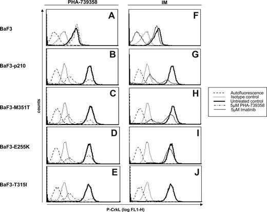
Next, the impact of the BCR-ABL mutational status on the efficacy of PHA-739358 to inhibit Bcr-Abl signaling was evaluated at concentrations (Cmax) that have previously been demonstrated in vivo.24 BaF3, BaF3-p210, and IM-resistant mutants M351T, E255K, and T315I were exposed to 5 μM PHA-739358 or 5 μM IM for 24 hours. Treatment with PHA-739358 resulted in strong inhibition of P-CrkL in BCR-ABL–positive BaF3 cells (Figure 6B-E), whereas no significant effects were observed in wt BaF3 cells (Figure 6A). The degree of inhibition of CrkL phosphorylation was within the same ranges for BaF3-p210, BaF3-M351T, and BaF3-T315I cells (Figure 6B,C,E). However, slightly weaker effects were accomplished in BaF3-E255K cells (Figure 6D). Changes of CrkL phosphorylation status under IM treatment reflected the level of IM resistance, with the strongest effects being observed in BaF3-p210 cells and a complete lack of activity in BaF3-T315I cells (Figure 6G-J).
PHA-739358 reduces phosphorylation of CrkL in BaF3-p210 cells and their IM-resistant mutants. Murine BaF3 and BaF3-p210 cells comprising the M351T, E255K, and T315I mutation were exposed to 5 μM PHA-739358 or 5 μM IM for 24 hours. Intracellular flow cytometry was used to measure CrkL phosphorylation status. See “PHA-739358 reduces phosphorylation of CrkL in BaF3-p210 wt cells and IM-resistant mutants” for details.
PHA-739358 reduces phosphorylation of CrkL in BaF3-p210 cells and their IM-resistant mutants. Murine BaF3 and BaF3-p210 cells comprising the M351T, E255K, and T315I mutation were exposed to 5 μM PHA-739358 or 5 μM IM for 24 hours. Intracellular flow cytometry was used to measure CrkL phosphorylation status. See “PHA-739358 reduces phosphorylation of CrkL in BaF3-p210 wt cells and IM-resistant mutants” for details.
PHA-739358 has a pronounced antiproliferative effect on CD34+ cells from CML patients, including those harboring the T315I mutation
We then evaluated the influence of PHA-739358 on proliferation of CD34+ cells from CML patients at different stages of disease and from healthy donors. CML-CD34+ cells were expanded in the presence of PHA-739358 (Figure 7A-C) or IM (Figure 7D-F) at concentrations ranging from 0.0047 μM to 1.25 μM or 0.038 μM to 20 μM, respectively. Cells were cultured for 9 days and cell numbers were assessed at days 3, 6, and 9. Plotting the relative cell number (ie, related to untreated control cells) against time and PHA-739358 concentration we observed a continuous, time- and dose-dependent inhibition of proliferation in CD34+ cells derived from patients in newly diagnosed chronic phase (Figure 7A), blast crisis (Figure 7B), and blast crisis with T315I mutation (Figure 7C). IC50 values for CD34+ cells from untreated patients in the chronic phase of CML (n = 4) were estimated to be approximately 0.005 μM for PHA-739358 (Figure 7A) and approximately 0.625 μM for IM at day 3 (Figure 7D).
Inhibition of CML progenitor cells growth after exposure to PHA-739358. CD34+ cells from patients diagnosed in the untreated chronic phase of CML (A,D; n = 4), in IM-resistant CML-blast crisis (B,E; n = 3), or from a patient with blast crisis with a confirmed T315I mutation (C,F; n = 1) were exposed to the indicated concentrations of PHA-739358 or IM for 9 days in serum-free medium supplemented with growth factors. Cell proliferation was assessed at day 3 (■), day 6 (▩), and day 9 (□). Bar graphs represent the mean percentage of cellular expansion plus or minus SD in relation to untreated control cells. The antiproliferative effects of PHA-739358 on CD34+ cells were found to be more pronounced in untreated CML patients at diagnosis compared with patients with IM-resistant CML-blast crisis; however, they seemed to be largely unaffected by the T315I BCR-ABL mutation.
Inhibition of CML progenitor cells growth after exposure to PHA-739358. CD34+ cells from patients diagnosed in the untreated chronic phase of CML (A,D; n = 4), in IM-resistant CML-blast crisis (B,E; n = 3), or from a patient with blast crisis with a confirmed T315I mutation (C,F; n = 1) were exposed to the indicated concentrations of PHA-739358 or IM for 9 days in serum-free medium supplemented with growth factors. Cell proliferation was assessed at day 3 (■), day 6 (▩), and day 9 (□). Bar graphs represent the mean percentage of cellular expansion plus or minus SD in relation to untreated control cells. The antiproliferative effects of PHA-739358 on CD34+ cells were found to be more pronounced in untreated CML patients at diagnosis compared with patients with IM-resistant CML-blast crisis; however, they seemed to be largely unaffected by the T315I BCR-ABL mutation.
In the cases of IM-resistant CML-blast crisis (n = 3), IC50 values for PHA-739358 at day 3 were marginally higher, reaching approximately 0.009 μM (Figure 7B). As expected, the increase of IC50 values for IM in those cases was substantially more pronounced, amounting to unphysiologic doses approximately 10.0 μM at day 3 (Figure 7E). Furthermore, whereas expansion of CD34+ cells from an IM-resistant patient in blast crisis harboring the T315I mutation remained virtually unaffected with IM treatment at day 3 (Figure 7F; IC50 > 20.0 μM), the IC50 value for PHA-739358 was estimated to be approximately 0.019 μM at day 3 (Figure 7C), which is only moderately higher than the IC50 values of CD34+ cells derived from untreated individuals in chronic phase of the disease. This suggests that the antiproliferative activity of the compound is not impaired by this particular mutation.
In the next set of experiments, CD34+ cells isolated from 2 healthy donors were cultured under the same conditions with increasing PHA-739358 concentrations ranging from 0.0024 μM to 0.625 μM for 12 days. In line with previous observations in IM-treated normal CD34+ cells,22 a dose-dependent decrease of proliferation was observed with IC50 values reaching approximately 0.014 μM at day 3 (data not shown).
PHA-739358 induces apoptosis and reduces phosphorylation of P-CrkL in CD34+ cells from an IM-resistant CML patient
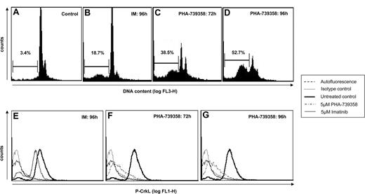
We next assessed the influence of PHA-739358 and IM on cell- cycle progression and CrkL phosphorylation status in CD34+ cells derived from an IM-resistant CML patient in blast crisis. PHA-739358 treatment predominantly caused an enhanced loss of viability compared with control (Figure 8A), reflected by an apoptotic fraction of 39% and 53% after 72 hours and 96 hours, respectively, compared with 19% after 96 hours of IM treatment (Figure 8B-D).
Effects of PHA-739358 on apoptosis and CrkL phosphorylation status of CD34+ cells from an IM-resistant CML patient. CD34+ cells derived from a patient with IM-resistant blast crisis were exposed to 5 μM PHA-739358 or 5 μM IM. Analysis of the cell cycle and the apoptotic fraction of propidium iodide–stained cells was assessed by flow cytometry in untreated cells serving as a control (A), cells treated for 96 hours with IM (B), and cells exposed to PHA-739358 for 72 hours (C) and 96 hours (D). Analysis of the expression of P-CrkL by intracellular flow cytometry. Strong reduction of CrkL phosphorylation was seen after exposure to PHA-739358 (72 hours and 96 hours; F and G, respectively) with no significant changes after 96 hours of IM treatment (E).
Effects of PHA-739358 on apoptosis and CrkL phosphorylation status of CD34+ cells from an IM-resistant CML patient. CD34+ cells derived from a patient with IM-resistant blast crisis were exposed to 5 μM PHA-739358 or 5 μM IM. Analysis of the cell cycle and the apoptotic fraction of propidium iodide–stained cells was assessed by flow cytometry in untreated cells serving as a control (A), cells treated for 96 hours with IM (B), and cells exposed to PHA-739358 for 72 hours (C) and 96 hours (D). Analysis of the expression of P-CrkL by intracellular flow cytometry. Strong reduction of CrkL phosphorylation was seen after exposure to PHA-739358 (72 hours and 96 hours; F and G, respectively) with no significant changes after 96 hours of IM treatment (E).
PHA-739358 effectively inhibits growth of K562 cells in vivo
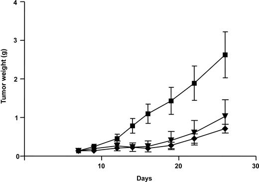
Efficacy and toxicity of PHA-739358 and IM were evaluated in vivo. Both treatment regimens were well tolerated and no major side effects were detected (data not shown). PHA-739358 and IM significantly inhibited proliferation of K562 cells andvirtually suppressed tumor growth during the 10-day treatment period (P < .01 compared with controls at day 19). However, when the application of the compounds was terminated, leukemic cells started to proliferate again in both treatment groups. Interestingly, tumor growth was now less pronounced in these groups compared with the control animals (Figure 9).
PHA-739358 effectively inhibits growth of K562 in vivo. K562 cells were injected subcutaneously in SCID mice. When tumors reached an estimated weight of 100 to 150 mg, the vehicle (■), PHA-739358 (♦), or IM (▾) was administered for 10 days. PHA-739358 and IM were well tolerated and significantly suppressed tumor growth compared with controls. Error bars represent SD.
PHA-739358 effectively inhibits growth of K562 in vivo. K562 cells were injected subcutaneously in SCID mice. When tumors reached an estimated weight of 100 to 150 mg, the vehicle (■), PHA-739358 (♦), or IM (▾) was administered for 10 days. PHA-739358 and IM were well tolerated and significantly suppressed tumor growth compared with controls. Error bars represent SD.
Discussion
IM has substantially changed the landscape of CML treatment, allowing cytogenetic response rates of more than 80%, translating into significantly improved survival of patients. However, this success is compromised to some degree by 3 major problems emerging in the course of treatment with this Bcr-Abl inhibitor: (1) limited therapeutic activity and durability in advanced disease stages,12 (2) primary or acquired resistance to IM, and (3) persistence of minimal residual disease because of the limited effects of the drug on immature hematopoietic stem cells.25 Resistance primarily caused by point mutations in the kinase domain of Bcr-Abl results in decreased or lost sensitivity to IM. Attempts to restore target inhibition of mutated Bcr-Abl led to the development of second-generation Bcr-Abl inhibitors, such as dasatinib,9 nilotinib,10 and bosutinib.11 However, despite promising clinical results for most mutations, the frequently observed mutant T315I is not effectively targeted by any of these compounds. As this mutation is predicted to become the major mechanism of resistance to second-generation Bcr-Abl inhibitors, the remaining challenge in the development of novel therapeutic strategies is to overcome resistance mediated by T315I.8,26-28 A first success in this direction was achieved by combining specific kinase inhibitors with agents impairing transcription and/or translation: decreased proliferation of leukemia cell lines and primary CML cells, including those harboring T315I mutant were observed in in vitro and ex vivo experiments.29-31 Embracing the idea of influencing tumor signaling pathways and cell-cycle check points at the same time, monotherapy with small molecule inhibitors targeting specific key enzymes simultaneously seems a desirable approach in the treatment of CML.
Here we describe a novel kinase inhibitor PHA-739358 that exhibits strong inhibitory effects on both Bcr-Abl and Aurora kinases. Efficacy of PHA-739358 was demonstrated against a broad panel of leukemia cell lines. Thus, treatment with PHA-739358 significantly decreased proliferation of BCR-ABL–positive and -negative human leukemia cell lines as well as of murine BaF3 and BaF3-p210 cells, expressing different IM-resistant BCR-ABL mutants, including T315I. Hence, antiproliferative activity of the small molecule inhibitor was at least partly independent of the presence of Bcr-Abl activity, and sensitivity to PHA-739358 did not depend on BCR-ABL mutational status or degree of resistance to IM as inhibition of proliferation was within the same ranges in all mutants conferring resistance to IM, including the highly IM-resistant T315I mutant. Pharmacologic synergism of IM and PHA-739358 was observed in both human and murine cell lines with a subcomplete resistance to IM, whereas, as expected, no synergistic effects were seen in BCR-ABL–negative cells or those harboring the T315I mutation.
To further elucidate the mechanism of action of PHA-739358, we evaluated the effects of the small molecule inhibitor on different functional downstream targets of Bcr-Abl and Aurora kinases. A highly significant decrease of phosphorylation of histone H3 at Ser10 was observed in cells exposed to PHA-739358, indicating strong inhibition of Aurora B activity. Thus, PHA-739358 ranks with other previously described Aurora kinase inhibitors, such as ZM447439,32 Hesperadin,33 and VX68018 suppressing phosphorylation of histone H3 at Ser10. In addition, in kinase assays PHA-739358 had been shown to potently inhibit Aurora kinases A, B, and C indicating a pan-Aurora kinases inhibitory activity of this compound.21 In analogy to other Aurora kinase inhibitors, PHA-739358 treatment resulted in accumulation of polyploid cells via endoreduplication. However, cells with a DNA content of greater than 4N were mainly found in BCR-ABL–negative BaF3 cells, whereas in BaF3-p210 cells, including their IM-resistant mutants, treatment with 0.2 μM PHA-739358 generated polyploid cells and produced a marked loss of viability resulting from apoptosis, probably because of a predominance of the inhibitory effect on Bcr-Abl in these cells. In contrast, treatment with higher doses of PHA-739358 primarily caused G2/M growth arrest, implicating a dose-dependent effect of PHA-739358 spanning from induction of polyploidy to G2/M arrest. In addition, PHA-739358 treatment led to a distinct inhibition of c-Abl autophosphorylation, indicating decreased Abl-kinase activity as well as to a pronounced inhibition of phosphorylation of the Bcr-Abl downstream target Stat5. Furthermore, phosphorylation of another well-known Bcr-Abl downstream target CrkL was significantly reduced after treatment with PHA-739358 in BCR-ABL–positive BaF3 cells, whereas no effects were observed in wild-type BaF3 cells. Interestingly, the decrease of phosphorylation of CrkL in cells with the highly resistant mutant T315I was significant and within the ranges of the other point mutations examined, indicating an effective inhibition of Bcr-Abl carrying this highly resistant mutation, whereas no inhibition of phosphorylation could be induced by IM treatment. In addition, in crystallographic studies of the T315I mutant binding of PHA-739358 to the active conformation of the kinase domain in the ATP-binding pocket was demonstrated.34 Hence, these data corroborate the hypothesis that PHA-739358 functions as an effective inhibitor of both Aurora kinases and Bcr-Abl kinase, including the T315I mutant.
To further expand our data to primary clinical material, efficacy of PHA-739358 was examined in CD34+ cells from patients with CML at different stages of disease, spanning from first diagnosis (previously untreated patients) to IM-resistant blast crisis, including one patient with IM- and dasatinib-resistant blast crisis harboring the T315I mutation. A both time- and dose-dependent inhibition of cell proliferation was observed in all CD34+ cells exposed to PHA-739358. Remarkably, whereas IC50 values of IM exceeded 10 μM and 20 μM in patients with blast crisis and the patient harboring the T315I mutation, respectively, IC50 values for PHA-739358 were less than 0.02 μM in these cases and thus only moderately higher than in previously untreated patients diagnosed in the chronic phase of the disease. When focusing on the mechanism of action of the small molecule inhibitor in primary clinical material, loss of viability was observed in PHA-739358-treated CD34+ cells from a patient with IM-resistant blast crisis with the apoptotic fraction being 34% higher compared with IM-treated cells. In addition, PHA-739358 decreased phosphorylation of CrkL significantly, whereas in IM treated CD34+ cells, the CrkL phosphorylation status remained unaffected. Thus, in line with the results of the in vitro experiments, these findings confirm high efficacy of the PHA-739358 in CML cells independent of the BCR-ABL mutational status, including T315I. As expected and in analogy to previous studies with IM,22 under maximum in vitro cytokine stimulation, a dose-dependent inhibition of proliferation of CD34+ cells derived from healthy donors was observed under PHA-739358.
To further evaluate efficacy and toxicity of PHA-739358 in vivo, a subcutaneous CML model was used. Treatment with both inhibitors, PHA-739358 or IM, efficiently and significantly suppressed growth of K562 chloromas at well-tolerated doses.
In conclusion, small molecule inhibitors, such as PHA-739358, targeting wild-type or mutant Bcr-Abl and Aurora kinases simultaneously represent a promising new treatment strategy for patients with IM-resistant CML, particularly for patients harboring the highly resistant T315I mutation. Indeed, PHA-739358 is currently being evaluated in a phase 2 clinical trial in CML, including patients with T315I mutation (NCT00335868, www.clinicaltrial.gov).
The publication costs of this article were defrayed in part by page charge payment. Therefore, and solely to indicate this fact, this article is hereby marked “advertisement” in accordance with 18 USC section 1734.
Acknowledgments
The authors thank Dr Phillipe Schafhausen for help with the collection of patient samples, Vera Bargsten and Joanna Schmid for technical assistance in preparation of CD34+ cells and help with MTT assays, and Dr Tessa L. Holyoake for the critical reading of the manuscript.
This work was supported in part by the “Eppendorfer Krebs- und Leukämiehilfe” and the European Leukemia Net (WP4).
Authorship
Contribution: A.G., S.B., E.P., R.C., A.G., and D.B. performed research and analyzed the data; G.K., C.B., and W.F. wrote the paper; J.M. contributed vital new reagents; T.H.B. and J.M. designed research and wrote the paper.
Conflict-of-interest disclosure: J.M., E.P., R.C., and A.G. are former or present full-time employees of Nerviano Medical Sciences Srl. The remaining authors declare no competing financial interests.
Correspondence: Tim H. Brümmendorf, Department of Oncology and Hematology, University Hospital Hamburg-Eppendorf, Martinistraβe 52, 20246 Hamburg, Germany; e-mail: t.bruemmendorf@uke.uni-hamburg.de.




This feature is available to Subscribers Only
Sign In or Create an Account Close Modal