Abstract
Programmed death-1 (PD-1)–PD-1 ligand (PD-L) signaling system is involved in the functional impairment of T cells such as in chronic viral infection or tumor immune evasion. We examined PD-L expression in lymphoid cell lines and found that they were up-regulated on Hodgkin lymphoma (HL) and several T-cell lymphomas but not on B-cell lymphomas. PD-L expression was also demonstrated in primary Hodgkin/Reed-Sternberg (H/RS) cells. On the other hand, PD-1 was elevated markedly in tumor-infiltrating T cells of HL, and was high in the peripheral T cells of HL patients as well. Blockade of the PD-1 signaling pathway inhibited SHP-2 phosphorylation and restored the IFN-γ–producing function of HL-infiltrating T cells. According to these results, deficient cellular immunity observed in HL patients can be explained by “T-cell exhaustion,” which is led by the activation of PD-1–PD-L signaling pathway. Our finding provides a potentially effective immunologic strategy for the treatment of HL.
Introduction
Programmed death-1 (PD-1), a member of the CD28 costimulatory receptor superfamily, inhibits T-cell activity by providing a second signal to T cells in conjunction with signaling through the T-cell receptor.1 To date, B7-H1 and B7-DC have been identified as ligands for PD-1 (PD-Ls). During chronic viral infection, PD-1 is selectively up-regulated by the exhausted T cells, and blockade of this pathway restores CD8+ T-cell function and reduces viral load.2 This signaling system has been recently highlighted in the research of human immunodeficiency virus (HIV) infection.3-6 In addition, PD-1 is indicated to be involved in the evasion of tumor immunity.7-10
Hodgkin lymphoma (HL) is characterized by massive reactive infiltrates surrounding Hodgkin/Reed-Sternberg (H/RS) cells. HL patients are well recognized as having defective cellular immunity; they are susceptible to bacterial, fungal, and viral infections, and in vitro studies show depressed T-cell proliferation and reduced synthesis of Th1 cytokines.11 We report here that PD-1–PD-L signaling system is operative in patients with HL, and tumor-infiltrating T cells around H/RS cells seem to be kept in balance by this inhibitory signaling. Our findings illuminate the mechanism for deficient cellular immunity observed in HL patients, and propose a potentially effective immunologic strategy for the treatment of HL.
Methods
Cell lines and clinical sample preparation
The following cell lines were described previously12,13 : HL cell lines KM-H2, L428, and HDLM-2; anaplastic large cell lymphoma (ALCL) cell line DEL; follicular lymphoma cell line FL-218; diffuse large B-cell lymphoma (DLBCL) cell line KIS-1; Burkitt lymphoma cell lines Daudi, Raji, Middle 91, Tree 92, and Ramos; adult T-cell leukemia/lymphoma cell line HUT 102; and acute T-cell leukemia cell line Jurkat. LCL-OHN is an Epstein-Barr virus (EBV)–transformed lymphoblastoid B-cell line (LCL). Peripheral blood samples were collected from 19 HL patients, 12 B-NHL patients, and 11 healthy volunteers after informed consent was obtained in accordance with the Declaration of Helsinki. This study is approved by the institutional review board of Kyoto University. After removing red blood cells using ACK lysis buffer, leukocytes were subjected to flow cytometry. For immunohistochemistry, tissue specimens were snap-frozen in OCT compound (TissueTek, Tokyo, Japan) and stored at −80°C.
Reverse transcription–polymerase chain reaction, flow cytometry, and immunohistochemistry
Total RNA was isolated from cells with Trizol (Invitrogen, Carlsbad, CA), and cDNA was synthesized using MultiScribe Reverse Transcriptase (Applied Biosystems, Foster City, CA). PCR assays were performed by the conventional method using Taq polymerase (TaKaRa Biotechnology, Shiga, Japan). For flow cytometry, cells were analyzed on a FACScan (Becton Dickinson, Mansfield, MS). The following antibodies were used: PE-conjugated B7-H1 and B7-DC (eBioscience, San Diego, CA), FITC-PD-1 (BD Pharmingen, San Diego, CA), PC5-CD4 and CD8 (Immunotech, Marseille, France), FITC-CD25 (Becton Dickinson), and PE-Foxp3 (eBioscience). Immunohistochemical staining was performed as described previously.14
Expression vector construction and luciferase assay
Fragments of the human B7-H1 and B7-DC promoter regions and Flag-tagged LMP1 and LMP2A were constructed by PCR amplification or by hybridization of a pair of complementary strands. The fragments of B7-H1 and B7-DC were subcloned into the pGL3-enhancer vector (Promega, Madison, WI) and pcDNA3-FLAG and CMV-FLAG-5a vectors (Sigma-Aldrich, St Louis, MO), respectively.
LMP1, LMP2A, or mock vector was transfected into 293T cells with each reporter vector using FuGene6 Transfection Reagent (Roche, Mannheim, Germany). Forty-eight hours later, cell lysates were measured for luciferase activity using the Dual Luciferase Assay System (Promega).
B7-H1/B7-DC blockade, IFN-γ production assay, immunoprecipitation, and Western blotting
Single-cell suspensions derived from HL and B-NHL tissue samples were cultured in RPMI 1640 medium supplemented with 10% autoserum in a 200-μL volume at a cell concentration of 2.5 × 10⁶/mL with B7-H1 and B7-DC blocking antibodies (eBioscience) or isotype control. After 48-hour incubation, the supernatants were analyzed for IFN-γ by an enzyme-linked immunosorbent assay (ELISA; Pierce Biotechnology, Rockford, IL). For the detection of SHP-2 phosphorylation, bulk HL cell suspensions were cultured with blocking antibodies, lysed, immunoprecipitated with anti–SHP-2 antibody (BD Pharmingen), and assessed for tyrosine phosphorylation by Western blotting as previously described.15 To determine which cell type is the main producer of IFN-γ, CD4+ or CD8+ T cells were depleted or enriched from HL cell suspensions by magnetic-activated cell sorting (MACS) using CD4 or CD8 MicroBeads (Miltenyi Biotec, Bergisch Gladbach, Germany).
Results and discussion
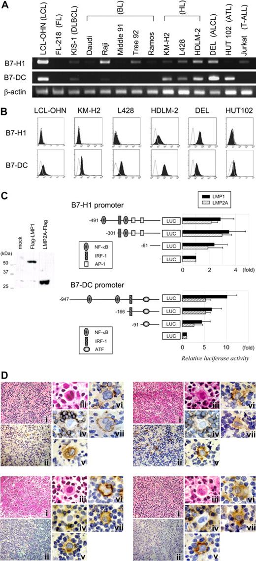
We first examined B7-H1 and B7-DC expression in several lymphoid cell lines (Figure 1A,B). Some HL and T-cell lines were positive for B7-H1, and B7-DC showed broader expression. In contrast, most B-cell lines except LCL-OHN lacked their expression.
Expression and gene regulation of B7-H1 and B7-DC. (A) Reverse transcription–polymerase chain reaction (RT-PCR) of human lymphoid cell lines for B7-H1 and B7-DC expression. β-Actin is shown as a cDNA control. (B) Histograms showing B7-H1 and B7-DC expression levels in human lymphoid cell lines. Open histograms represent isotype controls. (C) Luciferase reporter assay detecting alterations in B7-H1 and B7-DC promoter activities by EBV latent membrane proteins. Either flag-tagged LMP1, LMP2A expression vector, or mock vector was transfected with one of the reporter vectors into 293T cells. LMP1 and LMP2A protein expression was confirmed by Western blotting (left). Schematic diagrams of promoter regions of B7-H1 and B7-DC cloned into the pGL3-enhancer vector are shown (right). Numbers are the nucleotide positions that refer to the 5′ boundary of exon 1 of each gene, and binding sites of representative transcription factors are indicated. Luciferase activities are expressed relative to the activity of the mock vector–transfected controls. Values are the means of 3 independent experiments; bars represent SD. (D) Immunohistochemical analysis of 4 HL tissue specimens. HE (i) and LMP1 staining (ii) (original magnification ×400); HE (iii), CD3 (iv), CD30 (v), B7-H1 (vi), and B7-DC (vii) (original magnification ×1000), focusing on H/RS cells and surrounding T cells. EBV is shown to be positive in the top 2 samples and negative in the bottom 2.
Expression and gene regulation of B7-H1 and B7-DC. (A) Reverse transcription–polymerase chain reaction (RT-PCR) of human lymphoid cell lines for B7-H1 and B7-DC expression. β-Actin is shown as a cDNA control. (B) Histograms showing B7-H1 and B7-DC expression levels in human lymphoid cell lines. Open histograms represent isotype controls. (C) Luciferase reporter assay detecting alterations in B7-H1 and B7-DC promoter activities by EBV latent membrane proteins. Either flag-tagged LMP1, LMP2A expression vector, or mock vector was transfected with one of the reporter vectors into 293T cells. LMP1 and LMP2A protein expression was confirmed by Western blotting (left). Schematic diagrams of promoter regions of B7-H1 and B7-DC cloned into the pGL3-enhancer vector are shown (right). Numbers are the nucleotide positions that refer to the 5′ boundary of exon 1 of each gene, and binding sites of representative transcription factors are indicated. Luciferase activities are expressed relative to the activity of the mock vector–transfected controls. Values are the means of 3 independent experiments; bars represent SD. (D) Immunohistochemical analysis of 4 HL tissue specimens. HE (i) and LMP1 staining (ii) (original magnification ×400); HE (iii), CD3 (iv), CD30 (v), B7-H1 (vi), and B7-DC (vii) (original magnification ×1000), focusing on H/RS cells and surrounding T cells. EBV is shown to be positive in the top 2 samples and negative in the bottom 2.
As PD-L genes were also expressed in another 3 LCLs, we examined the effect of EBV-latent membrane proteins on their promoter activities (Figure 1C). The result showed that both LMP1 and LMP2A enhanced the transcriptional activity of B7-H1 and B7-DC. This finding implies that in cases of EBV+ HL, the expression of PD-L genes is possibly led by latent membrane proteins.
In immunohistochemical analysis, H/RS cells were shown to be positive for PD-Ls in both EBV+ and EBV− samples (Figure 1D). Thus the expression of PD-Ls was assumed to be a common feature of H/RS cells.
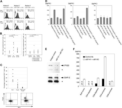
We next analyzed PD-1 expression of tumor-infiltrating T cells of HL tissue samples. PD-1+ cells were obviously increased in all 3 samples tested (Figure 2A), although PD-1 expression was previously described as negative in HL tissues.16 PD-1 expression level was also significantly elevated in the peripheral blood T cells of HL patients, in comparison with those of healthy controls and B-NHL patients (Figure 2B left). Expression levels tended to be higher in patients with active disease and declined along with treatment. In contrast, T cells of CD4+CD25+ regulatory phenotype were not apparently increased (Figure 2B right), whereas they are reported to play an important role in the pathogenesis of HL.17-21 We evaluated PD-1 and Foxp3 expression concurrently in 9 other HL patients (Figure 2C), and they were shown to be mostly independent of each other. This result is similar to the result of tumor-infiltrating T cells studied in B-NHL.22
PD-1 expression in T cells of HL patients and functional analysis of PD-1–PD-L signaling. (A) Histograms showing PD-1 expression in tumor-infiltrating T cells of 3 HL patients. CD4+ and CD8+ T cells in bulk HL cell suspensions were analyzed for PD-1. MFI indicates mean fluorescent intensity. (B) Proportion of PD-1+ and CD4+CD25+ cells in peripheral blood T cells of HL and B-NHL patients compared with those of healthy volunteers. Patients' characteristics are shown in Table 1. One HL patient was not analyzed for CD4+CD25+ cells. MC indicates mixed cellularity; NC, nodular sclerosis; LR, lymphocyte rich; MALT, extranodal marginal zone B-cell lymphoma of mucosa-associated lymphoid tissue; HBV, hepatitis B virus infection; HCV, hepatitis C virus infection; and MAC, mycobacterium avium complex infection. Patients classified as “during treatment” include those who are undergoing chemotherapy or radiotherapy. (C) Proportion of PD-1+ cells, Foxp3+ cells, and those positive for both PD-1 and Foxp3, in peripheral CD4+ T cells of 9 tumor-bearing HL patients. Representative dot-plot data of 2 patients: one had low and the other relatively high Foxp3 expression (Table 2). (D) Blockade of PD-Ls restores IFN-γ–producing function of HL-infiltrating T cells. Bulk tumor cell suspensions of 2 HL patients (left) and 1 DLBCL patient (right) were cultured with blocking antibodies, and IFN-γ levels in the supernatants were measured as described in “Methods.” Samples were analyzed in duplex. Error bars indicate standard deviation of duplicate measurements. (E) Blockade of PD-Ls inhibits tyrosine phosphorylation of SHP-2. After bulk HL cell suspensions were cultured with PD-L blocking antibodies or isotype control antibody, cell lysates were immunoprecipitated with anti–SHP-2 antibody and its tyrosine phosphorylation was examined by immunoblotting with PY20 antibody (top panel). The blot was subsequently reprobed with anti–SHP-2 antibody (bottom panel). The position of SHP-2 is indicated by arrowheads. Asterisks denote the immunoglobulin heavy chain. (F) Blockade of PD-Ls revive mainly the IFN-γ–producing function of CD4+ T cells. T-cell subsets were depleted or enriched from HL cell suspensions by MACS. IFN-γ production by each cell fraction was evaluated as in panel D.
PD-1 expression in T cells of HL patients and functional analysis of PD-1–PD-L signaling. (A) Histograms showing PD-1 expression in tumor-infiltrating T cells of 3 HL patients. CD4+ and CD8+ T cells in bulk HL cell suspensions were analyzed for PD-1. MFI indicates mean fluorescent intensity. (B) Proportion of PD-1+ and CD4+CD25+ cells in peripheral blood T cells of HL and B-NHL patients compared with those of healthy volunteers. Patients' characteristics are shown in Table 1. One HL patient was not analyzed for CD4+CD25+ cells. MC indicates mixed cellularity; NC, nodular sclerosis; LR, lymphocyte rich; MALT, extranodal marginal zone B-cell lymphoma of mucosa-associated lymphoid tissue; HBV, hepatitis B virus infection; HCV, hepatitis C virus infection; and MAC, mycobacterium avium complex infection. Patients classified as “during treatment” include those who are undergoing chemotherapy or radiotherapy. (C) Proportion of PD-1+ cells, Foxp3+ cells, and those positive for both PD-1 and Foxp3, in peripheral CD4+ T cells of 9 tumor-bearing HL patients. Representative dot-plot data of 2 patients: one had low and the other relatively high Foxp3 expression (Table 2). (D) Blockade of PD-Ls restores IFN-γ–producing function of HL-infiltrating T cells. Bulk tumor cell suspensions of 2 HL patients (left) and 1 DLBCL patient (right) were cultured with blocking antibodies, and IFN-γ levels in the supernatants were measured as described in “Methods.” Samples were analyzed in duplex. Error bars indicate standard deviation of duplicate measurements. (E) Blockade of PD-Ls inhibits tyrosine phosphorylation of SHP-2. After bulk HL cell suspensions were cultured with PD-L blocking antibodies or isotype control antibody, cell lysates were immunoprecipitated with anti–SHP-2 antibody and its tyrosine phosphorylation was examined by immunoblotting with PY20 antibody (top panel). The blot was subsequently reprobed with anti–SHP-2 antibody (bottom panel). The position of SHP-2 is indicated by arrowheads. Asterisks denote the immunoglobulin heavy chain. (F) Blockade of PD-Ls revive mainly the IFN-γ–producing function of CD4+ T cells. T-cell subsets were depleted or enriched from HL cell suspensions by MACS. IFN-γ production by each cell fraction was evaluated as in panel D.
Characteristics of HL and B-NHL patients compared with healthy controls
| . | Healthy controls . | HL patients . | B-NHL patients . |
|---|---|---|---|
| N | 11 | 10 | 12 |
| Sex, no. M:F | 7:4 | 4:6 | 3:9 |
| Median age, y (range) | 33 (31-36) | 37.5 (23-85) | 57 (38-74) |
| Histology | 5 MC, 5 NS(EBV+ in 3) | 5 FL, 5 DLBCL, 1 MALT, 1 MLBCL | |
| Stage at onset | 4 stage II, 6 stage IV | 5 stage II, 2 stage III, 5 stage IV | |
| Disease status, no. | |||
| Active disease | 2 | 6 | |
| During treatment | 4 | 2 | |
| Tumor free | 4 | 4 | |
| Chronic infectious disease | None | 1 HCV | 2 HBV, 1 HCV, 1 MAC |
Characteristics of tumor-bearing HL patients
| . | HL patients, n = 9 . |
|---|---|
| Sex, no. M:F | 7:2 |
| Median age, y (range) | 72 (14-80) |
| Histology | 3 MC, 5 NS, 1 LR (EBV+ in 3) |
| Stage at onset | 2 stage I, 1 stage II, 3 stage III, 3 stage IV |
| Disease status | |
| Active disease | 2 |
| During treatment | 7 |
| Chronic infectious disease | None |
We finally examined the effect of blockade of this pathway. After culturing bulk HL tumor cells in the presence of anti–PD-L blocking antibodies, IFN-γ production was measured by ELISA. Blockade of PD-Ls augmented IFN-γ production in 2 HL samples (Figure 2D left). In contrast, among 5 B-NHL tissues tested, we did not detect IFN-γ in 4 samples (1 DLBCL, 2 follicular lymphoma, and 1 marginal zone B-cell lymphoma), irrespective of PD-L blockade. IFN-γ was barely measurable in 1 DLBCL sample, but blockade of PD-Ls did not influence the IFN-γ production (Figure 2D right). Blockade of PD-Ls was accompanied with the inhibition of SHP-2 phosphorylation (Figure 2E), which has been reported as a mediator of the PD-1 signaling pathway.23 Depletion or enrichment of T-cell subsets from HL cell suspension indicated that PD-L blockade revived mainly the function of CD4+ T cells of HL (Figure 2F). Natural killer cell population was less than 0.5%, and they seemed unlikely to be the major source of IFN-γ (data not shown). We concluded that antitumor activity of HL-infiltrating T cells is inhibited via the PD-1–PD-L signaling pathway, and this inhibition can be successfully relieved by PD-L blockade. Our observations indicate that “T-cell exhaustion” is essential to the pathogenesis of HL, which is in line with the recent study of RNA fingerprints.24 Adoptive immunotherapy with tumor antigen–specific T cells has lately been attempted for the treatment of HL.25 Our findings provide a potentially effective and clinically applicable strategy for the immunotherapy of HL.
The online version of this article contains a data supplement.
The publication costs of this article were defrayed in part by page charge payment. Therefore, and solely to indicate this fact, this article is hereby marked “advertisement” in accordance with 18 USC section 1734.
Acknowledgments
We are grateful to the following physicians for providing blood samples of the patients: Dr H. Ohno, Takeda General Hospital; Dr C. Ueda, Kyoto Katsura Hospital; Drs K. Yamashita, T. Kitano, and H. Matsubara, Kyoto University; Drs T. Takeoka, M. Tsuji, and W. Kishimoto, Otsu Red-Cross Hospital; Dr Y. Maesako, Tenri Hospital; Drs M. Watanabe, H. Kaneko, and M. Nishizawa, Osaka Red-Cross Hospital. We also thank Dr Y. Kobashi, Tenri Hospital, and Dr H. Haga and Mr Y. Toda, Kyoto University, for histologic examination.
This work was supported in part by grants-in-aid from the Ministry of Education, Culture, Sports, Science and Technology (18790650) of Japan.
Authorship
Contribution: R.Y., M.N., and T. Kitawaki performed research; M.N. designed research and wrote the paper; T.S. and M.T. contributed vital new reagents; M.H, T. Kondo, K.O., M.K., and T.H. provided clinical samples and analyzed data; T.U. supervised research.
Conflict-of-interest disclosure: The authors declare no competing financial interests.
Correspondence: Momoko Nishikori, 54 Shogoin Kawahara-cho, Sakyo-ku, Kyoto, 606-8507, Japan; e-mail: nishikor@kuhp.kyoto-u.ac.jp.


This feature is available to Subscribers Only
Sign In or Create an Account Close Modal