Abstract
FcϵRI-activation–induced survival of mast cells is dependent on the expression and function of the prosurvival protein A1. The expression of A1 in lymphocytes and monocytes has previously been described to be transcriptionally regulated by NF-κB. Here we demonstrate that the expression of A1 in mast cells is not dependent on NF-κB but that NFAT plays a crucial role. FcϵRI-induced A1 expression was not affected in mast cells overexpressing an IκB-α super-repressor or cells lacking NF-κB subunits RelA, c-Rel, or c-Rel plus NF-κB1 p50. In contrast, inhibition of calcineurin and NFAT by cyclosporin A abrogated the expression of A1 in mast cells on FcϵRI-activation but had no effect on lipopolysaccharide-induced expression of A1 in J774A.1 monocytic cells. Cyclosporin A also inhibited luciferase expression in an A1 promoter reporter assay. A putative NFAT binding site in the A1 promoter showed inducible protein binding after FcϵRI crosslinking or treatment with ionomycin as detected in a band shift assay or chromatin immunoprecipitation. The binding protein was identified as NFAT1. Finally, mast cells expressing constitutively active NFAT1 exhibit increased expression of A1 after FcϵRI-stimulation. These results indicate that, in FcϵRI stimulated mast cells, A1 is transcriptionally regulated by NFAT1 but not by NF-κB.
Introduction
Mast cells are potent effector cells displaying versatile functions during immune responses and as regulators of inflammation.1-3 Many of these functions are executed via activation of the high affinity IgE receptor (FcϵRI) and subsequent release of regulatory factors stored in granules.4 In addition, receptor stimulation initiates signaling cascades, which result in activation of specific genes encoding cytokines and growth factors.5 In some cases, the activated transcription has been shown to be mediated by members of the NF-κB and/or NFAT transcription factor families.6 These transcription factors are sequestered in an inactive state in the cytosol of resting cells, and after cell stimulation they are translocated to the nucleus where they bind target DNA sequences and activate transcription.
In contrast to granulocytes and certain other hematopoietic cells, mature mast cells are not recruited from the bloodstream in response to inflammatory signals. Instead, long-lived mast cells are located in the tissues, and their relative abundance and increase during inflammation are regulated at the level of cell migration within the tissue and the control of survival/apoptosis.7,8 We and others have previously demonstrated that, after stimulation of the high affinity IgE receptor, FcϵRI, mast cell survival is substantially enhanced.9-11 These cells can therefore undergo a new round of activation and thus contribute again to an inflammatory response.12-14 The activation-induced survival effect is attributed to the specific up-regulation of the antiapoptotic Bcl-2 family member A1/Bfl-1 gene.10,15 Accordingly, mast cells from A1-deficient mice do not exhibit activation-induced survival after FcϵRI crosslinking.10
A1 is expressed and exerts its antiapoptotic function not only in mast cells but also in endothelial cells, T and B lymphocytes, neutrophils, and macrophages.16-21 In these cell types, expression of the A1 gene is induced by diverse stimuli, such as inflammatory cytokines, lipopolysaccharide (LPS), CD40-activation, and antigen receptor (TCR or sIg) receptor activation. The increased transcription of the A1 gene in lymphocytes has been demonstrated to be dependent on the NF-κB transcription factor pathway.22-25 It was shown that antigen receptor crosslinking-mediated A1 induction is abolished in NF-κB–deficient cells and that enforced NF-κB overexpression increases A1 levels. Moreover, a functional NF-κB binding site has been mapped within the A1 promoter.
Knowledge of the mechanisms leading to A1 induction after FcϵRI activation could identify possible ways to interfere with this pathway and thereby control mast cell survival and its downstream effects. In this report, we examine these signaling pathways in mast cells and show that, in contrast to other cell types and stimuli, NF-κB is not responsible for the IgE receptor activation-mediated induction of A1. Instead, this study indicates that in mast cells, a member of the NFAT class of transcription factors is responsible for the induced expression of A1.
Methods
Cells
Bone marrow–derived mouse mast cells (BMMCs) were obtained by culturing single cell suspensions from bone marrow of 3- to 4-month-old C57BL/6 mice (Bommice, Ry, Denmark) for 4 to 5 weeks in 15% WEHI-3 enriched RPMI 1640 medium (containing interleukin-3 [IL-3]) (Sigma-Aldrich, St Louis, MO), supplemented with 10% fetal bovine serum (Invitrogen, Carlsbad, CA), 10 mM N-2-hydroxyethylpiperazine-N′-2-ethanesulfonic acid buffer, MEM nonessential amino acid solution, 1 mM sodium pyruvate, 50 μM 2-mercaptoethanol, 2 mM l-glutamine, 100 IU/mL penicillin G, and 50 μg/mL streptomycin (all supplements from Sigma-Aldrich). The medium was changed once a week. Mast cell differentiation was confirmed by toluidine blue staining.
Bone marrow cells from mice deficient for RelA (Rela−/−),26 c-Rel (c-Rel−/−),27 or doubly deficient for c-Rel and NF-κB1 p50 (c-Rel−/−Nfκb1−/−)28 were differentiated into BMMCs as described in the previous paragraph. The C57 mast cells stably expressing a human IκB-α super-repressor (pHED IRES IκB-α-SR) were kindly provided by Dr Marc Pellegrini (Walter and Eliza Hall Institute, Melbourne, Australia). The murine mast cell line C57 and macrophage cell line J774A.1 were maintained in RPMI 1640 medium, supplemented with 10% fetal bovine serum,l-glutamine, and penicillin/streptomycin.
Analysis of mast cell survival
Mast cells were starved by culturing in RPMI without the addition of IL-3 or serum for the indicated time. To measure cell viability, the cell suspension was mixed with the vital dye trypan blue (Sigma-Aldrich), and the number of living cells counted.
In vitro cell activation
BMMCs or C57 mast cells were suspended at 1 × 10⁶ cells/mL. For FcϵRI-dependent activation, mast cells were sensitized for 90 minutes, using a monoclonal murine IgE anti-TNP antibody (IgE1-b4, American Type Culture Collection, Manassas, VA), supplied as a 15% hybridoma supernatant. The cells were then washed twice with phosphate-buffered saline (PBS) and challenged with 100 ng/mL TNP-BSA (coupling ratio 9, Biosearch Technologies, Novato, CA) for the time periods indicated. Alternatively, mast cells were activated using 1 μM ionomycin (Sigma-Aldrich). The calcineurin inhibitor cyclosporin A (Sigma-Aldrich) was added at 2 μg/mL 10 minutes before activation. J774A.1, a murine macrophage cell line, was seeded at approximately 50% confluence in 10 cm Petri dishes and activated with 10 μg/mL LPS (Escherichia coli serotype 026:B6, Sigma-Aldrich).
RNA expression analysis
Mast cells or J774A.1 cells were harvested 6 hours after activation (FcϵRI crosslinking or treatment with LPS, respectively) and total RNA prepared using TriPure Isolation reagent (Roche Diagnostics, Mannheim, Germany). The RNA was then quantitated and 10 μg per sample analyzed by RNase Protection Assay (RPA) using the multiprobe template set mAPO-2 (BD Biosciences PharMingen, San Diego, CA) and the RiboQuant system (BD Biosciences PharMingen), following the recommendations of the supplier. For reverse transcriptase polymerase chain reaction (RT-PCR) analysis, 2 μg RNA was used for cDNA synthesis using oligo-p(dT)15 primer and the First Strand cDNA synthesis kit (Roche Diagnostics) according to the supplier's instructions. One-tenth of the cDNA reaction was then used for PCR amplification using the PCR Core kit (Roche Diagnostics) according to the instructions of the manufacturer and using the following primers: A1 5′-AATTCCAACCGCCTCCAGATATG-3′, 5′-GAAACAAAATATCTGCAACTCTGC-3′, and Gapdh 5′-ACCACAGTCCATGCCATCAC-3′, 5′-TCCACCACCCTGTTGCTGTA-3′. The following PCR protocol was used: 94°C for 30 seconds; 57°C for 45 seconds; 72°C for 1 minute; A1, 30 cycles; GAPDH, 15 cycles. The PCR products were visualized on an agarose gel stained with ethidium bromide.
Real-time PCR
Real-time quantitative PCR analysis was performed using one tenth of the cDNA reaction together with iTaq SYBR Green supermix with ROX (Bio-Rad, Hercules, CA). Primers for quantitative RT-PCR were as follows: for A1 5′-GATTGCCCTGGATGTATGTGCTTAC-3′, 5′-AGCCATCTTCCCAACCTCCATTC-3′ and RNA-pol II 5′-GCCTCGAGAATGATTTACTTTCTT-3′, 5′-CTTCACCCAGTTGCAGAGTAA-3′. For the quantitative analysis the following protocol was used: 40 cycles of 95°C for 10 seconds, 57°C for 30 seconds, and 72°C 45 seconds. Amplification and detection of specific products were performed using an ICycler iQ (Bio-Rad). Melting curve analysis and agarose gel electrophoresis were performed to verify that the amplified products constituted discrete molecular species (data not shown).
Western blot analysis
C57 (2 × 10⁶ cells) stably transfected with a human IκB-α expression vector or an empty vector were activated by FcϵRI crosslinking (IgER CL), harvested after 10 minutes, washed in PBS, and lysed in 2 × sodium dodecyl sulfate (SDS) gel sample buffer (20 mM dithiothreitol, 6% SDS, 0.25 M Tris, pH 6.8, 10% glycerol, and bromophenol blue). The cell lysates were sonicated (2 × 7 seconds), and the samples heated for 5 minutes at 95°C; 10 μL protein was resolved on SDS-10% NuPAGE polyacrylamide gels (Novex, San Diego, CA). Nuclear extracts were prepared in the same way as for electrophoretic mobility shift assay (EMSA) and run on 4% to 12% NuPAGE Bis-Tris Western gels (Novex). Proteins were electroblotted onto nitrocellulose membranes (Hybond ECL; GE Healthcare, Little Chalfont, United Kingdom). IκB-α and Erk proteins were detected using rabbit anti-IκB-α or rabbit anti-Erk antibodies (Cell Signaling Technology, Danvers, MA), respectively, followed by horseradish peroxidase-conjugated donkey antirabbit antibodies (GE Healthcare) at a 1:2000 dilution. Probing with rabbit anti-NF-κB p50 (Santa Cruz Biotechnology, Heidelberg, Germany) antibodies followed by horseradish peroxidase-conjugated donkey antirabbit IgG antibodies (Cell Signaling Technology) was used for detection of the NF-κB subunit NF-κB1 p50. Western blots were visualized by enhanced chemoluminoscence (ECL) (LumiGLO; Cell Signaling Technology) and exposed to Hybond ECL film (GE Healthcare).
Construction of A1 promoter-luciferase reporter constructs and NFAT vectors
The A1 upstream region encompassing nucleotides −1996 to +84 relative to the transcription start site was amplified by PCR from BALB/c genomic tail DNA using the following primers: 5′-GGGAGCGTGAAGAAGATATGG-3′, 5′-GCAAAGACTGCAAAGATGG-3′. The PCR product was cloned into the pGEM-T Easy vector (Promega, Madison, WI), confirmed by automated sequencing and finally subcloned into the pGL3-Basic vector (Promega) upstream of the luciferase reporter gene, generating the 1996pA1 Luc plasmid. The −389pA1 vector was constructed by deleting nucleotides −1996 to −390 from the −1996pA1 Luc plasmid. Reporter plasmids were confirmed by sequencing and prepared for transfection using Plasmid Midi Kit (QIAGEN, Hilden, Germany). To overexpress members of the NFAT transcription factor family, we used vectors encoding constitutively active NFAT1 or NFAT2. The cloned vectors CA NFAT1 pEGFP, CA NFAT2 pIRES2-EGFP, and empty vector N1 pEGFP were kindly provided by Dr Anjana Rao, Harvard Medical School.29
Transient transfections and luciferase assays
C57 mast cells were transfected by electroporation. Briefly, 107 cells were mixed with 10 μg plasmid in 0.4 mL RPMI in a 4-mm cuvette. The cells were then electroporated in a Gene Pulser II apparatus (Bio-Rad), with the settings 250 V, 960 μF, transferred to dishes containing 10 mL complete growth medium and allowed to recover for 48 hours. Transfected cells were then split into 2 groups, with one being activated with IgE receptor crosslinking, thereby eliminating the effect of differences in transfection efficiencies between the control cells and activated cells. At 24 hours after activation, the cells were harvested and lysed in a buffer containing 25 mM Tris (pH 7.8), 2 mM ethylenediaminetetraacetic acid (EDTA), 2 mM dichlorodiphenyltrichloroethane (DTT), 10% glycerol, and 1% Triton X-100. Proteins were extracted by a freeze thaw cycle, and cellular debris was removed by centrifugation. Luciferase activity was measured using a Luciferase Assay System (Promega) and Lumat LB 9507 Luminometer according to the manufacturer's instructions. Reporter activity is presented as fold induction of luciferase activity of activated cells compared with control cells.
Electrophoretic mobility shift assay
Nuclear proteins were extracted from 5 × 10⁶ C57 cells, activated by ionomycin (1 μM, Sigma-Aldrich) or IgE receptor crosslinking (sensitization 15% IgE-lb4 90 minutes; activation 100 ng/mL TNP-BSA) for 2 hours. Cyclosporin A (2 μg/mL, Sigma-Aldrich) was added 10 minutes before activation. Cells were washed and resuspended in 0.5 mL lysis buffer containing a cocktail of proteinase inhibitors (Roche Diagnostics; 20 mM N-2-hydroxyethylpiperazine-N′-2-ethanesulfonic acid, pH 7.8, 10 mM KCl, 100 μM EDTA, 1 mM DTT, 0.5 mM phenylmethylsulfonyl fluoride) for 15 minutes on ice. After addition of 10% Nonidet NP40 (Sigma-Aldrich) and quick centrifugation, the cell pellet was resuspended in 0.1 mL extraction buffer containing a cocktail of protease inhibitors (Roche Diagnostics) and placed on a shaker for 30 minutes, followed by centrifugation for 5 minutes. Protein content of the nuclear extracts was quantified using a Bradford assay (Bio-Rad). The binding reaction (20 μL total volume) contained nuclear extract (5 μg), poly dI:dC (2 μg/mL, GE Healthcare), DTT (2 mM), PU-1 buffer (20 mM Tris, 100 mM NaCl, 2 mM DTT, 2 mM EDTA, 5% glycerol), and 32P-labeled (GE Healthcare) double stranded wild-type NFAT oligonucleotide (5′-TGCTCTGAAACATTTTCCTCCTTCACAGT-3′), mutated NFAT oligonucleotide (5′-TGCTCTGAAACATTTTGGACCTTCACAGT-3′) or constructed RelA oligonucleotide (5′-TGCTGTTTCGGGAATTTCCAGGTTTCGTC-3′) (TAG Copenhagen, Copenhagen, Denmark) corresponding to nucleotides −89/−61 of the A1 promoter. Competing unlabeled oligonucleotide probes were added 5 minutes prior labeled probes. NFAT c1 (K-18) specific rabbit polyclonal IgG Ab (200 μg/mL), NFAT c2 (4G6-G5) specific mouse monoclonal IgG2a Ab (200 μg/mL) or RelA (NF-κB p65 sc-372x) specific rabbit polyclonal IgG Ab (2 mg/mL) (Santa Cruz Biotechnology), were added 15 minutes before addition of oligonucleotides and incubated on ice. After incubation for 30 minutes at room temperature, the DNA-protein complexes were resolved on 6% Novex DNA retardation gel (Novex). Gels were dried and exposed to X-ray Kodak film (Eastman Kodak, Rochester, NY) with an intensifying screen at −70°C.
Chromatin immunoprecipitation
Chromatin immunoprecipitation (ChIP) assays were conducted using the EpiQuik kit (Epigentek, Brooklyn, NY) with some minor modifications. In short, J774A.1 cells or C57 cells (2 × 10⁶ per antibody), divided into 3 groups, nonactivated, ionomycin activated 3 hours (1 μM, Sigma-Aldrich) ± cyclosporin A (2 μg/mL, Sigma-Aldrich) 10 minutes before 3 hours stimulation with ionomycin, were fixed for 15 minutes at room temperature with 1% preheated formaldehyde (65°C, Scharlau Chemie, Barcelona, Spain). After incubation, glycine was added to a final concentration of 125 mM for 5 minutes. Cells were rinsed once in cold PBS and resuspended in lysis buffer containing a cocktail of protease inhibitors and incubated for 20 minutes on ice. Cell suspensions were sonicated 3 times for 20 seconds plus once for 10 seconds with 1 minute cooling on ice in between the pulses, using a Soniprep 150 (Sanyo, Gallenkamp, United Kingdom) at 20% power. The chromatin complexes were collected by centrifugation at 4°C, 5 μL was collected as “Input DNA.” Strip wells were incubated with 3 μg α-RelA (NF-κB p65) antibody (sc-372x, Santa Cruz Biotechnology) or 2 μg α-NFAT1 antibody (IG-209, ImmunoGlobe Antikörpertechnik, Himmelstadt, Germany), α-RNA polymerase II antibody, or α-IgG antibody (Epigentek) for 90 minutes before 90-minute incubation with chromatin complexes at room temperature. Wells were washed 6 times with wash buffer and twice with TE buffer (pH 8.0). The captured immunocomplexes were treated with proteinase K at 65°C for 15 minutes and incubated at 65°C for 90 minutes to reverse the crosslinks. Chromatin was washed and eluted according to the EpiQuik kit instructions. One tenth of immunoprecipitated DNA and input DNA were analyzed by PCR (33 or 35 cycles) with primer pairs for the NFAT1 binding site in the A1 promoter f: 5′-CTTTGTCTCCTCTTAACCTGGTC-3′ and r: 5′-TATGTGCATGGGCGGTC-3′; NF-κB binding site in the A1 promoter f: 5′-CCCCTCTTCTCGGTTCTGTG-3′ and r: 5′-GACCAGGTTAAGAGGAGACAAAG-3′; IL-13 promoter f: 5′-ACCAAAGTGATGACGCCTCA-3′ and r: 5′-CCTGCCCAAAGGGTGACA-3′ (TAG Copenhagen). PCR products were separated on a 2% agarose gel and stained with ethidium bromide.
Results
IgE receptor crosslinking induces A1 expression and mast cell survival
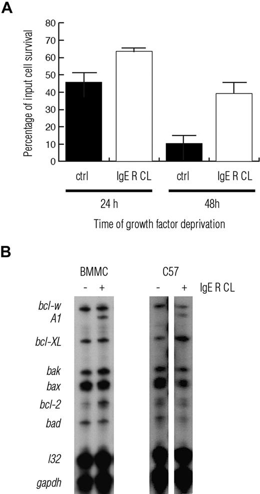
Murine bone marrow-derived mast cells (BMMCs) are normally IL-3 dependent and undergo apoptosis when deprived of this growth factor. We10 and others8,11,30 have previously demonstrated that activation via the high-affinity receptor for IgE rescues BMMCs from IL-3 deprivation-induced apoptosis. As shown in Figure 1A, the numbers of BMMCs surviving in the absence of IL-3 were increased when the cells were sensitized with TNP specific IgE followed by crosslinking with TNP-BSA. The increase in BMMC survival after activation was accompanied by a specific induction of A1 mRNA (Figure 1B). RNase protection assays showed that the levels of A1 mRNA were specifically up-regulated. The specific induction of A1 mRNA expression was also observed after FcϵRI-stimulation of the growth factor-independent mast cell line C57 (Figure 1B). Moreover, A1 induction was also apparent after activation with the calcium ionophore ionomycin (data not shown).
IgE receptor crosslinking induces mast cell survival and A1 expression. (A) Murine BMMCs, cultured under IL-3 deprivation, show enhanced survival when activated by IgE receptor crosslinking (IgER CL). (B) RNase protection assay (RPA) shows an increased expression of A1 in both BMMC and the mouse mast cell line C57 on IgER CL. Data are presented as mean plus or minus SEM of 3 independent experiments.
IgE receptor crosslinking induces mast cell survival and A1 expression. (A) Murine BMMCs, cultured under IL-3 deprivation, show enhanced survival when activated by IgE receptor crosslinking (IgER CL). (B) RNase protection assay (RPA) shows an increased expression of A1 in both BMMC and the mouse mast cell line C57 on IgER CL. Data are presented as mean plus or minus SEM of 3 independent experiments.
NF-κB is not required for the induction of A1 after IgE receptor activation
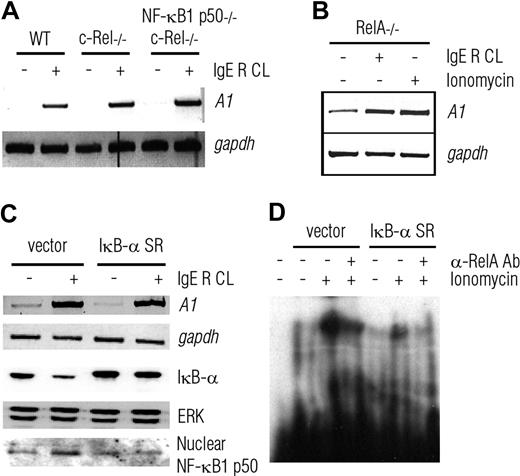
Because the NF-κB transcription factor pathway has been shown to be necessary and sufficient for A1 induction in other cell types, we analyzed its role in A1 expression after IgE receptor crosslinking. NF-κB binds DNA as homo- or hetero-dimers of related subunits (c-Rel, RelA, RelB, NF-κB1 p50, and NF-κB2 p52).31 RelA, c-Rel, and RelB contain a transcriptional activation domain, whereas NF-κB1 p50 and NF-κB2 p52 lack intrinsic transactivation potential and function as modulators of the transactivating partners within the dimer.31 It has been shown that c-Rel is critical for A1 induction and survival during mitogenic activation of B cells.22 We therefore activated bone marrow derived mast cells from RelA−/−, c-Rel−/−, or c-Rel−/−NF-κB1 p50−/− mice by IgE receptor crosslinking and analyzed A1 mRNA induction by RT-PCR. As demonstrated in Figure 2A, the induced expression of A1 was equivalent in wild-type, c-Rel−/− and c-Rel−/−NF-κB1 p50−/− mast cells. Moreover, the increased expression of A1 on IgE receptor activation or ionomycin stimulation was also seen in RelA−/− mast cells as shown in Figure 2B. These results indicate that in contrast to mitogen stimulated B cells, c-Rel, NF-κB1 p50, and RelA are not required for A1 induction after FcϵRI stimulation.
A1 induction is intact in NF-κB–deficient mast cells. (A,B) Reverse transcriptase PCR (RT-PCR) analysis of A1 expression in BMMCs deficient for 3 NF-κB subunits c-Rel (c-rel−/−) or c-Rel plus NF-κB1 p50 (c-rel−/−nfκb1−/−) (A) or RelA (rela−/−) (B) shows strong induction of A1 after IgER CL or ionomycin stimulation. (C) IgER CL increases A1 mRNA levels in C57 cells stably transfected with the nondegradable NF-κB super-repressor, IκB-α SR. Western blot analysis (WB) of cytosolic and nuclear extracts reveal intact IκB levels after IgER CL and an active IκB-α SR. The IκB-α SR is of human origin and runs at a slower mobility. (D) EMSA shows less protein-DNA complex of nuclear RelA and the oligonucleotide probe containing an RelA binding site, from C57 stable transfected with an active IκB-α SR compared with C57 cell transfected with an empty vector. Probing for GAPDH and ERK was used as loading controls in RT-PCR and Western blot analyses, respectively. RT-PCR experiments in panels A, B, and C were performed 3 times with similar results, whereas the Western blots and EMSA in panels C and D were performed twice and once, respectively.
A1 induction is intact in NF-κB–deficient mast cells. (A,B) Reverse transcriptase PCR (RT-PCR) analysis of A1 expression in BMMCs deficient for 3 NF-κB subunits c-Rel (c-rel−/−) or c-Rel plus NF-κB1 p50 (c-rel−/−nfκb1−/−) (A) or RelA (rela−/−) (B) shows strong induction of A1 after IgER CL or ionomycin stimulation. (C) IgER CL increases A1 mRNA levels in C57 cells stably transfected with the nondegradable NF-κB super-repressor, IκB-α SR. Western blot analysis (WB) of cytosolic and nuclear extracts reveal intact IκB levels after IgER CL and an active IκB-α SR. The IκB-α SR is of human origin and runs at a slower mobility. (D) EMSA shows less protein-DNA complex of nuclear RelA and the oligonucleotide probe containing an RelA binding site, from C57 stable transfected with an active IκB-α SR compared with C57 cell transfected with an empty vector. Probing for GAPDH and ERK was used as loading controls in RT-PCR and Western blot analyses, respectively. RT-PCR experiments in panels A, B, and C were performed 3 times with similar results, whereas the Western blots and EMSA in panels C and D were performed twice and once, respectively.
To exclude the possibility that the redundancy among the NF-κB transcription factors could still account for the induced expression of A1 in mast cells from the various NF-κB mutant mice, we used C57 cells expressing a super-repressor, which potently inhibits the activity of all NF-κB subunits. Normally, the different NF-κB subunits are sequestered in the cytoplasm via binding to IκB, but on activation IκB is phosphorylated and degraded, allowing the NF-κB dimers to enter the nucleus where they regulate expression of their target genes. The NF-κB super-repressor consists of a mutated form of IκB-α that is resistant to phosphorylation and degradation. As shown in Figure 2C, overexpression of the IκB-α SR had no effect on FcϵRI crosslinking-mediated A1 induction in C57 cells. The expression and activity of the IκB super-repressor in transfected C57 cells were confirmed by Western blotting (Figure 2C) and EMSAs (Figure 2D). On IgE receptor activation, nuclear translocation of NF-κB could be observed in control vector transfected cells but not in cells expressing the IκB-α SR (Figure 2C). Moreover, ionomycin activation increased complex formation of nuclear proteins and an oligonucleotide probe containing an NF-κB binding site in cells transfected with the empty control vector but not in IκB-α SR-transfected cells (Figure 2D). The enhanced complex formation was quenched when using a RelA specific antibody. These results confirm that NF-κB is not required for FcϵRI stimulation-induced transcriptional up-regulation of A1 in mast cells.
Cyclosporin A inhibits A1 induction in mast cells but not in macrophages
Because our results suggest that the induction of A1 in mast cells does not require NF-κB, other regulatory pathways and transcription factors were considered. The observation that A1 is strongly induced by treatment with the calcium ionophore ionomycin (Figure 2B) suggested that calcium flux may be involved. One of the downstream factors activated by calcium flux is the transcription factor NFAT, which is regulated by the phosphatase calcineurin. Dephosphorylation of NFAT by calcineurin enables nuclear translocation of NFAT.32 We therefore analyzed the levels of A1 mRNA (using RPA) in BMMCs and C57 cells that had been activated by IgE receptor crosslinking in the presence or absence of the calcineurin inhibitor cyclosporin A. As shown in Figure 3A,B, the induction of A1 was abrogated by pretreatment with cyclosporin A, consistent with the notion that this process requires NFAT. In contrast, cyclosporin A had no effect on LPS-induced A1 expression in the macrophage cell line J774A.1, a process that has been shown to be NF-κB dependent33 (Figure 3). These results indicate that NFAT is decisive for FcϵRI stimulation induced up-regulation of A1 mRNA in mast cells.
A1 induction is abrogated by the calcineurin inhibitor cyclosporin A specifically in mast cells. BMMCs (A) and the mast cell line C57 (B) show increased expression of A1 mRNA when activated by IgER CL. The calcineurin inhibitor cyclosporin A abrogated the induction of A1. (C) The inhibitory effect of cyclosporin A on A1 induction was not observed in the macrophage cell line J774A.1 activated by LPS. Expression of mRNA was analyzed by RNase protection assays (RPA). Probing for L32 and Gapdh was used as a control. The data are from one (B) and 2 (A, C) independent experiments. Results have also been confirmed by RT-PCR analysis, which was repeated 3 times (data not shown).
A1 induction is abrogated by the calcineurin inhibitor cyclosporin A specifically in mast cells. BMMCs (A) and the mast cell line C57 (B) show increased expression of A1 mRNA when activated by IgER CL. The calcineurin inhibitor cyclosporin A abrogated the induction of A1. (C) The inhibitory effect of cyclosporin A on A1 induction was not observed in the macrophage cell line J774A.1 activated by LPS. Expression of mRNA was analyzed by RNase protection assays (RPA). Probing for L32 and Gapdh was used as a control. The data are from one (B) and 2 (A, C) independent experiments. Results have also been confirmed by RT-PCR analysis, which was repeated 3 times (data not shown).
The A1 promoter is activated by IgE receptor crosslinking or ionomycin treatment
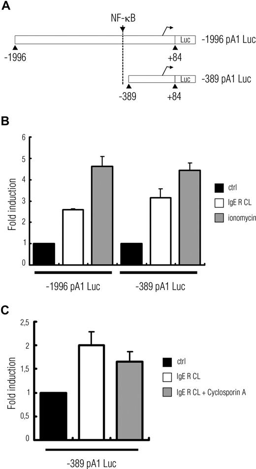
The A1 promoter contains a consensus NF-κB binding site required for antigen receptor crosslinking-mediated induction in lymphocytes22 (Figure 4A). To investigate whether this binding site was also involved in IgE receptor-mediated induction of A1 in mast cells, a DNA fragment extending upstream of the 5′-untranslated region of the mouse A1 gene (−1996 + 84) was inserted 5′ of the luciferase gene in a promoter-less reporter plasmid (designated −1996pA1Luc) and transiently transfected into C57 cells. We also generated a shorter A1 promoter construct in which the NF-κB binding site was deleted (plasmid designated −389pA1Luc; Figure 4A). The plasmids −1996pA1Luc, as well as −389pA1Luc, exhibited increased promoter activity in mast cells activated either by FcϵRI crosslinking or treatment with ionomycin (Figure 4B). Pretreatment with cyclosporin A decreased FcϵRI stimulation-induced activation of this reporter (Figure 4C). These results further support the notion that NF-κB is not critical for FcϵRI stimulation induced transcriptional up-regulation of A1 in mast cells. Instead, a calcineurin regulated target, most probably NFAT, appears to be required.
An A1 promoter, lacking the NF-κB site, can still be activated by IgE receptor crosslinking or treatment with the calcium ionophore ionomycin. (A) Schematic view of the 2 vectors with luciferase as a reporter, containing upstream regions of A1. The −389 pA1 Luc does not contain the NF-κB binding site. (B) C57 cells transiently transfected with −1996 pA1 Luc or −389 pA1 Luc show a 3- to 5-fold increase in luciferase activity after IgE receptor crosslinking or treatment with ionomycin (1 μM). (C) The increased luciferase activity was abrogated by the addition of cyclosporin A. Data represent mean plus or minus SEM from 2 or 3 independent experiments.
An A1 promoter, lacking the NF-κB site, can still be activated by IgE receptor crosslinking or treatment with the calcium ionophore ionomycin. (A) Schematic view of the 2 vectors with luciferase as a reporter, containing upstream regions of A1. The −389 pA1 Luc does not contain the NF-κB binding site. (B) C57 cells transiently transfected with −1996 pA1 Luc or −389 pA1 Luc show a 3- to 5-fold increase in luciferase activity after IgE receptor crosslinking or treatment with ionomycin (1 μM). (C) The increased luciferase activity was abrogated by the addition of cyclosporin A. Data represent mean plus or minus SEM from 2 or 3 independent experiments.
An IgER crosslinking inducible nuclear protein complex binds a putative NFAT site within the A1 promoter
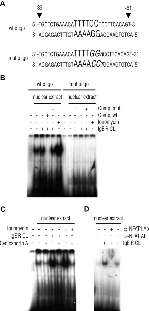
We next performed analysis of the nuclear protein-binding properties of a putative NFAT binding site, between −89 and −61 in the A1 promoter (Figure 5A), using EMSA. A single major nuclear complex was detected in resting cells. This was strongly up-regulated on FcϵRI crosslinking or treatment with ionomycin (Figure 5B,C). Addition of an unlabeled wild-type but not unlabeled mutated probe competitively reduced the complex formation. Mutating the putative NFAT binding site (Figure 5A) totally abrogated protein binding (Figure 5B). The increased binding of the wild-type probe to the nuclear extract was prevented by pretreatment with cyclosporin A (Figure 5C).
An NFAT-containing protein complex binds to a putative NFAT site in the A1 proximal promoter. (A) Sequences of the wild-type (wt) and mutated (mut) oligonucleotide probes used in the EMSA with the putative binding site for NFAT indicated in bold and the 3 base pair mutation in italics. (B) EMSA on nuclear extracts from C57 cells activated by IgE receptor crosslinking or treatment with ionomycin (1 μM). Activation of C57 cells results in an increased binding of a nuclear protein complex to the oligonucleotide probe shown in panel A. This complex formation is abrogated by pretreatment with unlabeled competitive wt but not mutated oligonucleotide probe. Mutated oligonucleotide probe does not form binding complex at all. (C) The augmented complex binding from activated C57 is inhibited by pretreatment of the cells with cyclosporin A. (D) NFAT-specific antibodies (anti-NFAT = NFATc1; anti-NFAT1 = NFATc2) cause a band shift in the EMSA, indicating the presence of NFAT within the DNA-protein complex. Representative results from 2 independent experiments are shown.
An NFAT-containing protein complex binds to a putative NFAT site in the A1 proximal promoter. (A) Sequences of the wild-type (wt) and mutated (mut) oligonucleotide probes used in the EMSA with the putative binding site for NFAT indicated in bold and the 3 base pair mutation in italics. (B) EMSA on nuclear extracts from C57 cells activated by IgE receptor crosslinking or treatment with ionomycin (1 μM). Activation of C57 cells results in an increased binding of a nuclear protein complex to the oligonucleotide probe shown in panel A. This complex formation is abrogated by pretreatment with unlabeled competitive wt but not mutated oligonucleotide probe. Mutated oligonucleotide probe does not form binding complex at all. (C) The augmented complex binding from activated C57 is inhibited by pretreatment of the cells with cyclosporin A. (D) NFAT-specific antibodies (anti-NFAT = NFATc1; anti-NFAT1 = NFATc2) cause a band shift in the EMSA, indicating the presence of NFAT within the DNA-protein complex. Representative results from 2 independent experiments are shown.
To confirm the identity of the protein complex bound to the A1 promoter probe, we performed super-shift assays. If the probe is bound to nuclear NFAT, a super-shift or diminished binding should be observed after addition of an NFAT-specific antibody. For this purpose, we used 2 different antibodies: one with a broad specificity recognizing several NFAT forms and another antibody specific for NFAT1 (also called NFAT2c or p). The broad spectrum antibody to NFAT diminished the shifted band in the EMSA, probably because it binds to and blocks the DNA binding region of NFAT (Figure 5D). A super-shift of the band was obtained using the NFAT1-specific antibody (Figure 5D). These results indicate that NFAT1 translocates to the nucleus on IgE receptor activation and binds to the A1 promoter.
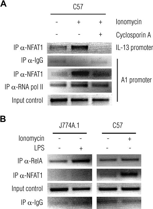
To detect binding of NFAT to the A1 promoter region within living cells, we performed ChIP (Figure 6A). Antibodies against NFAT1 pulled down more chromatin-bound protein complex containing NFAT1 from mast cells being stimulated with ionomycin compared with unstimulated cells. Pretreatment with cyclosporin A before stimulation with ionomycin inhibited chromatin/protein complex formation. In contrast to mast cells, the NFAT1-specific antibody could not pull down any chromatin bound protein either in unstimulated or LPS-activated macrophage cell line J774A.1 cells. Instead, an increased complex formation was detected in LPS activated J774A.1 cells compared with unstimulated, using an NF-κB–specific antibody (Figure 6B). The immunoprecipitated DNA was analyzed by PCR using primer pairs for the A1 promoter covering either a putative NF-κB (RelA) binding site or the putative NFAT binding site shown in Figure 5A. As a control, we included primer pairs for the IL-13 promoter, which recently has been reported to be regulated by NFAT1 in mast cells.34
NFAT1-containing protein complexes bind a promoter region of A1 on activation in mast cells but not the macrophage cell line J774A.1. (A) ChIP reveals an increase of NFAT1-containing protein complexes bound to the A1 promoter in C57 cells stimulated with ionomycin for 3 hours compared with unstimulated cells. The binding is inhibited by pretreatment (10 minutes) of the cells with cyclosporin A before stimulation with ionomycin. Chromatin fragments were prepared and immunoprecipitated with antibodies against NFAT1, control IgG (negative control), and RNA polymerase II (positive control). The immunoprecipitated DNA and input DNA were analyzed by PCR using specific primer pairs for A1 resulting in PCR products covering the sequence shown in Figure 5A, and for the promoter of IL-13. Shown is a representative of 3 independent experiments. (B) Activation of the macrophage cell line J774A.1 by LPS for 3 hours increases complex formation of the NF-κB subunit RelA and chromatin of the A1 promoter region. On the contrary, the NFAT1-specific antibody does not pull down any chromatin bound protein complexes in J774A.1 but in ionomycin-activated C57 cells.
NFAT1-containing protein complexes bind a promoter region of A1 on activation in mast cells but not the macrophage cell line J774A.1. (A) ChIP reveals an increase of NFAT1-containing protein complexes bound to the A1 promoter in C57 cells stimulated with ionomycin for 3 hours compared with unstimulated cells. The binding is inhibited by pretreatment (10 minutes) of the cells with cyclosporin A before stimulation with ionomycin. Chromatin fragments were prepared and immunoprecipitated with antibodies against NFAT1, control IgG (negative control), and RNA polymerase II (positive control). The immunoprecipitated DNA and input DNA were analyzed by PCR using specific primer pairs for A1 resulting in PCR products covering the sequence shown in Figure 5A, and for the promoter of IL-13. Shown is a representative of 3 independent experiments. (B) Activation of the macrophage cell line J774A.1 by LPS for 3 hours increases complex formation of the NF-κB subunit RelA and chromatin of the A1 promoter region. On the contrary, the NFAT1-specific antibody does not pull down any chromatin bound protein complexes in J774A.1 but in ionomycin-activated C57 cells.
Constitutively active NFAT1 increases the expression of A1
The results from the EMSA and ChIP assay indicated that NFAT1 might be a regulator of A1 expression in FcϵRI stimulated mast cells. To further investigate this hypothesis, we transfected C57 cells with expression vectors encoding constitutively active nuclear forms of either NFAT1 or NFAT2 and measured the activation of A1 after IgER CL. Analysis of A1 expression by RT-PCR revealed an augmented expression in cells transfected with NFAT1, whereas cells transfected with NFAT2 or mock-transfected cells exhibited similar expression levels (Figure 7A). The increased expression of A1 in NFAT1-transfected cells could be confirmed by real-time quantitative PCR (Figure 7B). These results demonstrate that NFAT1 plays a critical role in FcϵRI stimulation-induced up-regulation of A1 in mast cells.
Expression of constitutively active NFAT1 increases A1 mRNA levels in FcϵRI-stimulated mast cells. (A) RT-PCR analysis was used to determine the levels of A1 mRNA in C57 cells transiently transfected with vectors expressing constitutively active NFAT1, NFAT2, or empty vector. The cells were left untreated or were activated by FcϵRI crosslinking. Amplification of GAPDH was used as control. Overexpression of NFAT1, but not NFAT2, resulted in increased levels of A1 mRNA. Representative results from 2 independent experiments are shown. (B) Real-time quantitative PCR analysis of A1 mRNA shows increased levels of A1 expression in C57 cells expressing constitutively active NFAT1 compared with cells transfected with empty vector. Data are presented as mean plus or minus SEM from 3 independent experiments.
Expression of constitutively active NFAT1 increases A1 mRNA levels in FcϵRI-stimulated mast cells. (A) RT-PCR analysis was used to determine the levels of A1 mRNA in C57 cells transiently transfected with vectors expressing constitutively active NFAT1, NFAT2, or empty vector. The cells were left untreated or were activated by FcϵRI crosslinking. Amplification of GAPDH was used as control. Overexpression of NFAT1, but not NFAT2, resulted in increased levels of A1 mRNA. Representative results from 2 independent experiments are shown. (B) Real-time quantitative PCR analysis of A1 mRNA shows increased levels of A1 expression in C57 cells expressing constitutively active NFAT1 compared with cells transfected with empty vector. Data are presented as mean plus or minus SEM from 3 independent experiments.
Discussion
The present study indicates that the prosurvival gene A1 is transcriptionally regulated by NFAT in mast cells activated through FcϵRI crosslinking. This stands in contrast to lymphocytes in which A1 transcription on B- or T-cell receptor activation is NF-κB dependent. Mast cells deficient in RelA, c-Rel, or c-Rel plus NF-κB1 p50 or those overexpressing the IκB-α SR super-repressor could still increase A1 levels after FcϵRI stimulation. However, on treatment with the calcineurin inhibitor cyclosporin A, the expression of A1 was diminished in FcϵRI stimulated mast cells but not in macrophages activated by LPS. These findings demonstrate that the expression of A1 is transcriptionally regulated in a lineage- and stimulus-specific manner. A1 is a NFAT target gene in mast cells stimulated through their FcϵRI but is regulated by NF-κB in lymphocytes triggered through their antigen receptors.
Our interest in the apparent cell-specific regulation of A1 in mast cells started when we first noticed that activation-induced mast cell survival was not diminished in BMMCs deficient for the NF-κB family members RelA, c-Rel, or even those lacking c-Rel plus NF-κB1 p50 (data not shown). Although NF-κB1 p50 has been shown to be a critical component of the NF-κB dimers expressed in mast cells,35 it was important to rule out possible involvement of other NF-κB family members. For this purpose, we used C57 mast cells expressing the IκB-α super-repressor. In quiescent cells, NF-κB proteins reside in the cytoplasm in an inactive form because of their association with IκB. On activation, IκB is phosphorylated and ubiquitylated, which targets it for proteasomal degradation, thereby allowing NF-κB dimers to translocate to the nucleus where they activate transcription of their target genes.36 The IκB-α SR is mutated at the specific serine residues that become phosphorylated on activation and therefore is not phosphorylated and degraded and remains bound to NF-κB, even after cellular activation. The normal induction of A1 in C57 cells stably expressing IκB-α SR therefore provides compelling evidence that NF-κB is not essential for the transcriptional up-regulation of A1 in mast cells.
The inhibitory effect of cyclosporin A on mast cell degranulation and cytokine secretion is well documented.37-39 When we first started our work on A1 in mast cells, we noticed that cyclosporin A had an affect on activation-induced mast cell survival.10 NFAT was therefore a probably candidate for regulating A1-induction after allergic activation of mast cells. Cyclosporin A is a specific inhibitor of calcineurin, a calcium-activated protein phosphatase that dephosphorylates NFAT and thereby promotes its nuclear import.40 IgE receptor crosslinking causes an increase in calcium influx, which leads to NFAT activation.41 The complete inhibition of A1 expression by cyclosporin A treatment in FcϵRI-stimulated mast cells indicates an involvement of NFAT in the transcriptional regulation of A1 in these cells. In contrast, the LPS-mediated induction of A1 in the J774A.1 macrophage cell line was not affected by cyclosporin A. Interestingly, it has recently been shown that the zinc finger transcription factor WT1 regulates the expression of A1 in granulocytes.42 Collectively, these results provide evidence for lineage-specific regulation of the prosurvival gene A1 in different hematopoietic cell types.
The promoter for A1 contains an NF-κB binding site that was demonstrated to be crucial for BCR stimulation-mediated A1 induction in B cells.22 In our experiments, using A1 promoter-luciferase constructs, we still obtained activation of this reporter after IgE receptor crosslinking or treatment with ionomycin, even when using a construct in which the NF-κB binding site was deleted. In contrast, reporter activation was abolished by pretreatment with cyclosporin A. The −389pA1 construct (which lacks the NF-κB binding site) contains a putative NFAT-binding site, and this was used for further studies using EMSA. The results from these experiments showed that cyclosporin A attenuates the formation of the DNA-protein complex, and antibodies against NFAT induced a mobility shift of the complex. Chromatin immunoprecipitation experiments demonstrated that NFAT directly binds to the promoter region of A1 in activated mast cells. Again, pretreatment with cyclosporin A before stimulation with ionomycin inhibited binding of NFAT to its target site within the A1 promoter. Together, these results further strengthen our conclusion that NFAT is an essential regulator of the transcription of A1 in mast cells. We have, however, not yet been able to pinpoint the precise NFAT binding site within the A1 promoter. Although mutation of the putative NFAT binding site in the A1 promoter, −89/−61, diminished the binding of the probe to the nuclear extract in the EMSA, we could still see an activity in the luciferase reporter assay (data not shown). There are several possible explanations for this, as NFAT has a remarkable versatility in its binding to DNA.43 NFAT proteins can bind to regulatory sites within target genes cooperatively with unrelated transcription factors, such as AP-1 (Fos-Jun),44 and they can also act synergistically with other transcription factors, such as GATA, EGR, and IRF-4, which bind to distinct sites within common target genes.40 Furthermore, in addition to the proximal promoter, distal regulatory elements often have profound effects on gene expression. An example is the NFAT-regulated expression of the IL-4 gene in T cells and mast cells, which contains enhancers within intronic sequences.45,46 It is also possible that NFAT induces A1 indirectly through activation of an intermediate transcription factor. Therefore, further work is needed to clarify how NFAT regulates A1 transcription in mast cells.
The NFAT family of transcriptional regulators consists of 5 proteins: NFAT1 to NFAT5,43 of which NFAT1, NFAT2, and NFAT4 are expressed in mast cells.29 The different NFAT proteins appear to have largely redundant functions. For instance, IL-13 transcription in mast cells is regulated by both NFAT1 and NFAT2.34 In this study, we found that expression of NFAT1, but not NFAT2, caused increased A1 expression in FcϵRI-stimulated C57 mast cells. We also observed a super-shift in the EMSA and an immunoprecipitated protein/chromatin complex in ChIP when using NFAT1-specific antibodies. Accordingly, these data suggest that NFAT1 is the critical transcription factor for A1 induction in mast cells.
The molecular mechanisms by which A1 protects mast cells from undergoing apoptosis remain to be fully determined. Because antiapoptotic and proapoptotic Bcl-2 family members form heterodimers and probably titrate each others function,47,48 the induction of A1 may serve to neutralize the increase in the level and/or activity of one of its proapoptotic relatives. A1 binds with high affinity to several of the proapoptotic BH3-only proteins, including Bim, PUMA, and NOXA.49 Of these BH3-only proteins, Bim was shown to be involved in regulating mast cell apoptosis, and, interestingly, Bim is also up-regulated on FcϵRI crosslinking.50,51 It has recently been reported that the interaction between A1 and Bim stabilizes the A1 protein, thereby slowing A1 turnover and amplifying its antiapoptotic effects.52 Collectively, these data suggest that the simultaneous induction of A1 and Bim might balance each other and determine whether the mast cell will survive or undergo apoptosis.
The publication costs of this article were defrayed in part by page charge payment. Therefore, and solely to indicate this fact, this article is hereby marked “advertisement” in accordance with 18 USC section 1734.
Acknowledgments
The authors thank the members of the Nilsson laboratory for stimulating discussions and critical reading of the manuscript, Dr M. Pellegrini for transfected cells, Dr D. Baltimore for Rela−/− mice, Dr A. Rao for the NFAT expression vectors, and Marie Holmquist for A1 promoter constructs.
This study was supported by the Swedish Cancer Foundation, the Swedish Research Council-Medicine, the Swedish Cancer and Allergy Fund, Consul Th C Berghs Foundation, Hans von Kantzow's Foundation, Ollie and Elof Ericsson's Foundation, King Gustav V's 80-years Foundation, Ellen, Walter and Lennart Hesselmans foundation, Karolinska Institutet, the National Health and Medical Research Council (Australia; program 257502), the Leukemia and Lymphoma Society (New York; SCOR grant 7015) and the National Cancer Institute (National Institutes of Health, CA 80188 and CA 43540).
National Institutes of Health
Authorship
Contribution: E.U. and M.K. designed and performed research, analyzed data, and wrote the paper; C.M.W. and J.A. performed research; S.G. designed research and contributed valuable reagents; A.S. designed research and wrote the paper; and G.N. designed research, analyzed data, and wrote the paper.
Conflict-of-interest disclosure: G.N. holds a patent related to the work that is described in the present study. The other authors declare no competing financial interests.
Correspondence: Gunnar Nilsson, Karolinska Institutet, Department of Medicine, Clinical Immunology and Allergy Unit, KS L2:04, SE-171 76 Stockholm, Sweden; e-mail: Gunnar.P.Nilsson@ki.se.
References
Author notes
E.U. and M.K. contributed equally to this study.



This feature is available to Subscribers Only
Sign In or Create an Account Close Modal