To the editor:
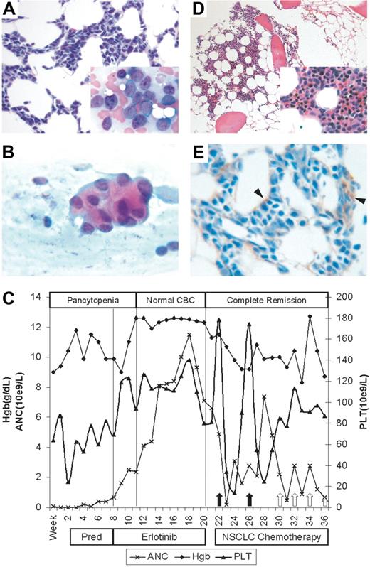
Newer agents with decreased toxicities are needed to treat elderly patients with acute myelogenous leukemia (AML).1,2 This is the first case report of AML responding to erlotinib, an epidermal growth factor receptor (EGFR) tyrosine kinase inhibitor. A 68-year-old Vietnamese man with a 50-pack-a-year smoking history presented with several weeks of dyspnea. Peripheral blood leukocyte count was 2.5 × 10⁹/L with 3% neutrophils, 1% bands, 66% lymphocytes, and 15% myeloblasts, hemoglobin of 100 g/L (10.0 g/dL), and platelet count of 72 × 109/L. Bone marrow biopsy revealed 86% myeloperoxidase-positive blasts, while the immunophenotype was 88% CD34, 78% HLA-DR, 20% CD13, 9% CD33, 0% CD10, 4% CD19, 0% CD20, 1% CD3, 2% CD5, 0% CD8, and 2% CD25 by flow cytometry, consistent with AML-M1 (Figure 1A). Cytogenetics were not performed. Computed tomography of the chest showed a 2.9 cm by 2.4 cm mass in the left upper lobe, and a fine-needle aspiration biopsy of the mass was consistent with well-differentiated adenocarcinoma (Figure 1B). Positron emission tomography scanning revealed uptake in the lung mass and mediastinum, consistent with clinical stage IIIB non–small-cell lung cancer (NSCLC).
Erlotinib-induced complete remission in AML lacking EGFR expression. (A) Pretreatment bone marrow with up to 90% of cellularity composed of myeloid blasts (hematoxylin-eosin; magnification, × 400). Insert: Wright-Giemsa stain, magnification × 1000 oil. (B) Fine-needle aspiration cytology of the lung mass reveals 3D cohesive clusters of atypical epithelial cells containing intracytoplasmic mucin vacuoles, consistent with adenocarcinoma (hematoxylin-eosin; magnification, × 1000 oil). (C) Graph of peripheral blood counts over time. From weeks 1 to 8, patient remained pancytopenic with AML. Three weeks after starting erlotinib therapy, patient's absolute neutrophil count (ANC) and hemoglobin (Hgb) normalized, while platelet counts exceeded 100 × 109/L. Complete remission was documented by bone marrow biopsy on week 20 (panel D). Peripheral counts show recovery after each round of combination chemotherapy for NSCLC (arrows), suggesting adequate marrow reserve while remaining in complete remission. (D) Posterlotinib bone marrow with variable (average 30%) cellularity and trilineage maturing hematopoiesis. Myeloblasts are less than 3% (hematoxylin-eosin; magnification, × 200). Insert: magnification, × 400. (E) Immunohistochemical staining of initial bone marrow biopsy for EGFR. Only stromal cells (arrowheads) stain positively. Myeloblasts are EGFR negative (magnification, × 1000 oil). Images were acquired using an Olympus BX51 microscope (Olympus, Tokyo, Japan) equipped with either a 20×/0.5 NA, 40×/0.65 NA, or 100×/1.3 NA oil objective and mounted DP12 digital camera, and were further processed (cut out and white balanced) with Adobe Photoshop CS2 (Adobe Systems, San Jose, CA).
Erlotinib-induced complete remission in AML lacking EGFR expression. (A) Pretreatment bone marrow with up to 90% of cellularity composed of myeloid blasts (hematoxylin-eosin; magnification, × 400). Insert: Wright-Giemsa stain, magnification × 1000 oil. (B) Fine-needle aspiration cytology of the lung mass reveals 3D cohesive clusters of atypical epithelial cells containing intracytoplasmic mucin vacuoles, consistent with adenocarcinoma (hematoxylin-eosin; magnification, × 1000 oil). (C) Graph of peripheral blood counts over time. From weeks 1 to 8, patient remained pancytopenic with AML. Three weeks after starting erlotinib therapy, patient's absolute neutrophil count (ANC) and hemoglobin (Hgb) normalized, while platelet counts exceeded 100 × 109/L. Complete remission was documented by bone marrow biopsy on week 20 (panel D). Peripheral counts show recovery after each round of combination chemotherapy for NSCLC (arrows), suggesting adequate marrow reserve while remaining in complete remission. (D) Posterlotinib bone marrow with variable (average 30%) cellularity and trilineage maturing hematopoiesis. Myeloblasts are less than 3% (hematoxylin-eosin; magnification, × 200). Insert: magnification, × 400. (E) Immunohistochemical staining of initial bone marrow biopsy for EGFR. Only stromal cells (arrowheads) stain positively. Myeloblasts are EGFR negative (magnification, × 1000 oil). Images were acquired using an Olympus BX51 microscope (Olympus, Tokyo, Japan) equipped with either a 20×/0.5 NA, 40×/0.65 NA, or 100×/1.3 NA oil objective and mounted DP12 digital camera, and were further processed (cut out and white balanced) with Adobe Photoshop CS2 (Adobe Systems, San Jose, CA).
Given the patient's poor performance status, no AML therapy was initiated. Prednisone at 20 mg daily was started for exacerbation of chronic obstructive pulmonary disease, while his only other medication was omeprazole. Six weeks later, the patient received palliative radiotherapy (800 cGy, single dose) to the lung mass and was started on erlotinib 150 mg orally once daily. Patient tolerated erlotinib well except for an acneiform rash that responded completely to oral tetracycline. After 3 months of erlotinib therapy, the patient had normal complete blood counts and no circulating blasts (Figure 1C). A repeat bone marrow biopsy was mildly hypocellular for age with maturing trilineage hematopoiesis, less than 3% myeloblasts, and normal cytogenetics (Figure 1D). Time to normal absolute neutrophil count, hemoglobin, and platelet counts were 20 days, 20 days, and 105 days, respectively. Due to progressive NSCLC, erlotinib was discontinued, and chemotherapy with paclitaxel and carboplatin was initiated. The patient's AML remained in remission following the discontinuation of erlotinib, but he ultimately died of progressive NSCLC 6 months later.
Spontaneous remissions of AML have occurred following sepsis,3-5 but this patient had no evidence of infection. Prednisone and omeprazole are not known to induce AML remission. Erlotinib is a small-molecule tyrosine kinase inhibitor (TKI) of human erythroblastic leukemia viral oncogene homolog 1 (ErbB1)/EGFR that is approved for the treatment of NSCLC6 and pancreatic cancer.7 Although erlotinib has not been studied in AML, gefitinib, another EGFR-TKI, can induce neutrophil differentiation in several EGFR− AML cell lines.8,9 In this patient's bone marrow biopsy, EGFR was expressed on stromal cells, including fibroblasts, but was undetectable on myeloblasts (Figure 1E). Erlotinib was well tolerated; complete remission was achieved within a month, and was durable for at least 6 months. In conclusion, this is the first report of a patient with AML who achieved complete remission following erlotinib. EGFR inhibitors may induce neutrophilic differentiation of AML blasts via effects on tyrosine kinases (or other targets) distinct from EGFR. Clinical trials of erlotinib in the treatment of AML are warranted.
Authorship
Conflict-of-interest disclosure: The authors declare no competing financial interests.
Correspondence: Geoffrey Chan, Assistant Professor of Medicine, Division of Hematology/Oncology, Tufts-New England Medical Center, 750 Washington St, Box no. 245, Boston, MA 02111; e-mail: gchan@tufts-nemc.org.


This feature is available to Subscribers Only
Sign In or Create an Account Close Modal