Abstract
Background: Telomeres are linear DNA structures located at the ends of chromosomes which undergo progressive shortening in somatic tissues with high replicative rate, including hematopoietic cells. Due to this peculiarity, telomere length (TL) has been used as a marker of cell senescence and of previous replicative stress. Following hematopoietic stem cell transplant (SCT) a variable shortening of TL has observed, this variability has been in part ascribed to differences in post-graft replicative stress. In a recent report (Ricca I et al, Leukemia 2005), we have analysed TL on peripheral blood progenitor cell (PBPC) harvested after 2 tightly-spaced hd-chemotherapy courses, i.e. hd-Cyclophosphamide (CY) and hd-Ara-C and found that TL of PBPC was markedly shortened at the second hd-course. Aim of this study was to investigate how TL of grafted cells may influence telomere status of post-SCT hematopoietic cells in both the autologous and allogeneic setting.
Patients and Methods: TL was monitored in 20 patients undergoing autograft with PBPC collected after hd-CY (10 patients) or hd-Ara-C (10 patients) and in 10 patients undergoing allograft with PBPC mobilized from healthy donors. Overall, patient median age was 48 years (range 19–64). The amount of CD34+ cells grafted was 7 × 10⁶ CD34+/Kg (range 2,03–22,6). TL was assessed both on the graft and on Bone Marrow (BM) samples taken at a median time of 14 months (range 12–17) after SCT. All patients were in continuous complete remission at the time of TL analisis. TL was evaluated by Southern blot, as previously described (Rocci A et al, Exp Hematol 2007).
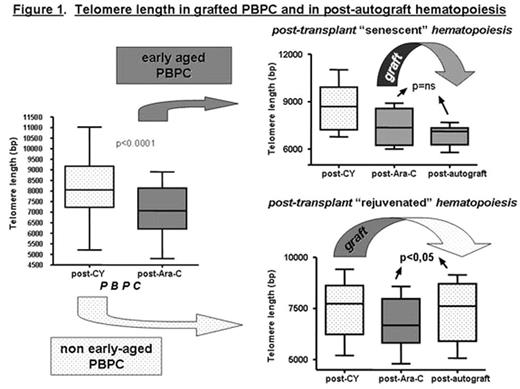
Results: As expected, TL was found markedly shortened in post-Ara-C, with median TL of 8051 bp (range 5209–11024 bp) and 7066 bp (range 4800–8906 bp) in PBPC post-CY and post-Ara-C, respectively (p<0.0001) (see Figure 1). TL analysis on post-autograft BM cells displayed a median TFR length of 7120 bp (range 5791–7689 bp) in patients autografted with post-Ara-C PBPC (p=NS compared to grafted cells); in contrast, patients autografted with post-CY PBPC had a significantly longer TL (7614 bp, range 5075–9141 bp) compared to pre-transplant value (6678 bp, range 4800–8576) (p<0.05) (see Figure 1). In the allograft setting, a similar behaviour was observed: median TL of post-graft BM cells was 7382 bp (range 6634–7755), quite similar to TL of donor grafted cells (7114 bp, range: 6836–9496 bp) (p=0.522), and markedly longer comapared to recipient pre-allogeneic SCT TL (6121 bp, range 5561–9036) (p=0.0005).
Conclusions: TL of post-SCT hematopoietic cells reflects that of the grafted cells; thus, the quality of reinfused cells rather than the replicative stress for engraftment may mostly influence the degree of telomere shortening observed following SCT. The results taken together suggest that SCT might be also considered for future approaches aimed to rejuvenate the hematopoietic system.
Telomere length in grafted PBPC and in post-autograft hematopoiesis
Author notes
Disclosure: No relevant conflicts of interest to declare.


This feature is available to Subscribers Only
Sign In or Create an Account Close Modal