Abstract
Immune thrombocytopenia (ITP) is classified as an autoimmune disease in, which antibody-coated platelets are phagocytosed by macrophages in the reticuloendothelial system (RES) through Fc gamma receptor-mediated or complement-mediated pathways. Based on the the pathophysiology of ITP, described above, and evidences that macrophages expresses mannose receptor on their surface and that mannose-terminated human glucocerebrosidase is highly effective in ameliorating many of the clinical manifestations of Gaucher’s disease, we devised mannose labeled liposome containing DXR to eliminate specifically macrophages. In this study, we evaluated the effects of mannose labeled liposome containing DXR (liposome-DXR) in a mouse model of ITP.
In vitro experiments: Murine peritoneal macrophages were obtained by the intra-peritoneal injection of thioglycolate (TGC) to ddY mice.
The uptake of fluorescence by peritoneal macrophages was measured using rhodamine-liposome by flow cytometry. The macrophages were incubated with mannose labeled rhodamine-liposome {mannose(+)-rhodamine-liposome} or mannose unlabeled rhodamine-liposome {mannose(−)-rhodamine-liposome} for 30 min at 37°C. After washing with PBS, they were applied for flow cytometry. Rhodamine fluorescence intensity in macrophages was higher in incubation with mannose(+)-rhodamine-liposome than in incubation with mannose(−)-rhodamine-liposome treatment.
The viability of peritoneal macrophages was measured by MTT assay after 30 min incubation with liposome-DXR. The viability in the incubation with mannose labeled liposome-DXR {mannose(+)-liposome-DXR} was much less than in the incubation with mannose unlabeled liposome-DXR {mannose(−)-liposome-DXR} (Figure 1).
In vivo experiment: Anti-mouse platelet serum (APS) was injected intra-peritoneally to ddY mice at 24 hr after intravenous injection of mannose(+)-liposome-DXR or mannose(−)-liposome-DXR.
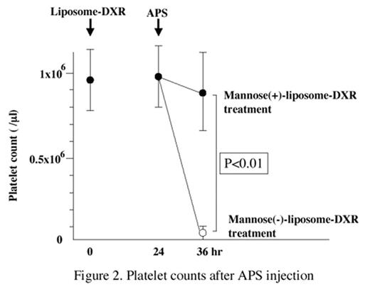
Platelet counts were measured at 12 hr after APS injection. They were 89.3±22.1 × 10⁴ /μl (n=6, p<0.01) in mannose(+)-liposome-DXR treatment, while 4.5±2.0 × 10⁴ /μl (n=8) in mannose(−)-liposome-DXR treatment (Figure 2). The platelet counts were 3.2±0.5 × 10⁴ /μl (n=4), 96.1±15.0 × 10⁴ /μl (n=4) or 99.3±26.2 × 10⁴/μl (n=4) in mice with only APS administration, mannose(+)-liposome-DXR+saline or mannose(−)-liposome-DXR+saline administrations instead of APS, respectively. These in vivo data indicated that APS induced thrombocytopenia was not observed in mice treated with mannose(+)-DXR-liposome, because of the destruction of macrophages by mannose(+)-DXR-liposome administration. These in vitro and in vivo data strongly suggest that mannose(+)-liposome-DXR may specifically delete the macrophages and and can be effective in the management of experimental ITP model mice.
The Viability of Murine Macrophages after Incubation with Liposome-Doxorubicine
The Viability of Murine Macrophages after Incubation with Liposome-Doxorubicine
Platelet counts after APS injection
Author notes
Disclosure: No relevant conflicts of interest to declare.


This feature is available to Subscribers Only
Sign In or Create an Account Close Modal