Hematopoiesis is a complex process which gives rise to all blood lineages in the course of an organism's lifespan. However, the underlying molecular mechanism governing this process is not fully understood. Here we report the isolation and detailed study of a newly identified zebrafish ugly duckling (Udu) mutant allele, Udusq1. We show that loss-of-function mutation in the udu gene disrupts primitive erythroid cell proliferation and differentiation in a cell-autonomous manner, resulting in red blood cell (RBC) hypoplasia. Positional cloning reveals that the Udu gene encodes a novel factor that contains 2 paired amphipathic α-helix–like (PAH-L) repeats and a putative SANT-L (SW13, ADA2, N-Cor, and TFIIIB–like) domain. We further show that the Udu protein is predominantly localized in the nucleus and deletion of the putative SANT-L domain abolishes its function. Our study indicates that the Udu protein is very likely to function as a transcription modulator essential for the proliferation and differentiation of erythroid lineage.
Introduction
Hematopoiesis in vertebrate occurs in 2 waves, primitive and definitive.1,–3 In the mouse, the primitive or embryonic wave of hematopoiesis occurs around embryonic day 7.5 in the yolk-sac blood island, and produces primitive erythrocytes and macrophages.4,5 This primitive wave of hematopoiesis lasts for a transient period of a few days and is subsequently replaced by the definitive program. Murine definitive hematopoiesis is believed to originate from a distinctive region known as the aorta-gonad-mesonephros at embryonic days 7.5 to 8.6,7 These definitive hematopoietic precursors, presumably the definitive hematopoietic stem cells, then migrate to the fetal liver, where they undergo rapid proliferation and differentiation, and finally colonize the bone marrow for adult hematopoiesis. In contrast to primitive hematopoiesis, the definitive hematopoietic program gives rise to all the mature blood cell types and remains active throughout the lifetime of the organism
In zebrafish, hematopoiesis also comprises both primitive and definitive programs and produces mature blood cell types similar to those found in mammals.8,9 Zebrafish primitive erythropoiesis begins at the 4-somite stage as a pair of bilateral stripes in the posterior lateral mesoderm.10 These stripes extend anteriorly and posteriorly, and then converge in the midline at the 20-somite stage to form the intermediate cell mass (ICM), where erythroid precursors further develop and enter the circulation by 24 to 26 hours after fertilization (hpf).10,11 On the other hand, primitive myelopoiesis originates from the rostral blood island in the anterior lateral mesoderm at around the 10-somite stage and produces mainly macrophages and possibly some neutrophils.12,,–15 Recent studies demonstrate that zebrafish definitive hematopoiesis initiates in the ventral wall of dorsal aorta between 26 and 48 hpf,16,17 and then establishes the adult hematopoietic organ in the kidney by 5 days after fertilization (dpf).11
The zebrafish mutant, ugly duckling (Udutu24), was first isolated from the 1996 Tuebingen large-scale screen as a mutant defective in morphogenesis during gastrulation and tail formation.18 In this article, we report the isolation and detailed study of a new udu allele, udusq1, as a mutant with a defect in blood cell development. Cell-cycle, cytologic, and transplantation analyses showed that the primitive erythroid cells in Udusq1−/− mutants were severely impaired in proliferation and differentiation in a cell-autonomous fashion. Positional cloning revealed that the udu gene encodes a novel zebrafish protein of 2055 amino acids (aa's) containing several conserved regions, including 2 paired amphipathic α-helix–like (PAH-L) repeats and a putative SANT-L (SW13, ADA2, N-Cor, and TFIIIB–like) or Myb-like DNA-binding domain (this domain is referred to as SANT-L hereafter). We further show that the Udu protein was predominantly localized in the nucleus, and that injection of a truncated Udu mutant RNA, in which the SANT-L domain was deleted, failed to rescue the erythroid cell defect in Udusq1−/− embryos. These data indicate that the zebrafish udu gene encodes a putative transcription modifier necessary for primitive erythroid cell proliferation and differentiation.
Materials and methods
Fish maintenance and genetic screen
WISH
Whole-mount in situ hybridization (WISH) was performed as described at the Zebrafish Model Organism Database (http://zf_in.org/zfinfo/zfbook/chapt9/9.82.html).19 Digoxigenin (Roche, Mannheim, Germany)–labeled antisense RNA probes were synthesized from linearized plasmids by in vitro transcription. Stained embryos were imaged under 5× plan apo objective (Leica, Wetzlar, Germany), with Spotadvance (Nikon, Tokyo, Japan).
TUNEL assay
Embryos were fixed in 4% paraformaldehyde (Fluka, Buchs, Switzerland) and dehydrated in PBST/methanol series (50%, 70%, 95%, and 100%), followed by incubation in 100% acetone at −20°C (10 minutes) and rinses in PBST. The fixed embryos were then permeabilized in fresh 0.1% sodium citrate in PBST (15 minutes) and followed by proteinase K treatment (10 μg/mL for 20 minutes). The embryos were fixed again in 4% PFA followed by PBST rinses and finally assayed using In Situ Cell Death Detection Kit, Fluorescein (Roche) according to the manufacturer's protocol.
FACS, cytology, and cell-cycle analysis
Udusq1−/− (24 hpf) and sibling embryos (300-400/pool) obtained from crossing adult Udusq1+/−/Tg(−5.0scl:EGFP) with Udusq1+/− fish were separated based on the morphologic phenotype and disaggregated in cold 0.9 × PBS with 5% FBS (Hyclone, Logan, UT). The cell suspensions were passed through a 70-μm pore-size filter and spun at 1000 rpm for 5 minutes. The pellets were resuspended in 1 mL Cell Dissociation Buffer-Free Hanks (Gibco, Carlsbad, CA) and incubated at 37°C for 20 minutes. After the addition of 1 mL washing buffer (Hanks buffered saline solution containing 20% calf serum, 5 mM CaCl2 and DNase [50 μg/mL]), the cell suspensions were spun at 1000 rpm for 5 minutes and resuspended in PBS. After passing through a 40-μm pore-size filter, cells (106/mL) were analyzed by fluorescence-activated cell sorter (FACS) analysis (Becton Dickinson, San Jose, CA).
For cytologic analysis, about 1 to 2 × 10⁵ sorted green fluorescent protein—positive (GFP+) cells were cytocentrifuged at 500 rpm for 5 minutes onto glass slides and subjected to May-Grunwald and Giemsa staining. Images were captured on an Olympus BX51 microscope (100×/1.4 NA 0.2) equipped with the Olympus DP70 digital camera (Olympus, Tokyo, Japan).
For cell-cycle analysis, cell suspensions (1 × 10⁶ cells) were spun at 1000 rpm for 5 minutes, resuspended in 1 mL warmed (37°C) DMEM (Gibco) supplemented with 2% fetal calf serum, 10 mM HEPES buffer (Gibco), and 5 μg/mL Hoechst 33342 (Sigma), and incubated at 37°C for 1 hour. The cells were then put on ice immediately, spun down at 1000 rpm for 5 minutes at 4°C, resuspended in ice-cold HBSS (Hanks balanced salt solution; Gibco) with 2% fetal calf serum (Hyclone) and 10 mM HEPES buffer, and finally subjected to cell-cycle analysis by FACS.
Positional cloning
Single-embryo genomic DNA was prepared by incubating a single embryo in 50 μL 1 × TE buffer (pH 8.0) with 0.5 mg/mL proteinase K (Finnzymes, Espoo, Finland) at 55°C overnight. After incubation at 98°C for 10 minutes, 1 μL of each sample was used as template for each polymerase chain reaction (PCR).
Initial mapping was done by the bulk segregation analysis as described in the Zon Lab guide to positional cloning in the zebrafish (http://134.174.23.167/zonrhmapper/positionCloningGuidenew/index.htm); and the udu mutation was found to link with 2 simple sequence-length polymorphism (SSLP) markers, z10036 and z1215, on linkage group 16. Two closer SSLP markers, z17246 (North marker) and SSLP-S16 (5′-atagtgtccagctgagggtc-3′/5′-gctgagattaggcaactgtc-3′ located at bacterial artificial Chromosome (BAC) clone zk30O16; South marker) were then used to analyze 2932 single Udusq1−/− embryos. The closest North and South SSLP markers are SSLP-N6 (5′-gcttactatcaaactgtatgggg-3′/5′-agttagaaatgatgatctcagtg-3′) and SSLP-S18 (5′-acatgtgtatatgagtacttgc-3′/5′-atgtgttaaaataatcttcactcc-3′), which map the Udu mutation to a region covered by 3 overlapping BACs, zC196P10, zK7E18, and zC113G11. The mutations in the udu gene were confirmed by sequencing the reverse transcription (RT)–PCR products of the total RNA extracted from the Udutu24−/− and Udusq1−/− embryos.
5′ and 3′ RACE
5′ and 3′ RACE (rapid amplification of cDNA ends) were performed using the BD SMART RACE cDNA amplification kit (Clontech, Palo Alto, CA) according to the protocol provided by the manufacturer.
udu RNA rescue experiments
The wild-type (udu-wt), T2976A mutant (udu-T2976A; nucleotide T2976 changed to A), and SANT-L domain–deficient mutant (udu-ΔSANT-L; deletion from aa 1963 to 2031) cDNAs were subcloned into pcDNA3.1 vector (Invitrogen, Carlsbad, CA). The capped RNA was synthesized and polyadenylated using Ambion's mMessage mMachine high-yield capped RNA transcription kit and Ambion's Poly(A) tailing kit, respectively, according to the manufacturer's protocols (Ambion, Foster City, CA). For the rescue experiment, each 1-cell–stage embryo derived from crossing adult udusq1+/− fish was injected with approximately 0.6 ng in vitro–synthesized RNA. The primer sequences for genotyping analysis are 5′-aaacacgctacccacagttcc-3′ and 5′-ttgtctgatgtctgttgctgc-3′.
MO knockdown
The udu morpholinos (MOs; Gene Tools, Philomath, OR) MO-udu-1 (5′-TAACACTACACTCACCACCCCTTTT-3′) and MO-udu-2 (5′-AAAAGGCTTGCTGACCGTCGTTGTC-3′) are designed to specifically block RNA splicing of the udu gene, whereas the p53 MO (5′-GCGCCATTGCTTTGCAAGAATTG-3′)22 (Gene Tools) is used to target the translational initiation site of the p53 gene. The standard control MO provided by Gene Tools was used as control. Approximately 0.5 to 1 pmol MO was injected into each 1-cell–stage embryo.
Cell transplantation analysis
Donor embryos derived from crossing adult udusq1+/− fish were injected at the 1-cell stage with rhodamine-dextran (Invitrogen). At around 3 hpf, the injected donor embryos were dechorionated with forceps in an agarose (2% agarose in 0.3 × Danieau buffer)–coated Petri dish covered with 1 × Danieau buffer, and 15 to 30 donor cells from each embryo were transplanted into the dechorionated host embryos of the same stage. The manipulated donor embryos were saved for genotyping analysis. Contribution of the rhodamine-dextran–labeled donor cells to the circulating blood cells in the host embryos was scored at around 30 hpf under a fluorescent microscope.
Immunohistochemistry staining
COS-7 cells were grown on 22 × 22 mm coverslips in 35-mm wells and transfected with pcDNA3.1/udu-wt using SuperFect Transfection Reagent (QIAgen, Hilden, Germany) as according to the manufacturer's protocol. After transfection (24 hours), cells were washed with PBS-CM (PBS with 1 mM CaCl2 and 1 mM MgCl2) and fixed with 3% paraformaldhyde (Fluka) in PBS-CM at 4°C for 30 minutes. The fixed cells were washed twice with PBS-CM followed by 0.26% NH4Cl in PBS-CM (2 times) and finally PBS-CM. The cells were permeabilized with 0.1% saponin (Sigma) in PBS-CM at room temperature for 15 minutes. The permeabilized cells were then incubated with rabbit anti-Udu antibody (against aa 1818-1941) in FDB (PBS-CM with 5% normal goat serum, 2% fetal bovine serum, and 2% BSA) at 4°C overnight. After rinsing 3 times with 0.1% saponin in PBS-CM, the cells were incubated with Alexa Fluor 488 donkey anti-rabbit IgG antibody (Molecular Probes, Eugene, OR) and washed with 0.1% saponin/PBS-CM (5 times). The coverslips were mounted with Vectashield mounting medium for fluorescence (Vector, Burlingame, CA) with propidium iodide (PI) and imaged on a Zeiss LSM 510 confocal system (Zeiss, Oberkochen, Germany).
Affymetrix array analysis
Total RNA was extracted from 1-dpf udusq1−/− and sibling embryos using TRIzol (Gibco-BRL, Gaithersburg, MD), treated with DNase I and purified using the RNeasy Mini kit (QIAgen). Hybridization was performed according to the manufacturer's instruction (Affymetrix, Santa Clara, CA). Data were analyzed using the Microarray Suite 5.0 software (Affymetrix).
Semiquantitative and real-time RT-PCR
Total RNA of embryos, extracted as previously described, was reversely transcribed using SuperScript III RNase H Reverse Transcriptase (Invitrogen). The amount of reversely transcribed cDNAs was normalized with the real-time LightCycler (Roche) using elongation factor 1 α (elf1α) as a reference. Semiquantitative RT-PCR was performed for either 25 or 30 cycles at 94°C for 30 seconds; 58°C for 10 seconds; or 72°C for 30 seconds. For real-time RT-PCR, total RNA of GFP+ cells sorted from 24-hpf udusq1−/− and wild-type sibling embryos was extracted using the RNeasy Micro kit (QIAgen). RT was performed using the Superscript III kit (Invitrogen), and about 10% of the RT products were amplified in the real-time PCR reaction using the LightCycler System (Roche) according to the manufacturer's instruction. elf1α was used as an internal reference to normalize the PCR reaction. Primer sequences are listed as follows: p53 (5′-gcttttagatttagtacaaccattg-3′/5′-gcaaatgcgtgtaaacagtaataag-3′), caspase8 (5′-ggattgatctggaagcctgg-3′/5′-tagcgtggttctggcatctg-3′), fos (5′-ccaaaacagagaaaagagcag-3′/5′-cagtgtatcggtgagttcacg-3′), gadd45αl (5′-attgaaaggatggactcggtg-3′/5′-tttcagttctgctcatcgctc-3′), tdag51 (5′-aaacagcgacgatgccttacaa3′/5′-ccaggcaacagcggtct-3′), mdm2 (5′-ctcgcagtgagggcagtgaag-3′/5′-tctaggcacgtagcgggaagg-3′), and elf1α (5′-cttctcaggctgactgtgc-3′/5′-ccgctagcattaccctcc-3′).
Results
Primitive erythrocyte hypoplasia in Udusq1−/− mutants
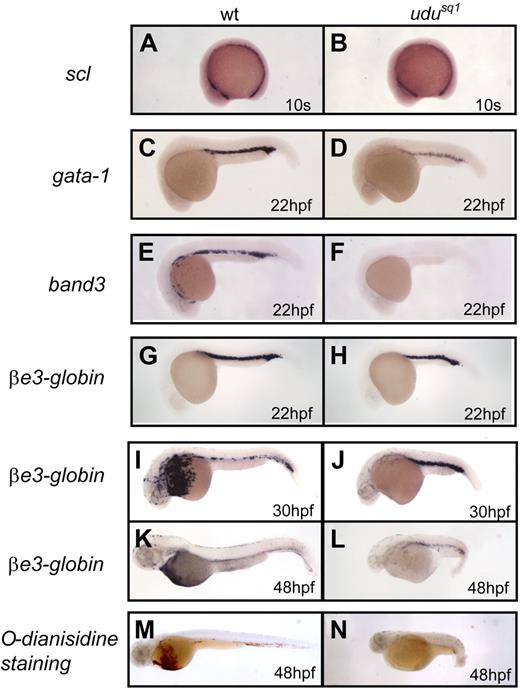
To study the genetic programs governing vertebrate hematopoiesis, we carried out a forward genetic screen in search of zebrafish mutants affecting hematopoiesis. Among the hematopoiesis-defective mutants recovered, wz260 exhibited a morphologic phenotype similar to that of zebrafish mutant ugly duckling (Udutu24) isolated from the 1996 Tuebingen screen.18 Complementation analysis confirmed that wz260 was a new allele of Udutu24; thus, this mutant was renamed Udusq1. As both udu alleles displayed similar phenotype, we used udusq1 for detailed analysis in this study. The zebrafish Udusq1−/− embryos began to show notable morphologic abnormalities at around 24 hpf, such as short body axis, bent-down tail, irregular somites, small head and eyes, and lack of blood circulation, and could not survive beyond 7 to 10 dpf (data not shown).18 In this report, we focused our study on the primitive erythroid defect of Udusq1. We first performed o-dianisidine staining to examine primitive erythropoiesis and found that hemoglobin levels were severely reduced in the 2-dpf Udusq1−/− mutant (Figure 1M-N). To explore the erythroid defects in depth, we examined the expression of 2 critical early hematopoietic markers, lmo2 and scl.23,24 WISH showed that neither lmo2 (data not shown) nor scl exhibited apparent expression differences between Udusq1−/− and wild-type embryos before the 10-somite stage (Figure 1A-B), indicating that the udu gene is dispensable for the initiation of primitive erythropoiesis. However, by 22 hpf, we found that the expression of gata1 (Figure 1C-D) was reduced and the band3 transcript, an erythrocyte-specific membrane protein critical for erythrocyte maturation,25 was almost undetectable in Udusq1−/− embryos (Figure 1E-F). Intriguingly, βe3-globin expression remained relatively normal or only slightly reduced in 22-hpf Udusq1−/− embryos (Figure 1G-H). These observations suggest that aberrance in erythrocyte differentiation is very likely to be the main reason that causes the dramatic decrease of band3 expression in Udusq1−/− mutants and eventually leads to red blood cell (RBC) hypoplasia at later stages of development as indicated by reduced βe3-globin expression from 30 hpf onwards (Figure 1I-L).
Primitive erythroid hypoplasia in Udusq1−/− mutants. (A-L) WISH of scl (A-B), gata1 (C-D), band3 (E-F), and βe3-globin (G-L) expression. (M-N) o-dianisidine staining of 2-dpf wild-type and Udusq1−/− embryos. All embryos are in lateral views with anterior to the left.
Primitive erythroid hypoplasia in Udusq1−/− mutants. (A-L) WISH of scl (A-B), gata1 (C-D), band3 (E-F), and βe3-globin (G-L) expression. (M-N) o-dianisidine staining of 2-dpf wild-type and Udusq1−/− embryos. All embryos are in lateral views with anterior to the left.
Primitive erythroid cells in Udusq1−/− mutants are impaired in proliferation and differentiation
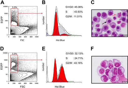
TUNEL staining of the Udusq1−/− embryos revealed that, while extensive cell death was occurred in the central nervous system (CNS), no increased apoptotic cells were observed in the ICM (Figure S1, available on the Blood website; see the Supplemental Materials link at the top of the online article). This suggests that aberrance in cell cycle rather than apoptosis contributes to the reduction of erythroid cells in Udusq1−/− mutants. To clarify this issue, we outcrossed the Udusq1+/− fish with the Tg(−5.0scl:EGFP) (referred to as Tg(+5.5 kb scl:EGFP in the original paper26 )–transgenic line,26 in which the expression of the enhanced GFP (EGFP) reporter gene is under the control of the scl promoter. The resulting Udusq1+/−/Tg(−5.0scl:EGFP) was then mated with Udusq1+/− fish. Finally, the GFP+ cells of the 24-hpf udusq1−/−/Tg(−5.0scl:EGFP) and sibling embryos were collected by the FACS (Figure 2A,D) and subjected to cell-cycle analysis. DNA content examination using Hoechst 33342 staining27 revealed that, while wild-type cells displayed 45.06%, 43.93%, and 11.01% of cells in G1/G0, S, and G2/M phases, respectively (Figure 2B), the Udusq1−/− mutant cells exhibited abnormal accumulation in G2/M phase (43.16%), and reduction in G1/G0 (32.13%) and S (24.71%) phases (Figure 2E), indicating that the Udusq1−/− mutant cells arrest at G2/M phase. Cytologic analysis showed that the wild-type GFP+ cells consisted of mainly erythroid cells in various stages of differentiation (cells with a higher degree of differentiation exhibited a round central nucleus, chromatin condensation, and were smaller in size) and some myeloid cells (characterized by their irregular nucleus; Figure 2C). In contrast, the Udusq1−/− mutant GFP+ cells, which were mainly erythroid cells, were much larger in cell size and lacked chromatin condensation (Figure 2F). Although these features resembled those erythroid progenitors found in the 16-hpf wild-type embryos (data not shown), the mutant GFP+ cells also displayed nuclear-cytoplasm asynchrony, which is a characteristic of abnormal megaloblastic erythroid cells found in human patients with megaloblastic anemia.28 Taken together, these data demonstrate that the Udusq1−/− primitive erythroid cells are defective in cell proliferation and differentiation abilities.
Primitive erythroid cells in Udusq1−/− mutants are defective in cell proliferation and differentiation. (A,D) FACS profile of the cell suspensions from 24-hpf wild-type (A) or Udusq1−/− mutant (D) embryos. The y-axis indicates the intensity of the GFP expression; x-axis represents cell size. (B,E) The GFP+ hematopoietic cells in panel A (total of 5394 cells) (B) and panel D (total of 6874 cells) (E) are subjected to DNA content analysis by Hoechst 33342 staining. The y-axis indicates the cell number, whereas the x-axis represents the DNA content. The percentages of each phase in cell cycle are given. (C,F) May-Grunwald and Giemsa staining analysis (magnification, × 1000) of sorted GFP+ cells in panels A (C) and D (F). Black and red arrows in panel C indicate erythroid and myeloid cells, respectively.
Primitive erythroid cells in Udusq1−/− mutants are defective in cell proliferation and differentiation. (A,D) FACS profile of the cell suspensions from 24-hpf wild-type (A) or Udusq1−/− mutant (D) embryos. The y-axis indicates the intensity of the GFP expression; x-axis represents cell size. (B,E) The GFP+ hematopoietic cells in panel A (total of 5394 cells) (B) and panel D (total of 6874 cells) (E) are subjected to DNA content analysis by Hoechst 33342 staining. The y-axis indicates the cell number, whereas the x-axis represents the DNA content. The percentages of each phase in cell cycle are given. (C,F) May-Grunwald and Giemsa staining analysis (magnification, × 1000) of sorted GFP+ cells in panels A (C) and D (F). Black and red arrows in panel C indicate erythroid and myeloid cells, respectively.
Identification of the Udusq1 gene
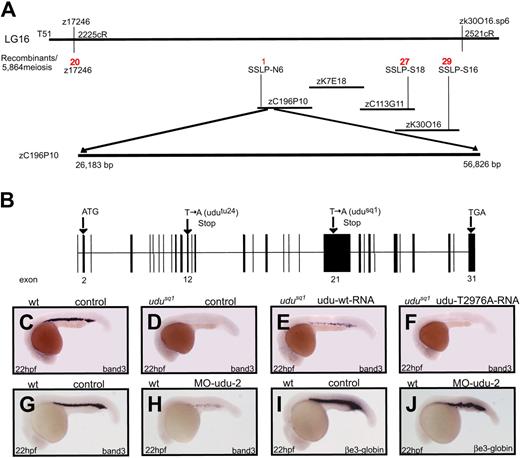
Positional cloning revealed that the predicted Ensemble gene ENSDARG00000005867 was the candidate for the Udusq1 mutant gene (Figure 3A). 5′ and 3′ RACE revealed that the full-length cDNA sequence of ENSDARG00000005867 were 6787 base pairs encoding a novel protein of 2055 aa (Figure 3B; Figure S2). Through sequencing, the mutations in Udutu24 and Udusq1 mutants were discovered at exon 12 (T1461 to A) and exon 21 (T2976 to A), respectively, resulting in the creation of a premature stop codon in each case (Figure 3B). To confirm that the Udusq1−/− mutant phenotype is indeed caused by mutation in the ENSDARG00000005867 gene, we first performed a rescue experiment with in vitro–synthesized ENSDARG00000005867 RNA. Primitive erythropoiesis was restored in 59 (92%) of the 64 Udusq1−/− embryos injected with wild-type RNA (udu-wt; Figure 3E; Table S1). In contrast, injecting the udusq1 mutant RNA (udu-T2976A) bearing a single-point nonsense mutation (nucleotide T2976 changes to A) failed to do so (Figure 3F; Table S1). As expected, embryos injected with 2 Udu MOs, MO-udu-1 (data not shown) and MO-udu-2, displayed a phenotype mimicking the Udusq1−/− mutant (Figure 3G-J; Table S2). These data demonstrate that the loss-of-function mutation in the ENSDARG00000005867 gene indeed causes the udusq1−/− mutant phenotype. We herein refer to the ENSDARG00000005867 gene as the udu gene.
Identification of the Udu gene. (A) The udu gene is mapped to LG16 within the region covered by 3 BACs, zC196P10, zK7E18, and zC113G11. The number in red represents the number of the recombinants over 5864 meiosis events for each SSLP marker. Sequence analysis confirms that the udu mutation is situated in the BAC zC196P10. (B) The udu gene consists of 31 exons (solid box) and encodes a protein of 2055 aa. Both Udutu24 and Udusq1 harbor a nonsense mutation in exons 12 and 21, respectively. (C-F) WISH of band3 expression of 22-hpf wild-type embryo (C), Udusq1−/− embryo (D), Udusq1−/− embryo injected with in vitro synthesized wild-type udu (udu-wt) RNA (E), and Udusq1−/− embryo injected with in vitro–synthesized T2976A mutant (udu-T2976A) RNA (F). (G-J) band3 (G-H) and βe3-globin (I-J) expression by WISH in the 22-hpf control morphants (G,I), and MO-udu-2 morphants (H,J). In panels C to J, embryos are in lateral views with anterior to the left.
Identification of the Udu gene. (A) The udu gene is mapped to LG16 within the region covered by 3 BACs, zC196P10, zK7E18, and zC113G11. The number in red represents the number of the recombinants over 5864 meiosis events for each SSLP marker. Sequence analysis confirms that the udu mutation is situated in the BAC zC196P10. (B) The udu gene consists of 31 exons (solid box) and encodes a protein of 2055 aa. Both Udutu24 and Udusq1 harbor a nonsense mutation in exons 12 and 21, respectively. (C-F) WISH of band3 expression of 22-hpf wild-type embryo (C), Udusq1−/− embryo (D), Udusq1−/− embryo injected with in vitro synthesized wild-type udu (udu-wt) RNA (E), and Udusq1−/− embryo injected with in vitro–synthesized T2976A mutant (udu-T2976A) RNA (F). (G-J) band3 (G-H) and βe3-globin (I-J) expression by WISH in the 22-hpf control morphants (G,I), and MO-udu-2 morphants (H,J). In panels C to J, embryos are in lateral views with anterior to the left.
Cell-autonomous erythroid defect in Udusq1−/− mutants
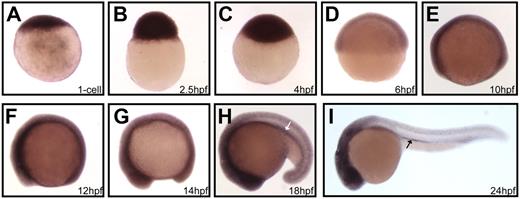
WISH showed that the Udu transcript was detected as early as the 1-cell stage in wild-type zebrafish embryos (Figure 4A). The maternal udu mRNA retained robust expression during blastula and diminished at the onset of gastrulation (approximately 6 hpf; Figure 4B-D). Around the time when segmentation began, the Udu transcript, presumably the zygotic mRNA, began to reappear in a ubiquitous manner, and was subsequently enriched in the CNS as well as the ICM from the 18-somite stage onwards (Figure 4E-H). At 24 hpf, udu mRNA was also detected in the anterior parts of kidney ducts (Figure 4I).
Temporal and spatial expression of the Udu gene during early zebrafish development. (A-I) Lateral views of WISH of udu expression in 1-cell (A), 2.5-hpf (B), 4-hpf (C), 6-hpf (D), 10-hpf (E), 12-hpf (F), 14-hpf (G), 18-hpf (H), and 24-hpf (I) embryos. White and black arrows indicate the ICM and the anterior region of kidney duct, respectively. Embryos in panels A-D are orientated with animal pole on top, whereas embryos in panels E-I are orientated with anterior to the left.
Temporal and spatial expression of the Udu gene during early zebrafish development. (A-I) Lateral views of WISH of udu expression in 1-cell (A), 2.5-hpf (B), 4-hpf (C), 6-hpf (D), 10-hpf (E), 12-hpf (F), 14-hpf (G), 18-hpf (H), and 24-hpf (I) embryos. White and black arrows indicate the ICM and the anterior region of kidney duct, respectively. Embryos in panels A-D are orientated with animal pole on top, whereas embryos in panels E-I are orientated with anterior to the left.
From the temporal and spatial expression of the udu gene, we believe that Udu possibly plays a cell-autonomous role during the primitive RBC development. To investigate this speculation, we performed a transplantation experiment. Around 15 to 30 donor cells from 3-hpf udusq1−/− or sibling embryos, preinjected with rhodamine-dextran, were transplanted into wild-type host embryos of the same stage. Contribution of the donor cells to the circulating blood cells in the host embryos was scored at around 30 hpf by counting the number of the rhodamine-dextran–labeled cells in the circulation under a fluorescent microscope. As summarized in Table 1, when sibling donor cells were transplanted, about 42% (50 of 119) of the recipients had the rhodamine-dextran–labeled donor cells in circulation, of which 20% (10 of 50) contained 10 to 30 circulating donor cells and 22% (11 of 50) had more than 30 circulating donor cells. In contrast, when the udusq1−/− mutant cells were transplanted, only 26.7% (16 of 60) of the host embryos had the donor cells contributing to the blood circulation. More important, none of these embryos contained more than 10 circulating rhodamine-dextran–labeled cells. The transplantation result strongly indicates that the udu gene acts in a cell-autonomous manner to affect primitive red blood cell development. However, non–cell-autonomous effects cannot be excluded.
Summary of cell transplantation analysis
| Donor genotype . | No. hosts . | No. (%) of hosts with donor-derived tissue . | ||||
|---|---|---|---|---|---|---|
| Muscle . | Blood . | |||||
| Total . | More than 30 blood cells . | 11-30 blood cells . | 1-10 blood cells . | |||
| Sibling | 119 | 86 (72.3) | 50 (42) | 11 (9.2) | 10 (8.4) | 29 (24.4) |
| Udusq1–/– | 60 | 46 (76.7) | 16 (26.7) | 0 (0) | 0 (0) | 16 (26.7) |
The Udu gene encodes a putative transcriptional modulator
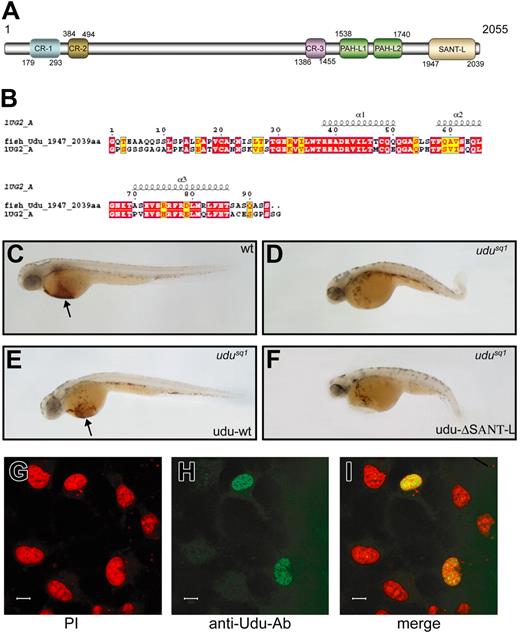
Blast searches in the National Center for Biotechnology Information (NCBI) database29 revealed that the Udu protein had the highest homology to the human and mouse GON4L in both protein sequence and gene structure (Figure S2).30 Sequence alignment reveals that there are several highly conserved regions (Figure 5A; Figure S2). The first 3 conserved regions (CR-1, CR-2, and CR-3) share no obvious similarity to any of the known domains. The fourth and fifth conserved regions (from aa 1538 to 1740), which are predicted to consist of 4 α-helixes each, are similar to the PAH repeats found in SIN3 proteins31,32 and are thus designated as PAH-L 1 and 2 domains (Figure 5A). Finally, the solution structure (IUG2 A) of the last conserved region (aa 1947 to aa 2039) has been solved in the mouse udu homolog GON4L33 (www.ebi.ac.uk/thornton-srv/databases/cgi-bin/pdbsum/GetPage.pl?pdbcode=1ug2). The structure analysis reveals that this last conserved region resembles the SANT domain found in several chromatin-remodeling molecules (Figure 5A-B),34 suggesting that the Udu protein may be involved in transcription regulation.
The Udu gene encodes a putative transcriptional modulator. (A) The Udu protein contains 6 conserved regions: CR-1, CR-2, CR-3, PAH-L1, PAH-L2, and SANT-L. (B) Protein sequence alignment of SANT-L domain between zebrafish Udu (top, fish-Udu, 1947-2039 aa) and mouse Udu homolog GON4L (1UG2 A). (C-F) Lateral views of o-dianisidine staining of 2-dpf wild-type (C), Udusq1−/− embryo (D), the Udusq1−/− embryo injected with udu-wt RNA (E), and the Udusq1−/− embryo injected with udu-ΔSANT-L RNA (F). The embryos in panels C-F are in lateral views with anterior to the left. Arrows in panels C and E indicate o-dianisidine–stained red blood cells. (G) PI staining of the pcDNA3.1/udu-wt–transfected COS7 cells. (H) Immunohistochemistry staining of the pcDNA3.1/udu-wt–transfected COS7 cells with the anti-Udu polyclonal antibody. (I) Superimposed image of panels G and H. Scale bars in panels G-I represent 10 μm.
The Udu gene encodes a putative transcriptional modulator. (A) The Udu protein contains 6 conserved regions: CR-1, CR-2, CR-3, PAH-L1, PAH-L2, and SANT-L. (B) Protein sequence alignment of SANT-L domain between zebrafish Udu (top, fish-Udu, 1947-2039 aa) and mouse Udu homolog GON4L (1UG2 A). (C-F) Lateral views of o-dianisidine staining of 2-dpf wild-type (C), Udusq1−/− embryo (D), the Udusq1−/− embryo injected with udu-wt RNA (E), and the Udusq1−/− embryo injected with udu-ΔSANT-L RNA (F). The embryos in panels C-F are in lateral views with anterior to the left. Arrows in panels C and E indicate o-dianisidine–stained red blood cells. (G) PI staining of the pcDNA3.1/udu-wt–transfected COS7 cells. (H) Immunohistochemistry staining of the pcDNA3.1/udu-wt–transfected COS7 cells with the anti-Udu polyclonal antibody. (I) Superimposed image of panels G and H. Scale bars in panels G-I represent 10 μm.
To provide additional evidence to support this argument, we carried out rescue experiments with the truncated udu–ΔSANT-L RNA, in which 3 α-helixes (from aa 1963 to 2031) of the SANT-L domain of the Udu protein were deleted. As shown in Figure 5, the RBC development was restored in the mutant embryos injected with the udu-wt RNA (Figure 5E). However, the truncated udu–ΔSANT-L RNA failed to do so (Figure 5F; Table S1), demonstrating that the SANT-L domain is critical for the function of the Udu protein. In addition, udu cDNA (pcDNA3.1/udu-wt) was transfected into COS7 cells to examine the subcellular localization of the Udu protein. Immunohistochemistry analysis showed that the Udu protein was predominantly localized in the nucleus (Figure 5G-I). These data strongly suggest that the Udu protein may function as a transcription regulator important for erythroid cell development.
Elevation of p53 activity partially contributes to the erythroid defect in Udusq1−/− mutants
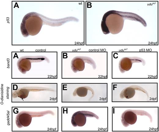
Microarray analysis was performed to identify potential target genes perturbed in udusq1−/− mutants. Results from 2 sets of independent experiments showed that expression of 87 and 57 genes were down- and up-regulated, respectively, with 2-fold and higher differences in the udusq1−/− mutant embryos (Tables S3-S4). As expected, most of the down-regulated genes are related to either hematopoiesis or neurogenesis. Notably, several up-regulated genes, including p53, fos, mdm2, gadd45αl, caspase 8, and tdag51, are known to be involved in cell-cycle control and apoptosis35,,–38 (Table S4; Figure S3). Considering the G2/M cell-cycle arrest of the udusq1−/− erythroid cells, we focused on p53 and its downstream target gadd45αl. WISH showed that in the 24-hpf Udusq1−/− embryos, the RNA levels of p53 (Figure 6A-B) and gadd45αl (Figure 6I-J) were significantly elevated in the CNS and ICM regions. To confirm that p53 transcript was indeed up-regulated in udusq1−/− mutant erythroid cells, real-time RT-PCR was performed using GFP+ cells sorted from 24-hpf wild-type or Udusq1−/− mutant embryos. As shown in Table 2, p53 transcript was significantly increased in udusq1−/− erythroid cells. These observations suggest that the up-regulation of p53 activity may contribute to the erythroid defect in udusq1−/− embryos. This argument was further supported by the findings that knock-down of the p53 protein by p53 MO22 rescued the erythroid phenotype in the Udusq1−/− embryos (Figure 6C-H; Table S1). As anticipated, elevated Gadd45αl expression was no longer detected in these p53 morphant mutants (Figure 6I-K). Taken together, these data suggest that up-regulation of p53 activity contributes, at least partially, to the erythroid cell hypoplasia in Udusq1−/− mutants.
Up-regulation of p53 activity partially contributes to the erythroid phenotype in Udusq1−/− mutants. (A-B) WISH shows that p53 expression is elevated in 24-hpf Udusq1−/− embryos (B) compared with wild-type (A). (C-E) WISH of band3 in 22-hpf wild-type embryos (C) and Udusq1−/− mutant embryos injected with control MO (D) or p53 MO (E). (F-H) o-dianisidine staining of 2-dpf wild-type embryo (F) and Udusq1−/− mutant embryos injected with control MO (G) or p53 MO (H). (I-K) WISH of gadd45αl in 24-hpf wild-type embryos (I) and Udusq1−/− mutant embryos injected with control MO (J) or p53 MO (K). All embryos are in lateral views with anterior to the left. Arrows in panels F and H indicate o-dianisidine–stained red blood cells.
Up-regulation of p53 activity partially contributes to the erythroid phenotype in Udusq1−/− mutants. (A-B) WISH shows that p53 expression is elevated in 24-hpf Udusq1−/− embryos (B) compared with wild-type (A). (C-E) WISH of band3 in 22-hpf wild-type embryos (C) and Udusq1−/− mutant embryos injected with control MO (D) or p53 MO (E). (F-H) o-dianisidine staining of 2-dpf wild-type embryo (F) and Udusq1−/− mutant embryos injected with control MO (G) or p53 MO (H). (I-K) WISH of gadd45αl in 24-hpf wild-type embryos (I) and Udusq1−/− mutant embryos injected with control MO (J) or p53 MO (K). All embryos are in lateral views with anterior to the left. Arrows in panels F and H indicate o-dianisidine–stained red blood cells.
Difference of p53 transcript levels in erythroid cells between wild-type and udusq1–/– mutants by real-time RT-PCR
| . | Crossing point . | Cycle no. difference* . | Relative fold difference . | |
|---|---|---|---|---|
| Wt sibling . | udusq1–/– . | |||
| Test 1 | 38.74 | 35.93 | 2.81 | 7.012845771 |
| Test 2 | 39.15 | 36.01 | 3.14 | 8.815240927 |
| Average | 2.975 | 7.914043349 | ||
| . | Crossing point . | Cycle no. difference* . | Relative fold difference . | |
|---|---|---|---|---|
| Wt sibling . | udusq1–/– . | |||
| Test 1 | 38.74 | 35.93 | 2.81 | 7.012845771 |
| Test 2 | 39.15 | 36.01 | 3.14 | 8.815240927 |
| Average | 2.975 | 7.914043349 | ||
*Elongation factor 1 alpha (elf1α) was used as an internal reference to normalize the reaction.
Discussion
In this article, we have shown that the udu gene, which encodes a novel nuclear protein containing 2 PAH-L repeats and a SANT-L domain, plays a critical role in regulating primitive erythroid lineage cell-cycle progression and differentiation. This was indicated by the lack of erythroid specific marker band3 expression and the accumulation of G2/M phase in erythroid cells in Udusq1−/− embryos. We noticed that, although expressed ubiquitously throughout early development, the udu gene appears to be dispensable for early embryogenesis as well as initiation of primitive hematopoiesis. Considering that robust maternal udu RNA is detected in fertilized embryos, the lack of early phenotype in Udusq1−/− mutant embryos is possibly due to the functional compensation of maternal udu RNA. The primordial germ-cell replacement approach described by Ciruna et al can be used to address this issue in the future.39
Sequence analysis reveals that the Udu protein contains several conserved regions, including 2 PAH-L repeats and a SANT-L or Myb-like DNA-binding domain. The PAH domain, originally defined in the yeast SIN3 protein,31 is distantly related to the helix-loop-helix motif and has been shown to mediate protein-protein interaction.32 The yeast SIN3 protein and its related mammalian homologs, Sin3A and Sin3B, interact through the PAH domain, with numerous sequence-specific transcription factors, and recruit histone deacetylases to suppress downstream target gene transcription.40 This implies that the Udu protein may form complexes with interaction partners via the PAH domain. The SANT and Myb-DNA–binding (Myb-DB) domains have a similar overall structure but confer distinct functions.41 The Myb-DB domain usually contains 2 to 3 tandem repeats and recognizes specific DNA sequence, whereas the SANT domain consists of 1 to 2 repeats and plays an important role in chromatin remodeling.41 Considering the fact that the Udu protein contains only 1 repeat of this domain that lacks positive electrostatic surface patch as predicted by structural modeling analysis, we believe that this conserved region resembles the SANT domain. As the Udu protein has both PAH-L and SANT-L domains, it is possible that Udu may function as a chromatin-remodeling molecule involved in transcription regulation of many target genes. Further experiments need to be performed to support our hypothesis.
p53 is well recognized as one of the most important tumor suppressors in preventing cancer by modulating downstream target gene expression in response to various cell stresses, such as DNA damage and oncogene activation, resulting in cell-cycle arrest or death of abnormal cells.35,36 Overexpression of p53 during embryogenesis causes various developmental defects in mouse, fly, and worm.42,,–45 More recently, the developmental defects caused by loss-of-function mutations in the zebrafish DNA polymerase delta1 or def gene have been shown to be associated with increased p53 activity.46,47 These observations imply that p53 plays an important role in maintaining normal cell growth and differentiation during animal development. Our findings show that MO knockdown of p53 protein expression partially rescues the primitive erythroid cell defect in Udusq1−/− embryos, which provides another example to demonstrate that p53 activity must be tightly controlled during embryogenesis in order to maintain normal cell growth and differentiation. At this moment, it is not clear how loss-of-function mutation in the udu gene leads to up-regulation of p53 activity. One possible explanation is that Udu protein may function as a general transcriptional modulator, and its loss-of-function causes a global change in gene expression. This remarkable change triggers a stress response in mutant cells, resulting in up-regulation of p53 expression. Alternatively, Udu protein may act as a suppressor that directly regulates transcription or protein activity of p53. Identifying the interaction partners or the direct target genes of Udu protein will be helpful to understand the relationship between udu and p53.
It has been recently shown that a new gene next to the human GON4L, the human counterpart of udu, is generated as a result of segmental duplication on human chromosome 1q22.30 Surprisingly, this segmental duplication does not exist in rat, mouse, chicken, or zebrafish, suggesting that this duplication may be associated with more recent evolutionary events specific for anthropoid primates.30 It has been known that human chromosome 1q22, where the GON4L gene is located, is amplified in several human cancer types, including ovarian and breast cancer,48 sarcomas,49 and hepatocellular carcinoma.50,51 A more detailed study has further confirmed that 3 loci covered by YAC955E11, YAC876B11, and YAC945D5 within 1q21-q22 have the highest amplified copy number in 5 of the 10 hepatocellular carcinoma cases examined.52 Intriguingly, the genomic fragment covered by YAC876B11 contains a marker D1S2140 that is 30 kb apart from the GON4L gene, suggesting that perhaps amplification of the GON4L gene is associated with tumor development and this warrants future study.
The online version of this article contains a data supplement.
The publication costs of this article were defrayed in part by page charge payment. Therefore, and solely to indicate this fact, this article is hereby marked “advertisement” in accordance with 18 USC section 1734.
Acknowledgments
We thank Dr Haiwei Song for protein domain analysis; Ms Meipei She and Dr Chengjin Zhang for technical advice; Drs Sudipto Roy, Vladimir Korzh, and Mei Huang for stimulating discussions; and Dr Dingxiang Liu for comments on the manuscript.
This work was supported by the Agency for Science, Technology, and Research, Singapore.
Authorship
Contribution: Y.M.L., L.S.D., and M.O. performed research and analyzed data; E.H.T., F.Q., H.J., F.H.Z., J.X., L.G., H.H.H., and J.C. performed experiments; R.G. and Y.J.J. provided critical reagents; J.R.P. analyzed data; and Y.M.L. and Z.L.W. designed the research, analyzed data, and wrote the paper.
Conflict-of-interest disclosure: The authors declare no competing financial interests.
Correspondence: Z. Wen, Laboratory of Molecular & Developmental Immunology, Institute of Molecular and Cell Biology, 61 Biopolis Dr, Proteos, Singapore 138673; e-mail: zilong@imcb.a-star.edu.sg; or J. Peng, Functional Genomics Laboratory, Institute of Molecular and Cell Biology, 61 Biopolis Dr, Proteos, Singapore 138673; e-mail: pengjr@imcb.a-star.edu.sg.

