Abstract
Receptor activator of nuclear factor κB ligand (RANKL) induces osteoclast formation from hematopoietic cells via regulation of various transcription factors. Here, we show that MafB negatively regulates RANKL-induced osteoclast differentiation. Expression levels of MafB are significantly reduced by RANKL during osteoclastogenesis. Overexpression of MafB in bone marrow-derived monocyte/macrophage lineage cells (BMMs) inhibits the formation of TRAP+ multinuclear osteoclasts, but phagocytic activity of BMMs is retained. Furthermore, overexpression of MafB in BMMs attenuates the gene induction of NFATc1 and osteoclast-associated receptor (OSCAR) during RANKL-mediated osteoclastogenesis. In addition, MafB proteins interfere with the DNA-binding ability of c-Fos, Mitf, and NFATc1, inhibiting their transactivation of NFATc1 and OSCAR. Furthermore, reduced expression of MafB by RNAi enhances osteoclastogenesis and increases expression of NFATc1 and OSCAR. Taken together, our results suggest that MafB can act as an important modulator of RANKL-mediated osteoclastogenesis.
Introduction
Bone is continuously remodeled by 2 specialized cells: osteoblasts and osteoclasts. Proper balance between bone matrix formation by osteoblasts and resorption by osteoclasts is essential for proper bone metabolism and these processes are tightly regulated by various hormones and cytokines in local microenvironments. An imbalance of these processes can lead to bone diseases such as osteoporosis.1-3
Current dogma dictates that macrophages, osteoclasts, and dendritic cells originate from bone marrow precursors. Cytokine conditions favoring differentiation into these cell types in vitro have been well defined. For example, granulocyte-macrophage colony-stimulating factor (GM-CSF) favors the outgrowth of dendritic cells from bone marrow progenitors. Macrophage colony-stimulating factor (M-CSF) regulates the growth, differentiation, and function of monocytes and macrophages, whereas RANKL (also called TRANCE, OPGL, and ODF) and M-CSF are essential cytokines for the differentiation of osteoclast precursors in bones.2,4,5
Binding of RANKL to its receptor, receptor activator of nuclear factor κB (RANK), activates transcription factors including c-Fos, Mi transcription factor (Mitf), PU.1, and nuclear factor of activated T cells (NFAT) c1, which are known to be important for osteoclastogenesis.1,3,6 Costimulatory signals mediated by immunoreceptor tyrosine-based activation motif (ITAM)–harboring adaptors, including DNAX-activating protein (DAP) 12 and Fc receptor common γ chain (FcRγ), cooperate with RANKL during osteoclastogenesis, and their activation enhances the induction of NFATc1 via calcium signaling.7-9 Osteoclast-associated receptor (OSCAR) is a member of the immunoglobulin-like surface receptor family and plays an important role as a costimulatory receptor for osteoclast differentiation by activating NFATc1 via association with the FcRγ chain.7,10-12 In addition, NFATc1 synergistically induces OSCAR gene expression with Mitf.13,14 Therefore, a positive feedback circuit involving RANKL, NFATc1, and OSCAR appears to be important for efficient differentiation of osteoclasts.13,15
Maf family proteins have been implicated in eye, hindbrain, and hematopoietic developement.16-19 Maf proteins share a conserved basic region and leucine zipper (bZIP) motif that mediate DNA binding and dimer formation. Members of the Maf family are divided into 2 subgroups: the large Maf proteins (MafA/L-Maf, MafB/Kreisler, c-Maf, and NRL), which contain an acidic transcription-activating domain (TAD) located at their N-terminus, and the small Maf proteins (MafK, MafF, and MafG), which contain only the bZIP region.17,20
MafB is expressed selectively in monocytes and macrophages, but not in other hematopoietic cells.21,22 During macrophage differentiation, MafB expression is undetectable in multipotent progenitors, but is expressed at moderate levels in myeloblasts, and strongly up-regulated in monocytes and macrophages.22 MafB contributes to the establishment and maintenance of the myelomonocytic phenotype by preventing erythroid-specific gene expression.22 Overexpression of MafB in transformed myeloblasts stimulates the rapid formation of macrophages,23 suggesting that MafB is important for macrophage differentiation.
In this study, we have attempted to elucidate the regulatory molecules at play during RANKL-mediated osteoclastogenesis by means of microarray analysis. We report that MafB negatively regulates RANKL-induced osteoclastogenesis by interfering with the DNA-binding domains of transcription factors including c-Fos, Mitf, and NFATc1, thereby inhibiting the transactivation of NFATc1 and OSCAR. Our data suggest that RANKL stimulation skews phagocytic monocytes/macrophages toward a nonphagocytic, bone-resorbing osteoclast lineage by overcoming negative regulation of the macrophage factor MafB.
Materials and methods
Reagents
SB203580, LY294002, PD98059, L-l-tosylamido-2-phenylethyl chloromethyl ketone (TPCK), and SP600125 were purchased from Calbiochem (San Diego, CA). Antibodies specific for Flag and actin were from Sigma-Aldrich (St Louis, MO); c-Fos from Calbiochem; MafB from Bethyl Laboratories (Montgomery, TX); NFATc1 from BD Biosciences (San Jose, CA); GST from Cell Signaling Technology (Beverly, MA); and hemagglutinin (HA) from Roche Applied Sciences (Indianapolis, IN). A polyclonal antibody for OSCAR was prepared as previously described.13
Constructs
MafB was prepared by reverse transcription–polymerase chain reaction (RT-PCR) using RNA from BMMs. The primer sequences are as follows: 5′-GGA TCC ACC ATG GCC GCG GAG CTG AGC ATG G-3′ (5′-MafB) and 5′-GCG GCC GCT TAC AGA AAG AAC TCA GGA GAG GAG G-3′ (3′-MafB). The amplified PCR fragments were cloned into the pMX-IRES-EGFP vector or pGEX-6p. All constructs were confirmed by DNA sequencing (Korea Basic Science Institute, Gwangju, Korea). The OSCAR reporter vector and expression vectors for Mitf, PU.1, NFATc1, and c-Fos were previously described.13,24
Osteoclast formation
Murine osteoclasts were prepared from bone marrow cells as previously described.14,25 In brief, bone marrow cells were cultured in α-MEM containing 10% FBS with M-CSF (5 ng/mL) for 16 hours. Nonadherent cells were harvested and cultured for 3 days with M-CSF (30 ng/mL). Floating cells were removed and the attached cells were used as osteoclast precursors (bone marrow-derived monocyte/macrophage lineage cells [BMMs]). To generate osteoclasts, BMMs were cultured with M-CSF (30 ng/mL) and RANKL (100 ng/mL) for 3 days. To generate osteoclasts from coculture with osteoblasts and bone marrow cells, primary osteoblasts were prepared from calvariae of newborn mice as previously described.26 Bone marrow cells and primary osteoblasts were cocultured for 6 days in the presence of 1,25(OH)2D3 (1 × 10⁻⁸ M) and PGE2 (1 × 10⁻⁶ M). Cultured cells were fixed and stained for tartrate-resistant acid phosphatase (TRAP) as previously described.14 TRAP-positive multinuclear cells (TRAP+ MNCs), containing more than 3 nuclei were counted. Cells were observed using a Leica DMIRB microscope equipped with an N Plan 10×/0.25 numerical aperture objective lens (Leica, Wetzlar, Germany). Images were obtained using a Leica IM50 camera and Leica IM 4.0 software (Leica, Cambridge, United Kingdom).
Retroviral infection
To generate retroviral stocks, retroviral vectors were transfected into the packaging cell line Plat E using FuGENE 6 (Roche Applied Sciences). Viral supernatant was collected from cultured media 24 to 48 hours after transfection. BMMs were incubated with viral supernatant for 8 hours in the presence of Polybrene (10 μg/mL). After removing the viral supernatant, BMMs were cultured with M-CSF (30 ng/mL) and RANKL (100 ng/mL) for 3 days.
Semiquantitative RT-PCR
RT-PCR was performed as previously described.27 Primers used were the following: 5′-MafB, 5′-AGT GTG GAG GAC CGC TTC TCT G-3′; 3′-MafB, 5′-CAG AAA GAA CTC AGG AGA GGA GG-3′; 5′-NFATc1, 5′-CTC GAA AGA CAG CAC TGG AGC AT -3′; 3′-NFATc1, 5′-CGG CTG CCT TCC GTC TCA TAG -3′; 5′-OSCAR, 5′-CTG CTG GTA ACG GAT CAG CTC CCC AGA -3′; 3′-OSCAR, 5′-CCA AGG AGC CAG AAC CTT CGA AAC T -3′; 5′-TRAP, 5′-CTG GAG TGC ACG ATG CCA GCG ACA -3′; 3′-TRAP, 5′-TCC GTG CTC GGC GAT GGA CCA GA -3′; 5′-HPRT, 5′-GTA ATG ATC AGT CAA CGG GGG AC -3′; 3′-HPRT, 5′-CCA GCA AGC TTG CAA CCT TAA CCA -3′; 5′-MafA, 5′-GCT TGG AGG AGC GCT TCT CCG ACG -3′; 3′-MafA, 5′-CTG CTC CAC CTG GCT CTG GAG CTG -3′; 5′-c-Maf, 5′-ATG GCT TCA GAA CTG GCA ATG AAC -3′; 3′-c-Maf, 5′-GTC TTC CAG GTG CGC CTT CTG TTC -3′; 5′-NRL, 5′-GCT GCA GAA CCA GGG TCC TGT CTC -3′; 3′-NRL, 5′-TTG CAG CCG CTT GGA GCG ACA AGC -3′.
Real-time PCR
Total RNA was extracted from cultured cells using TRIzol (Invitrogen, Carlsbad, CA). First-strand cDNA was transcribed from 1 μg RNA using Superscript RT (Invitrogen) following the protocol provided by supplier. To determine the expression levels of specific genes and HPRT (for endogenous reference), PCRs were done with the QuantiTect SYBR Green PCR kit (Qiagen, Valencia, CA) in triplicates in Rotor-Gene 3000 (Corbett Research, Mortlake, Australia). The thermal cycling conditions were as follows: 15 minutes at 95°C, followed by 40 cycles of 95°C for 30 seconds, 58°C for 30 seconds, and 72°C for 30 seconds. All quantitations were normalized to an endogenous control HPRT. The relative quantitation value for each target gene compared to the calibrator for that target is expressed as 2−(Ct−Cc) (Ct and Cc are the mean threshold cycle differences after normalizing to HPRT). The relative expression levels of samples are presented by semilog plot.
Northern hybridization and Western blot analysis
Northern blot analysis was performed as previously described.13 For immunoblotting analysis, BMMs were transduced with pMX or pMX-MafB for the indicated times in the presence of M-CSF and RANKL. Cells were washed with ice-cold PBS and lysed in extraction buffer (50 mM Tris-HCl, pH 8.0, 150 mM NaCl, 1 mM EDTA, 0.5% Nonidet P-40, and protease inhibitors). Cell lysates were subjected to sodium dodecyl sulfate-polyacrylamide gel electrophoresis (SDS-PAGE) and Western blotting. Signals were detected and analyzed by LAS3000 luminescent image analyzer (Fuji, Tokyo, Japan).
Transfection and luciferase assay
For transfection of reporter plasmids, 293T cells were plated on 6-well plates at a density of 2 × 10⁵ cells/well 1 day before transfection. Plasmid DNA was mixed with FuGENE and transfected into the cells following the manufacturer's protocol. After 48 hours of transfection, the cells were washed twice with PBS and then lysed in reporter lysis buffer (Promega, Madison, WI). Luciferase activity was measured with a luciferase assay system (Promega) according to the manufacturer's instructions. Luciferase activity was measured in triplicate, averaged, and then normalized to β-galactosidase activity using o-nitrophenyl-β-d-galactopyranoside (Sigma-Aldrich) as a substrate.
Preparation of GST fusion proteins and protein interaction assay
MafB was subcloned into the GST fusion vector pGEX-6P. GST-MafB fusion proteins were expressed in Escherichia coli BL21(DE) and purified with glutathione-Sepharose 4B beads (Amersham Biosciences, Piscataway, NJ) according to the manufacturer's instructions. Equal amounts of GST or GST-MafB proteins (2 μg) immobilized on the beads were used for protein interaction assays. Cell lysates transfected with Flag-Mitf, HA-NFATc1, or HA-c-Fos DNA were incubated with either GST or GST-MafB proteins for 3 hours at 4°C in 0.5% Nonidet P-40 lysis buffer. Samples were washed 4 times with lysis buffer, and the bound proteins were subjected to SDS-PAGE and Western blotting.
EMSA
Electrophoretic mobility shift assay (EMSA) was performed as previously described.13 Wild- and mutant-type sequences of Mitf-, NFATc1-, and AP1-binding sites were as follows: wild-type Mitf-binding site, 5′-CAG GAC TCT CAC ATG GCT TTC TG-3′ (sense) and 5′-CA GAA AGC CAT GTG AGA GTC CTG-3′ (antisense), mutant-type Mitf-binding site, 5′-CAG GAC TCT GAG ATG GCT TTC TG-3′ (sense) and 5′-CA GAA AGC CAT CTC AGA GTC CTG-3′ (antisense); wild-type NFATc1-binding site, 5′-CTT GCA ACT TTT TCC AAG ACA ATG-3′ (sense) and 5′-CAT TGT CTT GGA AAA AGT TGC AAG-3′ (antisense), mutant-type NFATc1-binding site, 5′-CTT GCA ACT TTA AGC AAG ACA ATG-3′ (sense) and 5′-CAT TGT CTT GCT TAA AGT TGC AAG-3′ (antisense); wild-type AP1-binding site, 5′-AGC CCG GCC CTG CGT CAG AGT GAG AC-3′ (sense) and 5′-GTC TCA CTC TGA CGC AGG GCC GGG CT-3′ (antisense), mutant-type AP1-binding site, 5′-AGC CCG GCC CAA CGT AGG AGT GAG AC-3′ (sense) and 5′-GTC TCA CTC CTA CGT TGG GCC GGG CT-3′ (antisense).
ChIP
A chromatin immunoprecipitation (ChIP) assay was performed with a ChIP kit (Upstate Biotechnology, Lake Placid, NY) according to the manufacturer's instructions, using antibodies against c-Fos, NFATc1, or control IgG (Santa Cruz Biotechnology, Santa Cruz, CA). The precipitated DNA was subjected to PCR amplification with specific primers for the NFATc1 promoter region containing AP1-binding sites or an OSCAR promoter region containing NFATc1-binding sites. The following primers were used for PCR: NFATc1 sense, 5′-CCG GGA CGC CCA TGC AAT CTG TTA GTA ATT-3′; NFATc1 antisense, 5′-GCG GGT GCC CTG AGA AAG CTA CTC TCC CTT-3′; OSCAR sense, 5′-GAA CAC CAG AGG CTA TGA CTG TTC-3′; OSCAR antisense, 5′-CCG TGG AGC TGA GGA AAA GGT TG-3′.
siRNA preparation and transfection
Predesigned mouse MafB siRNAs (ID nos. 156037 and 156036) were purchased from Ambion (Austin, TX). Only siRNA no. 156036 (100 nM) had a significant inhibitory effect on MafB expression in BMMs; therefore, we used this siRNA for all studies. The siRNAs were transfected into BMMs using the X-tremeGENE siRNA transfection reagent (Roche Applied Sciences).
Results
RANKL down-regulates MafB expression
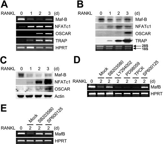
RANKL induces activation and induction of transcription factors, including NF-κB, c-Fos, Mitf, and NFATc1, and supports osteoclast differentiation from precursors.1,3,6 To identify important genes for osteoclast differentiation, we performed a microarray analysis between RAW264.7 cells and the same cells treated with RANKL for 16 hours. We found that MafB expression was lower in RAW264.7 cells treated with RANKL as compared to untreated cells. To confirm the differential expression of MafB, we examined the expression pattern of MafB during RANKL-mediated osteoclastogenesis by RT-PCR. MafB is abundantly expressed in the RAW264.7 cell line, which is derived from a monocyte/macrophage lineage and is capable of differentiating into bone-resorbing osteoclasts. One day after RANKL treatment, MafB expression was significantly reduced and remained relatively low until slightly increased levels of MafB expression were observed at the end of osteoclastogenesis. Expression levels of NFATc1, OSCAR, and TRAP, important markers of osteoclastogenesis, increased as the cells differentiated (Figure 1A). Northern blot and Western blot analysis (Figure 1B-C) obtained from BMMs during RANKL-induced osteoclastogenesis produced a similar pattern of results. In contrast to MafB, the expression levels of other large Maf members including Maf-A, c-Maf, and NRL were not changed by RANKL stimulation during osteoclastogenesis (Figure S1, available on the Blood website; see the Supplemental Figures link at the top of the online article).
RANKL down-regulates the expression of MafB during osteoclastogenesis. (A) RAW264.7 cells were cultured for the indicated times in the presence of RANKL. RT-PCR was performed for the expression of MafB, NFATc1, OSCAR, TRAP, and HPRT. (B-C) BMMs were cultured for the indicated times in the presence of M-CSF and RANKL. Northern blot analysis (B) and Western blot analysis (C) were performed to assess the expression of the indicated genes. (D) RAW264.7 cells were cultured with RANKL for 2 days in the presence of various inhibitors: mock (DMSO), SB203580 (20 μM), LY294002 (10 μM), PD98059 (20 μM), TPCK (5 μM), or SP600125 (5 μM). RT-PCR was performed for the expression of MafB and HPRT. (E) BMMs were cultured with M-CSF and RANKL for 2 days in the presence of various inhibitors: mock (DMSO), SB203580 (20 μM), or SP600125 (5 μM). RT-PCR was performed for the expression of MafB and HPRT.
RANKL down-regulates the expression of MafB during osteoclastogenesis. (A) RAW264.7 cells were cultured for the indicated times in the presence of RANKL. RT-PCR was performed for the expression of MafB, NFATc1, OSCAR, TRAP, and HPRT. (B-C) BMMs were cultured for the indicated times in the presence of M-CSF and RANKL. Northern blot analysis (B) and Western blot analysis (C) were performed to assess the expression of the indicated genes. (D) RAW264.7 cells were cultured with RANKL for 2 days in the presence of various inhibitors: mock (DMSO), SB203580 (20 μM), LY294002 (10 μM), PD98059 (20 μM), TPCK (5 μM), or SP600125 (5 μM). RT-PCR was performed for the expression of MafB and HPRT. (E) BMMs were cultured with M-CSF and RANKL for 2 days in the presence of various inhibitors: mock (DMSO), SB203580 (20 μM), or SP600125 (5 μM). RT-PCR was performed for the expression of MafB and HPRT.
Because RANKL activates NF-κB, c-Jun N-terminal kinase (JNK), p38 mitogen-activated protein kinase (MAPK), extracellular signal-related kinase (ERK), and AKT, we used various inhibitors to determine which signaling cascades are essential for RANKL-induced MafB down-regulation. Treatment of RAW264.7 cells with PI3K inhibitor (LY294002), MEK inhibitor (PD98058), or NF-κB inhibitor (TPCK), did not affect down-regulation of MafB by RANKL, whereas p38 MAPK inhibitor (SB203580) and JNK inhibitor (SP600125) strongly blocked RANKL-induced MafB down-regulation (Figure 1D). Previous studies have shown that the concentrations used in these experiments are specific for kinase inhibition.28-31 In addition, we confirmed that the viability of the cells was not affected by treatment with inhibitors (data not shown). To determine whether both pathways are involved in RANKL-mediated MafB down-regulation, we treated BMMs for 2 days with SB203580 or SP600125. Consistent with RAW264.7 cells, both inhibitors strongly blocked RANKL-induced MafB down-regulation (Figure 1E). These results suggest that RANKL down-regulates MafB expression through JNK and p38 MAPK pathways.
Overexpression of MafB inhibits osteoclast formation
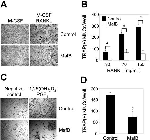
To investigate the role of MafB in RANKL-mediated osteoclastogenesis, we retrovirally overexpressed MafB in BMMs. Transduced BMMs were cultured with M-CSF alone or M-CSF and RANKL and were stained for TRAP (Figure 2A). RANKL treatment of control vector-infected BMMs increased the number of TRAP+ MNCs in a dose-dependent manner, whereas overexpression of MafB in BMMs largely inhibited the formation of TRAP+ MNCs mediated by M-CSF and RANKL (Figure 2A-B). However, an MTT assay revealed that overexpression of MafB has a marginal effect on the proliferation and survival of BMMs (Figure S2). When transduced BMMs were cocultured with primary osteoblasts in the presence of 1,25(OH)2D3 and PGE2, the number of TRAP+ MNCs was significantly reduced by overexpression of MafB as compared to control (Figure 2C-D). These data suggest that MafB plays a critical role in RANKL-mediated osteoclast differentiation.
Overexpression of MafB in BMMs inhibits osteoclastogenesis. (A-B) BMMs were transduced with pMX-IRES-EGFP (control) or MafB retrovirus and cultured for 3 days with M-CSF alone or M-CSF and various concentrations of RANKL as indicated. (A) Cultured cells were fixed and stained for TRAP. (B) Numbers of TRAP+ multinucleated cells were counted (#P < .001 versus control vector; *P < .005 versus control vector). (C-D) BMMs were transduced with pMX-IRES-EGFP (control) or MafB retrovirus and cocultured for 6 days with osteoblasts in the presence of 1,25(OH)2D3 and PGE2. (C) Cultured cells were fixed and stained for TRAP. (D) Numbers of TRAP+ multinucleated cells were counted (#P < .001 versus control vector). Results are representative of at least 3 independent sets of similar experiments (A-D). (B,D) Data represent mean ± SD of triplicate experiments.
Overexpression of MafB in BMMs inhibits osteoclastogenesis. (A-B) BMMs were transduced with pMX-IRES-EGFP (control) or MafB retrovirus and cultured for 3 days with M-CSF alone or M-CSF and various concentrations of RANKL as indicated. (A) Cultured cells were fixed and stained for TRAP. (B) Numbers of TRAP+ multinucleated cells were counted (#P < .001 versus control vector; *P < .005 versus control vector). (C-D) BMMs were transduced with pMX-IRES-EGFP (control) or MafB retrovirus and cocultured for 6 days with osteoblasts in the presence of 1,25(OH)2D3 and PGE2. (C) Cultured cells were fixed and stained for TRAP. (D) Numbers of TRAP+ multinucleated cells were counted (#P < .001 versus control vector). Results are representative of at least 3 independent sets of similar experiments (A-D). (B,D) Data represent mean ± SD of triplicate experiments.
Overexpression of MafB attenuates expression of NFATc1 and OSCAR
Given our observation that overexpression of MafB inhibits RANKL-mediated osteoclast differentiation, we examined the expression levels of NFATc1 and OSCAR, which are known to be important modulators of osteoclastogenesis.11,32 As compared to control, exogenous overexpression of MafB attenuated the expression of NFATc1 as well as OSCAR during RANKL-mediated osteoclastogenesis (Figure 3A). When we examined the effect of MafB on the protein level of these genes, similar results were obtained by Western blot analysis (Figure 3B).
Overexpression of MafB in BMMs attenuates expression of NFATc1 and OSCAR. BMMs were transduced with pMX-IRES-EGFP (control) or MafB retrovirus and cultured with M-CSF and RANKL for the indicated times. (A) Total RNA was collected from each time point. RT-PCR (top panel) and real-time PCR analysis (bottom panel) were performed to detect the indicated genes in control (▪) or MafB-overexpressed (□) samples (#P < .05 versus control vector; *P < .005 versus control vector). (B) Cells were harvested at each time point and lysates were analyzed by Western blot analysis using antibodies specific for MafB, NFATc1, OSCAR, and actin. Arrow indicates the band representing MafB.
Overexpression of MafB in BMMs attenuates expression of NFATc1 and OSCAR. BMMs were transduced with pMX-IRES-EGFP (control) or MafB retrovirus and cultured with M-CSF and RANKL for the indicated times. (A) Total RNA was collected from each time point. RT-PCR (top panel) and real-time PCR analysis (bottom panel) were performed to detect the indicated genes in control (▪) or MafB-overexpressed (□) samples (#P < .05 versus control vector; *P < .005 versus control vector). (B) Cells were harvested at each time point and lysates were analyzed by Western blot analysis using antibodies specific for MafB, NFATc1, OSCAR, and actin. Arrow indicates the band representing MafB.
MafB attenuates the binding ability of transcription factors
C-fos, an AP-1 transcription factor, induces expression of NFATc1 mediated by RANKL. Because overexpression of MafB attenuates RANKL-induced NFATc1 expression, we investigated whether MafB affects expression of c-Fos. However, the expression of c-Fos was not affected by MafB overexpression (Figure 4A). Next, we examined whether MafB can regulate the ability of c-Fos to induce NFATc1 gene expression. We used a reporter assay involving transient transfection into a highly transfectable 293 human embryonic kidney cells (293T). When a reporter plasmid containing a 6.2-kb NFATc1 promoter region was cotransfected with c-Fos, relative luciferase activity was increased (Figure 4B). However, MafB decreased the induction of luciferase activity by c-Fos in a dose-dependent manner.
MafB interacts with c-Fos and inhibits transactivation of NFATc1. (A) BMMs were transduced with pMX-IRES-EGFP (control) or MafB retrovirus and cultured with M-CSF and RANKL for the indicated times. Cells were harvested from each time point and lysates were analyzed for c-Fos and actin by Western blot analysis. (B) 293T cells were cotransfected with NFATc1 6.2-kb promoter luciferase reporter and c-Fos (80 ng) together with the indicated amounts of MafB. Each well was also cotransfected with 20 ng of a β-galactosidase expression vector to control for transfection efficiency. Luciferase activity was normalized to β-galactosidase activity as expressed by the cotransfected plasmid. Data represent the mean and the SE of triplicate samples. Results are representative of at least 3 independent sets of similar experiments. (C) 293T cells were transfected with HA-tagged c-Fos plasmid. After 36 hours of transfection, cell lysates were incubated with glutathione S-transferase (GST) or GST-MafB fusion proteins (2 μg of each) immobilized on glutathione-Sepharose beads. The beads were washed and the bound proteins were resolved by SDS-PAGE and detected by Western blotting (WB) using anti-HA (top panel) and anti-GST (middle panel) antibodies. Whole-cell extracts (WCEs) were also subjected directly to Western blot analysis with the anti-HA antibody to show that equal amounts of c-Fos were expressed (bottom panel). (D) EMSA analysis was performed with 32P-labeled probes spanning AP1-binding sites in the mouse NFATc1 promoter and c-Fos prepared using TNT rabbit reticulocyte lysate. Specific binding was determined by cold competition using unlabeled wild-type and mutant probes at 1.5-fold and 5-fold molar excess concentrations (lanes 3-6). C-fos lysate and probe were incubated with the indicated amounts of GST or GST-MafB proteins (lanes-7-10). (E) ChIP assay of c-Fos binding to NFATc1 promoter region. BMMs were treated with or without RANKL for 1 day before cross-linking. Samples were immunoprecipitated with control IgG or anti–c-Fos antibody and subjected to PCR amplification with specific primers for AP1-binding sites in the NFATc1 promoter region.
MafB interacts with c-Fos and inhibits transactivation of NFATc1. (A) BMMs were transduced with pMX-IRES-EGFP (control) or MafB retrovirus and cultured with M-CSF and RANKL for the indicated times. Cells were harvested from each time point and lysates were analyzed for c-Fos and actin by Western blot analysis. (B) 293T cells were cotransfected with NFATc1 6.2-kb promoter luciferase reporter and c-Fos (80 ng) together with the indicated amounts of MafB. Each well was also cotransfected with 20 ng of a β-galactosidase expression vector to control for transfection efficiency. Luciferase activity was normalized to β-galactosidase activity as expressed by the cotransfected plasmid. Data represent the mean and the SE of triplicate samples. Results are representative of at least 3 independent sets of similar experiments. (C) 293T cells were transfected with HA-tagged c-Fos plasmid. After 36 hours of transfection, cell lysates were incubated with glutathione S-transferase (GST) or GST-MafB fusion proteins (2 μg of each) immobilized on glutathione-Sepharose beads. The beads were washed and the bound proteins were resolved by SDS-PAGE and detected by Western blotting (WB) using anti-HA (top panel) and anti-GST (middle panel) antibodies. Whole-cell extracts (WCEs) were also subjected directly to Western blot analysis with the anti-HA antibody to show that equal amounts of c-Fos were expressed (bottom panel). (D) EMSA analysis was performed with 32P-labeled probes spanning AP1-binding sites in the mouse NFATc1 promoter and c-Fos prepared using TNT rabbit reticulocyte lysate. Specific binding was determined by cold competition using unlabeled wild-type and mutant probes at 1.5-fold and 5-fold molar excess concentrations (lanes 3-6). C-fos lysate and probe were incubated with the indicated amounts of GST or GST-MafB proteins (lanes-7-10). (E) ChIP assay of c-Fos binding to NFATc1 promoter region. BMMs were treated with or without RANKL for 1 day before cross-linking. Samples were immunoprecipitated with control IgG or anti–c-Fos antibody and subjected to PCR amplification with specific primers for AP1-binding sites in the NFATc1 promoter region.
To investigate the inhibitory mechanism of MafB on c-Fos transcription activity, we used a GST pull-down assay to determine the interaction between MafB and c-Fos. GST alone or GST-MafB fusion proteins immobilized on glutathione-Sepharose beads were incubated with cell lysates containing HA-tagged c-Fos. As shown in Figure 4C, c-Fos was retained by GST-MafB, but not by GST alone.
To determine whether MafB proteins can modulate the binding of c-Fos to AP1-binding sites in the NFATc1 promoter region, we carried out an EMSA in the presence of GST-MafB fusion proteins. We observed a shift in the c-Fos–specific band resulting from the reaction mixture containing c-Fos translated in vitro with the probe containing AP1-binding site. The specificity of this binding was confirmed by competition studies using cold wild-type and mutant competitor probes. When the purified GST-MafB fusion proteins were added to the reaction mixture, we observed a significant, dose-dependent decrease in c-Fos binding to labeled probe caused by increasing doses of GST-MafB proteins, but not by GST alone (Figure 4D). Consistent with EMSA data, ChIP assays showed that overexpression of MafB in BMMs strongly attenuated an increase of c-Fos binding to the NFATc1 promoter region mediated by RANKL treatment as compared to control (Figure 4E). Collectively, these results suggest that MafB proteins decrease c-Fos binding to AP1-binding sites in the NFATc1 promoter region by association with c-Fos.
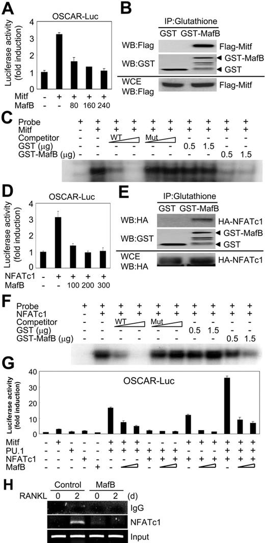
To investigate the underlying mechanism by which MafB regulates the expression of OSCAR, we performed similar experiments with Mitf and NFATc1 (Figure 5). Consistent with previous results,13,14 Mitf and NFATc1 increased OSCAR luciferase activity (Figure 5A–5D). However, overexpression of MafB abolished the induction of luciferase activity by these transcription factors. Similar to NFATc1 results (Figure 4C-D), we show that MafB interacts with Mitf and NFATc1 and inhibits the binding of these transcription factors to their respective target sites (Figure 5B-C,E-F). Consistent with EMSA data, ChIP assays showed that overexpression of MafB in BMMs markedly inhibited NFATc1 binding to the OSCAR promoter region mediated by RANKL treatment as compared to control (Figure 5H).
MafB associates with Mitf and NFATc1 and inhibits their transactivation. (A,D) 293T cells were cotransfected with OSCAR 1.7-kb promoter luciferase reporter and 80 ng of Mitf (A) or 100 ng of NFATc1 (D) together with the indicated amounts of MafB. The data were normalized and presented as described in Figure 4. Results are representative of at least 3 independent sets of similar experiments. (B,E) 293T cells were transfected with Flag-tagged Mitf (B) or HA-tagged NFATc1 (E) plasmid. Western blot analysis using anti-Flag (B) or anti-HA (E) antibodies was performed as described in Figure 4. (C,F) EMSA analysis was performed with 32P-labeled probes spanning E-box (C) or NFATc1-binding sites (F) in the mouse promoter regions for OSCAR and Mitf (C) or NFATc1 (F) prepared using TNT rabbit reticulocyte lysate as described in Figure 4. Specific binding was determined by cold competition using unlabeled wild-type and mutant probes at 5-fold and 50-fold molar excess concentrations (C,F lanes 3-6). (G) 293T cells were cotransfected with an OSCAR reporter plasmid together with the indicated plasmids expressing Mitf, PU.1, NFATc1, or MafB (150-300 ng). The data were normalized and presented as described in Figure 4. Results are representative of at least 3 independent sets of similar experiments. (H) ChIP assay of NFATc1 binding to the OSCAR promoter region. BMMs were treated with or without RANKL for 2 days before cross-linking. Samples were immunoprecipitated with control IgG or anti-NFATc1 antibodies and subjected to PCR amplification with primers specific for the NFATc1 binding sites of the OSCAR promoter region. Data represent mean ± SD of triplicate experiments.
MafB associates with Mitf and NFATc1 and inhibits their transactivation. (A,D) 293T cells were cotransfected with OSCAR 1.7-kb promoter luciferase reporter and 80 ng of Mitf (A) or 100 ng of NFATc1 (D) together with the indicated amounts of MafB. The data were normalized and presented as described in Figure 4. Results are representative of at least 3 independent sets of similar experiments. (B,E) 293T cells were transfected with Flag-tagged Mitf (B) or HA-tagged NFATc1 (E) plasmid. Western blot analysis using anti-Flag (B) or anti-HA (E) antibodies was performed as described in Figure 4. (C,F) EMSA analysis was performed with 32P-labeled probes spanning E-box (C) or NFATc1-binding sites (F) in the mouse promoter regions for OSCAR and Mitf (C) or NFATc1 (F) prepared using TNT rabbit reticulocyte lysate as described in Figure 4. Specific binding was determined by cold competition using unlabeled wild-type and mutant probes at 5-fold and 50-fold molar excess concentrations (C,F lanes 3-6). (G) 293T cells were cotransfected with an OSCAR reporter plasmid together with the indicated plasmids expressing Mitf, PU.1, NFATc1, or MafB (150-300 ng). The data were normalized and presented as described in Figure 4. Results are representative of at least 3 independent sets of similar experiments. (H) ChIP assay of NFATc1 binding to the OSCAR promoter region. BMMs were treated with or without RANKL for 2 days before cross-linking. Samples were immunoprecipitated with control IgG or anti-NFATc1 antibodies and subjected to PCR amplification with primers specific for the NFATc1 binding sites of the OSCAR promoter region. Data represent mean ± SD of triplicate experiments.
We have previously shown that transcription factors such as Mitf, PU.1, and NFATc1 cooperate and synergistically induce OSCAR expression.13,14 Therefore, we examined whether MafB affects the synergistic activation of OSCAR by the aforementioned transcription factors. As shown in Figure 5G, MafB abrogated the synergistic activation of OSCAR by Mitf, PU.1, and NFATc1. Taken together, these results suggest that MafB inhibits RANKL-mediated expression of NFATc1 and OSCAR by associating with the key transcription factors c-Fos, Mitf, and NFATc1, thereby precluding binding of these factors to the promoter regions of NFATc1 and OSCAR.
Down-regulation of MafB enhances osteoclastogenesis and expression of NFATc1 and OSCAR
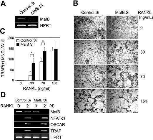
Because MafB acts as a negative regulator of osteoclastogenesis, we investigated its physiologic role in osteoclastogenesis by means of siRNAs. A net decrease in RNA expression was observed in BMMs transfected with MafB-specific siRNAs compared to control (Figure 6A). Down-regulation of MafB significantly enhanced RANKL-mediated osteoclastogenesis at a relatively low concentration range of RANKL (30-70 ng/mL; Figure 6B-C). In addition, the silencing of MafB in BMMs resulted in a significant increase in the induction of NFATc1 and OSCAR in response to RANKL stimulation (Figure 6D). These results suggest that MafB plays an important role in RANKL-induced osteoclastogenesis.
Enhancement of RANKL responses by MafB siRNA in BMMs. (A) Control or MafB siRNAs were transfected into BMMs. Total RNA was harvested from cultured cells 48 hours after transfection, and RT-PCR was performed with the primers specific for MafB and HPRT (control). (B) BMMs transfected with control or MafB siRNA were cultured for 3 days in the presence of M-CSF and various concentrations of RANKL. Cultured cells were fixed and stained for TRAP. (C) TRAP+ MNCs having more than 3 nuclei were counted as osteoclasts (*P < .01 versus control Si). Data represent mean ± SD of triplicate experiments. (D) BMMs transfected with control or MafB siRNA were cultured with M-CSF and RANKL (50 ng/mL) for the indicated times. Total RNA was harvested from cultured cells and RT-PCR was performed with primers specific for MafB, NFATc1, OSCAR, TRAP, and HPRT (control).
Enhancement of RANKL responses by MafB siRNA in BMMs. (A) Control or MafB siRNAs were transfected into BMMs. Total RNA was harvested from cultured cells 48 hours after transfection, and RT-PCR was performed with the primers specific for MafB and HPRT (control). (B) BMMs transfected with control or MafB siRNA were cultured for 3 days in the presence of M-CSF and various concentrations of RANKL. Cultured cells were fixed and stained for TRAP. (C) TRAP+ MNCs having more than 3 nuclei were counted as osteoclasts (*P < .01 versus control Si). Data represent mean ± SD of triplicate experiments. (D) BMMs transfected with control or MafB siRNA were cultured with M-CSF and RANKL (50 ng/mL) for the indicated times. Total RNA was harvested from cultured cells and RT-PCR was performed with primers specific for MafB, NFATc1, OSCAR, TRAP, and HPRT (control).
Discussion
RANKL induces osteoclast formation from monocyte/macrophage precursors. It has been shown that positive regulators, such as NF-κB, c-Fos, Mitf, and NFATc1, are important for RANKL-mediated osteoclastogenesis.1,3,6 Here, we show a novel role for MafB as a negative regulator of osteoclast differentiation. We found that RANKL down-regulates MafB expression via JNK and p38 MAPK, which are important signaling cascades for osteoclast differentiation. Furthermore, MafB negatively regulates RANKL-induced osteoclastogenesis by down-regulation of NFATc1 and OSCAR, which are important modulators for osteoclast differentiation. In addition, we found that MafB associates with the transcription factors c-Fos, Mitf, and NFATc1 and inhibits their ability to transactivate NFATc1 and OSCAR by blocking binding of these transcription factors to the NFATc1 and OSCAR promoter regions. Our data suggest that MafB plays an important role in regulating RANKL-induced osteoclastogenesis.
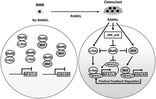
Here, we propose a possible schematic model for the role of MafB in RANKL-mediated osteoclast differentiation based on past and present results (Figure 7). MafB, which is abundantly expressed in osteoclast precursors (BMMs), interacts with the transcription factors c-Fos and Mitf, which in turn prevents binding of these transcription factors to the NFATc1 and OSCAR promoter regions, thereby abrogating expression of these important modulators of osteoclastogenesis. During osteoclastogenesis, RANKL strongly stimulates c-Fos gene induction and also activates Mitf.33 On the other hand, RANKL reduces MafB expression via JNK and p38 MAPK pathways. Reduction of MafB by RANKL accelerates the binding of c-Fos to the NFATc1 promoter region, thereby inducing NFATc1 gene expression. Later, expression of NFATc1 is strongly up-regulated by autoamplification.34 Mitf and NFATc1 synergistically activate OSCAR.13 OSCAR signaling enhances RANKL-mediated induction of NFATc1 via calcium activation.7,11 Therefore, a positive feedback circuit involving RANKL, NFATc1, and OSCAR appears to be important for efficient differentiation of osteoclasts. Thus, MafB acts as a key modulator of RANKL-induced osteoclastogenesis by means of its ability to attenuate the expression of NFATc1 and OSCAR.
A schematic model of role of MafB in osteoclastogenesis. MafB, which is abundantly expressed in BMMs, associates with c-Fos and Mitf, thereby suppressing NFATc1 and OSCAR expression. RANKL stimulation down-regulates MafB expression via JNK and p38 MAPK and also up-regulates c-Fos expression; therefore, NFATc1 expression is induced by c-Fos and NFATc1 autoamplification. Lower levels of MafB enable Mitf and NFATc1 to bind to the OSCAR promoter region, subsequently leading to the up-regulation of OSCAR during osteoclastogenesis. A positive feedback loop between NFATc1 and OSCAR appears to be important for efficient differentiation of osteoclasts.
A schematic model of role of MafB in osteoclastogenesis. MafB, which is abundantly expressed in BMMs, associates with c-Fos and Mitf, thereby suppressing NFATc1 and OSCAR expression. RANKL stimulation down-regulates MafB expression via JNK and p38 MAPK and also up-regulates c-Fos expression; therefore, NFATc1 expression is induced by c-Fos and NFATc1 autoamplification. Lower levels of MafB enable Mitf and NFATc1 to bind to the OSCAR promoter region, subsequently leading to the up-regulation of OSCAR during osteoclastogenesis. A positive feedback loop between NFATc1 and OSCAR appears to be important for efficient differentiation of osteoclasts.
Bakri et al reported that dendritic cells have high levels of PU.1 expression but undetectable MafB expression, whereas monocyte-derived macrophages express MafB and only relatively moderate levels of PU.1.35 PU.1 binds directly to MafB, and inhibits its transcriptional activity in macrophages. Therefore, the authors proposed that tipping the scales of MafB and PU.1 expression specifies alternative macrophage or dendritic-cell fates.35 During osteoclast differentiation from monocyte/macrophage precursor cells, the expression level of PU.1 is not changed by RANKL stimulation.14 These results suggest that although down-regulation of MafB favors osteoclasts or dendritic-cell fate from progenitors, the down-regulation mechanisms during both processes are somewhat different from those ascribed to PU.1 by Bakri et al. RANKL reduces MafB expression without affecting PU.1 expression during osteoclastogenesis, whereas increased levels of PU.1 expression might be involved in MafB down-regulation in dendritic cells.
MafB binds to Maf recognition elements (MAREs) and induces target genes such as F4/80.36 Recently, Moriguchi et al showed that MafB is essential for F4/80 expression in macrophages.37 MafB-induced macrophages have normal phagocytic activity and nitric oxide release after activation by lipopolysaccharide.23 In addition, a dominant-negative form of MafB inhibits macrophage differentiation.23 These data suggested that MafB is important for maintaining macrophage phenotypes such as phagocytic ability. We have previously shown that the phagocytic activity of BMMs is abolished by RANKL stimulation.25 However, overexpression of MafB in BMMs did not affect phagocytic activity even in the presence of RANKL (Figure S3). Herein, we present evidence that MafB has dual roles as a repressor of osteoclastogenesis as well as an inducer of macrophage differentiation, suggesting that expression levels of MafB are important for determining cell fate between macrophages and osteoclasts from common precursors, and that RANKL skews phagocytic BMMs toward bone-resorbing osteoclasts via down-regulation of the macrophage factor MafB.
Similar to our data, other studies have shown that MafB binds to the DNA-binding domain of Ets-1 and represses Ets-1 transactivation of the transferrin receptor, which is essential for erythroid differentiation.22 These data suggest that MafB might be involved in the development of multiple hematopoietic lineages including erythrocytes, macrophages, dendritic cells, and osteoclasts.
Taken together, our results demonstrate that RANKL induces osteoclast formation by activating positive regulators as well as repressing negative regulators such as MafB. Recently, we have shown that RANKL down-regulates Id genes, which act as repressors of osteoclastogenesis.24 Hence, our work reveals an additional layer of negative regulation, as well as a mechanism by which RANKL signaling overcomes it, allowing osteoclastogenesis to proceed. Therefore, further elucidation of the detailed mechanism of MafB gene regulation should provide additional therapeutic approaches to various bone diseases.
Authorship
Contribution: K.K., J.H.K., J.L., and H.M.J. performed research and analyzed data; H.K. contributed vital reagents; K.K.K. contributed vital analytic tools; S.Y.L. contributed vital reagents and analytic tools; and N.K. designed research and wrote the paper.
Conflict-of-interest disclosure: The authors declare no competing financial interests.
Correspondence: Nacksung Kim, Medical Research Center for Gene Regulation, Chonnam National University Medical School, Hak-Dong 5, Dong-Ku, Gwangju 501-746, Korea; e-mail: nacksung@jnu.ac.kr.
The online version of this article contains a data supplement.
The publication costs of this article were defrayed in part by page charge payment. Therefore, and solely to indicate this fact, this article is hereby marked “advertisement” in accordance with 18 USC section 1734.
This work was supported in part by grant R13-2002-013-03001-0 from the Korea Science and Engineering Foundation through the Medical Research Center for Gene Regulation at Chonnam National University and the Korea Health 21 R&D Project (A060164) from Ministry of Health and Welfare.
We thank T. Kitamura for Plat-E cells and J. Y. Kwon and S. I. Park for assistance.



This feature is available to Subscribers Only
Sign In or Create an Account Close Modal